
We use cookies to ensure our website works properly and to personalise your experience. Cookies policy
Rajarambapu College of Pharmacy, Kasegaon, Walwa, Sangli, Maharashtra, India 415404
This review critically assesses the role of microorganisms and plants in synthesizing nanoparticles, highlighting the potential of chemical approaches in nanotechnology. Green synthesis of nanoparticles is a promising area in nanoscience and technology, focusing on developing clean, biocompatible, non-toxic, and eco-friendly methods. Various types of nanoparticles with specific chemical compositions, sizes, and morphologies have been synthesized using various microorganisms. These nanoparticles have applications in various technological areas, including diagnosis, treatment, drug delivery, medical device coating, wound dressings, medical textiles, contraceptive devices, anti-fungal, and anti-inflammatory applications. Future prospects for synthesis using bacteria are also discussed. This paper discusses the recent developments in biosynthesis of inorganic nanoparticles, including metallic, oxide, and sulfide nanoparticles, using microorganisms. Nanotechnology advancements focus on synthesis of nanoparticles using eco-friendly, reliable techniques. Numerous microorganisms, such as viruses, microalgae, fungus, yeast, bacteria,and have been explored for metal, oxide of metal, and additional nanomaterials. Genetic engineering can enhance their capacity for nanoparticle synthesis making them quicker, safer, and more environmentally friendly.
Richard Feynman introduced the main idea of nanotechnology in 1959. Professor Norio Taniguchi of Tokyo Science University first used the term nanotechnology. The Greek word "nano" means "extremely small," and it is synonymous with dwarf. Typically, nanoparticles range in size from 0.1 to 1000 nm in every spatial dimension and are created through two main methods: top-down and bottom-up. In the first, materials are molded and etched into smaller components, and in the second, structures are put together to create functional devices. Due to the numerous uses of nanomaterials in the biomedical, agricultural, catalysis, optical, and electronic domains, nanotechnology is currently a frontier of research. Because of their unique biological, chemical, and physical characteristics relative to the corresponding bulk materials, inorganic nanoparticles (NP) have garnered a lot of attention recently. The physical, chemical, biological, and other hybrid methods are used to create the nanoparticles. The physical and chemical methods are very useful for production of mono dispersed nanoparticles. Because the chemicals used in these procedures are toxic, flammable, and difficult to remove from the environment, they are harmful in one way or another. These techniques cause harmful substances to become adsorbed on the exterior of nanoparticles, which could be harmful in medical applications. Thus, it is worthwhile to develop safe, biocompatible, non-toxic, and environmentally friendly processes in order to create nanoparticles. Biological methods are considered to be safe, economical, environmentally friendly, and sustainable processes. Prokaryotes and eukaryotes are the principal biological components used in green approaches in order to create nanoparticles. Numerous biological processes involve the direct or indirect participation of microbes [1]. The process of creating NPs can include done in two main ways. First, NPs are made chemically, which includes physiochemical (c-radiation), photochemical (using light's photons), chemical reduction, electrochemical, micro emulsion, microwave radiation, and laser ablation. All of these techniques produce a large number of NPs with limitations because of their expensive, labor-intensive equipment, and toxic ingredients. The method of chemical reduction is one of these techniques that is frequently used to create NPs. In an aqueous medium, a variety of substances, including tollens, polyethylene glycol, sodium borohydride, sodium citrate, and elemental hydrogen, function as reducing agents. Clusters of NPs, or nanoparticles are created when these reducing compounds convert metal ions (M+) into their metallic form (M0) Second, green, or biosynthesis, is a productive process that substitutes affordable, hazardous chemicals with natural compounds as capping, stabilizing, and reducing agents. Because of this, researchers Adapt techniques for biosynthesis. This preference has multiple justifications. First off, NPs produced biologically have different physicochemical characteristics from NPs produced chemically. Second, compared to the chemical method, which uses pricey chemicals, this method is relatively less expensive and more human. These substances are hazardous to people, pets, and the environment in particular. As a result, the majority of experts refer to the bottom-up strategy of green synthesis, which substituted plant extract for chemicals [2]. The morphology, size, and shape of the nanoparticles are used to categorize them into various types. This article mentions a few significant classes of nanoparticles.
Classification of Nanoparticles:
1] Organic nanoparticles:
Ferritin, micelles, dendrimers, and liposome’s are among the organic nanoparticles depicted in Figure. The organic nanoparticles are biodegradable, non-toxic, and some Micelles and liposome's are two examples of organic nanoparticles with hollow spheres.
2] Inorganic nanoparticles:
Inorganic nanoparticles do not contain carbon. It is safe to handle the inorganic nanoparticles. The inorganic nanoparticles exhibit hydrophilicity and biocompatibility.
a] Metal nanoparticles:
Metallica nanoparticles are synthesized from metals through either constructive or destructive methods. The pure metal is created using the metal precursors, tiny particles.
b] Metal oxide nanoparticles:
The goal of creating metal oxide nanoparticles is to alter the characteristics of the corresponding metals. For example, iron nanoparticles are oxidized to oxide nanoparticles of iron. When compared to iron nanoparticles, iron oxide nanoparticles exhibit higher reactivity [3].
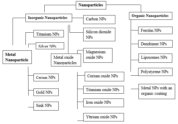
Fig 1. Classification of Nanoparticles [3]
Sources of nanoparticles:
Nanomaterials can generally be grouped into three primary categories based on their origin: (i) incidental nanomaterials, which are unintentionally generated as byproducts of various industrial activities—such as particles released from vehicle exhaust, welding operations, combustion, or even natural events like forest fires; (ii) engineered nanomaterials, which are intentionally created by humans to exhibit specific properties for particular applications; and (iii) naturally occurring nanomaterials, which are present in nature and can be found within living organisms, including plants, animals, insects, and humans. However, the boundaries between these categories natural, incidental, and engineered are not always clearly defined. In fact, incidental nanomaterials are sometimes regarded as a subset of naturally occurring ones [4].
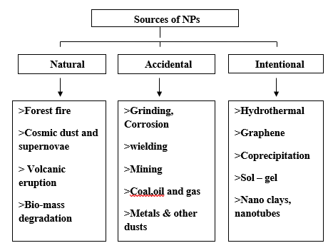
Fig 2. Sources of Nanoparticles [4]
Bacteria used in Nanotechnology:
1] Pseudomonas:
The silver mine Pseudomonas stutzeri AG259 is the bacterium produced silver-based single nanocrystals in the space between cells of the bacterium. Moreover, P. stutzeri exhibited the capacity to aerobically reduce selenite into insoluble elemental selenium. As of late, cell Gold ions were reduced using P. aeruginosa supernatant, leading to the extracellular biosynthesis of gold nanoparticles.
2] Magnetotactic bacteria:
Particles known as magnetic (Fe3O4) and greigite (Fe3S4) nanoparticles are produced by magneto tactic bacteria, such as Magneto spirillum magneticum.
3] sulphate- reducing bacterium:
The sulphate-reducing bacteria NCIMB 8307 Desulfovibrio desulfuricans has been demonstrated to synthesize palladium nanoparticles on the exterior of cells in the presence of an exogenous electron donor.
4] Lactobacillus:
Frequent strains of Lactobacillus identified in Within the cell, buttermilk promoted the growth of microscopic crystals of gold, silver, and gold-silver alloys with distinct morphologies without compromising their viability.
5] Bacillus:
Au3+ ions can be reduced by Bacillus subtilis 168 to create nanoscale octahedral gold particles (5–25 nm) Ag+ ions have also recently been found to be reduced to Ag0 by an airborne Bacillus species.
6] Fe [III] reducing bacteria:
Gold was precipitated intracellularly in the area around the periplasm of the Fe (III) reducing bacterium Geobacter ferrireducens.
7] Staphylococcus:
Within a few minutes of subjecting the S. aureus to AgNO3, the reaction began, and the solution's color changed to a yellowish-brown hue, signifying the extracellular formation of AgNPs.
8] Entrobacteriacea:
Silver nanoparticles were produced quickly by the supernatants of culture Enterobacteriaceae, which includes Enterobacter cloacae, Escherichia coli, and Klebsiella pneumonia, by reducing Ag+ to Ag0.There have been reports of E. coli DH5-mediated bioreduction of chloroauric acid to nanoparticles [5].
Applications of bacteria used in nanotechnology:
1) Applications of Magnetotactic bacteria:
Magnetotactic Bacteria (MTB) in Drug Delivery:
In a recent review, Martel (2017) explores the potential of using drug-loaded magnetotactic bacteria (MTB) as "smart therapeutic agents" for precise drug delivery to specific organs or tissues. A key advantage of employing MTB or their magnetosomes is the ability to guide them to target sites using an external magnetic field, thereby minimizing effects on healthy, non-target tissues. For instance, ‘Magnetococcus marinus’ strain MC-1 was used to transport drug-loaded nanoliposomes into oxygen-poor regions of colorectal tumors in mice. The results showed enhanced therapeutic efficiency when nanocarriers were combined with MTB. Remarkably, after being injected near the tumor, the ‘Mc. Marinus’ cells remained alive and motile, demonstrating characteristics that are both aerotactic and magnetotactic. This allowed them to penetrate deeper into the tumor compared to passive delivery agents like microspheres or dead ‘Mc. Marinus’ cells. Although ‘Mc. Marinus’ is a marine bacterium not naturally suited to survive inside mammals, it unexpectedly remained active in the host environment. Even more surprising were the findings by Felfoul and colleagues, who reported that Mc. marinus cells were clinically “safe” and did not trigger adverse effects in mice. This outcome was unanticipated given the usual immune responses elicited by Gram-negative bacteria due to their lipopolysaccharide (LPS)-rich outer membranes, which are known endotoxins [6].
Magnetotactic Bacteria (MTB) in Bioremediation:
Innovative strategies utilizing live magnetotactic bacteria (MTB) and their unique metabolic functions have been explored for advancing bioremediation technologies. For instance, Shimoshige et al. (2017) identified a novel strain of Magnetospirillum magneticum (RSS-1) that produces magnetite magnetosomes coated with a thin layer of samarium oxide [7]. The co-precipitation of additional minerals within the magnetosome vesicle indicates that magnetotactic bacteria (MTB) could be utilized for magnetic recovery of transition metals and for creating structures that combine magnetic particles with these metals. In a separate study, Zhou et al. employed genetic engineering techniques to enhance the ability of Magnetospirillum gryphiswaldense to accumulate phosphate, improving its effectiveness in wastewater treatment [8].
2)Applications of Magnetosomes:
Magnetosomes in Cell Separation:
Magnetic cell separation has also been accomplished using magnetosomes in combination with specific antibodies. For instance, Yoshino (2008) engineered modified magnetosomes that displayed the antibody-binding protein A on their surface to facilitate cell separation [9].
Magnetosomes in Antigen recovery/detection assays and DNA:
Magnetosomes have been effectively utilized in tests for protein detection. By attaching biotin groups to their membranes, the magnetosomes were able to bind specifically to the protein streptavidin. These engineered composite particles, with their targeted biotin-binding ability, can serve as versatile platforms for attaching various Biotinylated antibodies and DNA oligonucleotides are examples of functional biomolecules. [10].
Magnetosomes in Immobilization of Enzyme:
Magnetosomes' protein display system can be utilized to express catalytic components, making them well-suited for supporting enzymes that are immobilized, as previously stated. Ginet and colleagues demonstrated this by expressing an organ phosphohydrolase enzyme from Flavobacterium species, fused to the magnetosome-associated protein MamC, for the purpose of breaking down paraoxon a widely used but toxic pesticide. The resulting protein-magnetosome complex exhibited paraoxon degradation activity similar to that of the purified enzyme. Furthermore, the study showed that the enzyme-magnetosome complex maintained 90% to 100% of its catalytic activity even after three reuse cycles, indicating high stability and reusability [11].
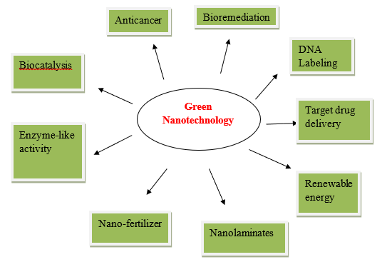
Fig 3. Applications of nanotechnology [12]
Methods of preparation of Nanoparticles:
Since the beginning of life on Earth, biological entities and inorganic materials have been in constant contact with each other. Because of these frequent interactions, life could continue to on this planet where the mineral deposits are orderedly. The relationship between inorganic molecules and biological species has drawn increasing attention from scientists recently. Numerous microbes have demonstrated themselves to be capable of producing inorganic nanoparticles through extracellular or intracellular pathways. This section covers the biological processes used to produce different types of nanoparticles. The categories covered are metallic nanoparticles, which include gold, silver, alloy, and other metal nanoparticles; oxide nanoparticles, which include sulfur dioxide, magnetic and nonmagnetic oxide nanoparticles; and other miscellaneous nanoparticles [13].
Common Methodologies for Metal Nanoparticle Synthesis using Microbes:
1] Extracellular Mechanism:
The test strain (culture) is raised in appropriate media and kept at 37°C in an orbital shaker set to 150 rpm. The broth is centrifuged after incubation, and the supernatant is utilized in the creation of nanoparticles. After being added to different reaction vessels that already contain the metal ions in the appropriate concentrations, the supernatant is incubated for a full 72 hours. The reaction mixture's color shift indicates the presence of nanoparticles, and a UV-visible spectrophotometer is used to sample the aqueous solution and measure the absorption spectrum to track the bioreduction of silver ions in the solution. X-ray diffraction (XRD) and scanning electron microscopy (SEM) are used to examine the shape and homogeneity of silver nanoparticles [14].
2] Intracellular Mechanism:
The culture is raised in suitable liquid media and kept at the ideal temperature in an incubator. The flask is left in a static position following incubation to allow the biomass to settle. After that, the supernatant is disposed of and sterile distilled water is added to wash the cells. After settling the biomass for thirty minutes, the flask is again sealed and the supernatant is thrown away. There are three iterations of this process. Centrifugation is subsequently employed for ten minutes to separate the biomass from the sterile distilled water. The moist biomass is placed in a shaker at an appropriate temperature and exposed to 50 milliliters of sterile metal aqueous solution at different dilutions until a visible color shift is observed noted. The development of silver nanoparticles is indicated by a change in color from light yellow to brownish, the formation of gold nanoparticles by light yellow to pinkish, and the formation of manganese and zinc nanoparticles by Pale yellow to yellow [15]
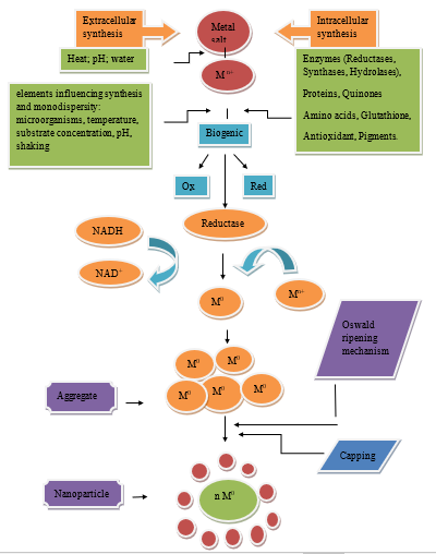
Fig 4. General scheme of synthesis of nanoparticles of metals by microorganisms [12]
Mechanism Used by Bacteria:
Since the beginning of life on Earth, Both inorganic materials and living beings have been in continual contact with one another. The phenomena that occurs through an enzymatic or biological response is known as biosynthesis. Inorganic compounds are produced by microbes through intra- or extracellular pathways, frequently at nanoscale dimensions with fine morphology [16]. Microorganisms are able to thrive in environments with elevated levels of harmful metals because of two mechanisms: chemical detoxification and energy-dependent membrane protein efflux from the cell, which can take the form of ATPase, chemo-osmotic or proton anti-transporters [17-19]. Since various biological agents utilize different mechanisms with different metals and because different biomolecules have varying responses to nanoparticle synthesis, the precise method for the production of utilizing biological agents has not yet been discovered. The efflux system, changes in solubility and toxicity through reduction or oxidation, bio-absorption, bioaccumulation, extracellular complexion or precipitation of metals, and the absence of a specific metal transportation system [20].
Evaluation of nanoparticles:
Evaluation of type of nanoparticles is done by following methods:
1] UV-Visible spectroscopy (UV-VIS):
Absorption spectroscopy is a powerful, non-destructive technique used to study the optical properties of semiconductor nanoparticles. The UV–Vis absorption spectrum varies with wavelength, and the nanoparticle size depends on the maximum absorption wavelength (λmax). The energy gap (Eg) represents the difference between the valence and conduction bands. In ZnO nanoparticles, the absorption peak related to surface plasmon resonance (SPR) is influenced by particle size a higher λmax indicates larger nanoparticles and lower energy, resulting in a red shift [21]. Moreover, UV-Vis spectroscopy can be easily integrated with other analytical techniques to assess a wide variety of parameters, thereby enhancing the overall quality of analysis. Owing to these advantages, UV-Vis spectroscopy is extensively applied in various fields — for instance, in the biopharmaceutical industry to analyze high-concentration protein solutions, in environmental monitoring to identify and compare contaminants and their by-products in real time, and in industrial wastewater treatment plants to evaluate color and compliance with regulatory standards. As technological advancements continue to refine spectrophotometry, its range of applications and measurable parameters is expected to expand even further. For example, in field-based studies, online UV-Vis spectrometry serves as an efficient tool for real-time monitoring of multiple parameters across different liquid samples, a capability that sets it apart from many other online sensor systems [22].
2] Transmission Electron Microscopy Analysis (TEM):
The TEM analysis of the gold nanoparticles used in this study was previously described. The 10 nm sample primarily contained individual nanoparticles along with a few clusters comprising up to 60 particles. These clusters were loosely packed, and the individual nanoparticles were clearly distinguishable, suggesting that they formed agglomerates held together by weak interactions. In contrast, samples containing nanoparticles of 50, 100, and 250 nm displayed mostly separate particles with smaller clusters consisting of 2–8 nanoparticles. Furthermore, Energy Dispersive X-ray (EDX) analysis confirmed the presence of 10 nm gold nanoparticles in the examined solutions [23].
3] Scanning Electron Microscopy (SEM):
Scanning Electron Microscopy (SEM) is a highly versatile and advanced tool widely used to examine the surface characteristics of various materials. In SEM, a sample is bombarded with high-energy electrons, and the emitted electrons or X-rays are then analyzed. This emitted signal provides valuable insights into a material’s surface topography, morphology, elemental composition, grain orientation, and crystallographic structure. Morphology refers to the shape and size of the particles, while topography describes the surface features such as texture, smoothness, or roughness. Composition relates to the elements and compounds present in the material, and crystallography reveals the atomic arrangement within it. SEM stands out as a leading technique capable of producing high-resolution images with spatial detail as fine as 1 nanometer [24].
4] X-Ray Diffraction (XRD):
X-ray diffraction (XRD) analysis reveals the broadening of diffraction peaks, which result from the constructive interference of monochromatic X-rays scattered at specific angles by the lattice planes within a sample. This peak broadening can be interpreted to estimate the average crystallite size of nanoparticles using the Scherrer equation. The equation incorporates several parameters, including the Scherrer constant (K), the wavelength of the X-ray (λ), the full width at half maximum (FWHM) of the diffraction peak, and the diffraction angle (θ) [25]. X-ray diffraction (XRD) is an effective method for analyzing nanostructured materials, as the shape and width of diffraction peaks provide valuable insights into their internal structure—such as microcrystallite size, lattice distortions, and dislocation arrangements. Several methods are frequently employed to interpret XRD line profiles, with the Scherrer, Williamson–Hall, and Warren–Averbach approaches being among the most widely employed [26]. XRD analysis is performed to examine the structure and crystallite size of synthesized nanoparticles. In their study, they characterized the synthesized silver nanoparticles using XRD and came to the conclusion that the observed 2θ positions correspond to silver crystalline particles. The identified hkl values confirmed that the sample has a face-centered cubic (FCC) silver crystal structure [27].
5] Fourier Transform Infrared (FTIR) Spectroscopy:
Fourier transform ainfrared (FTIR) spectroscopy is a technique used to detect changes in the overall composition of microorganisms by analyzing alterations in the functional groups of their biomolecules. It works by measuring the vibrations and rotations of molecules when they are exposed to infrared radiation at particular wavelengths. This method helps identify structural changes in the molecular bonds between microorganisms and metal atoms, providing insights into the nature of their interactions [28, 29]. FTIR or Fourier Transform The spectroscopy of infrared, is the most commonly used technique in infrared (IR) spectroscopy. In this method, infrared radiation is directed through a sample, where part of the radiation is absorbed and the rest is transmitted. The resulting spectrum displays how the molecules in the sample absorb and transmit IR radiation, effectively generating a unique molecular fingerprint. Just as no two fingerprints are alike, no two distinct molecular structures produce identical IR spectra. This uniqueness makes IR spectroscopy highly valuable for a variety of analytical applications [30,31].
FUTURE PROSPECTS:
Nanoparticle synthesis using microorganisms is a slow process compared to physical and chemical methods. Reducing synthesis time and controlling particle size and monodispersity is crucial for its effectiveness. Biological processes with strict particle morphology offer advantages. Variating parameters like microorganism type, growth stage, growth medium, synthesis conditions, pH, substrate concentrations, source compound, temperature, reaction time, and non-target ions can achieve sufficient control. Understanding cellular and molecular synthesis mechanisms can lead to short reaction times and high synthesis efficiency.
CONCLUSION:
The focus of this study is regarding the production of nanoparticles by bacteria and possible uses for them. The field of microorganism-produced applications of nanoparticles over the past ten years has seen enormous advancements, as this study shows. To increase the effectiveness of synthesis and the control of particle size and morphology, however, a great deal of work remains. The study of nanomedicine is a rapidly expanding field that holds great promise for enhancing human disease detection and treatment. The process of nanoparticle biosynthesis regarded as safe, nontoxic, and environmentally acceptable "green chemistry" processes carried out by microbes. Depending on where the nanoparticles are formed, using microorganisms such as bacteria, yeast, fungi, and actinomycetes can be done.
CONFLICT OF INTEREST:
The authors declare no conflict of interest.
REFERENCES

Omkar Ranjane, Sanika Chapane, Amruta Nikam, Sonal Kapse, Sagar Patil*, A Brief Review on Biosynthesis of Nanoparticles by Using Bacteria, Int. J. Med. Pharm. Sci., 2025, 1 (11), 89-99. https://doi.org/10.5281/zenodo.17582110
 10.5281/zenodo.17582110