

We use cookies to ensure our website works properly and to personalise your experience. Cookies policy
KYDSCT’s College of Pharmacy, Sakegaon, Bhusawal, Dist. Jalgaon, MS, India
Curcumin, a natural polyphenolic compound derived from Curcuma longa (turmeric), possesses significant anticancer, antioxidant, and anti-inflammatory properties. However, its clinical application is limited due to poor aqueous solubility, low bioavailability, rapid metabolism, and limited systemic circulation. The present study aimed to formulate and evaluate curcumin-loaded solid lipid nanoparticles (SLNs) to enhance its therapeutic efficacy in cancer management. Curcumin SLNs were prepared using the hot homogenization and ultrasonication method with suitable lipids and surfactants. The formulated nanoparticles were characterized for particle size, polydispersity index (PDI), zeta potential, drug entrapment efficiency, drug loading, and in vitro drug release behavior. Morphological analysis was performed using scanning electron microscopy (SEM) or transmission electron microscopy (TEM). The optimized formulation exhibited nanoscale particle size, high entrapment efficiency, good stability, and sustained drug release over an extended period. In vitro cytotoxicity studies against cancer cell lines demonstrated enhanced anticancer activity of curcumin SLNs compared to free curcumin, attributed to improved cellular uptake and prolonged drug retention. The findings suggest that solid lipid nanoparticles represent a promising nanocarrier system for improving the delivery and therapeutic effectiveness of curcumin in cancer treatment. Therefore, curcumin-loaded SLNs may serve as an effective and safe approach for targeted cancer management and warrant further in vivo and clinical investigations.
Cancer is one of the leading causes of morbidity and mortality worldwide and remains a major public health challenge. It is characterized by uncontrolled cell proliferation, invasion of surrounding tissues, and the potential to metastasize to distant organs [1]. Conventional treatment modalities such as chemotherapy, radiotherapy, and surgery have significantly improved patient outcomes; however, these approaches are often associated with severe side effects, drug resistance, lack of selectivity, and limited therapeutic efficacy. Therefore, the development of safer and more effective anticancer therapies has become an important area of pharmaceutical research. Curcumin, a naturally occurring polyphenolic compound obtained from the rhizomes of Curcuma longa (turmeric), has gained considerable attention due to its diverse pharmacological activities, including anticancer, antioxidant, anti-inflammatory, antimicrobial, and immunomodulatory effects. Numerous studies have demonstrated that curcumin can modulate multiple cellular signaling pathways involved in cancer initiation, progression, angiogenesis, and metastasis. It exerts its anticancer effects by inducing apoptosis, inhibiting cell proliferation, suppressing tumor growth, and regulating various molecular targets [2]. Despite its promising therapeutic potential, the clinical application of curcumin is significantly limited by its poor aqueous solubility, low oral bioavailability, rapid metabolism, chemical instability, and poor absorption. These limitations result in insufficient drug concentrations at the target site, thereby reducing its therapeutic effectiveness. To overcome these challenges, novel drug delivery systems have been explored to improve the pharmacokinetic and pharmacodynamic properties of curcumin. Nanotechnology-based drug delivery systems have emerged as a promising strategy for enhancing the delivery of poorly soluble drugs [3]. Among these systems, Solid Lipid Nanoparticles (SLNs) have attracted considerable interest due to their biocompatibility, biodegradability, controlled drug release, high drug loading capacity, physical stability, and ability to improve bioavailability. SLNs are submicron-sized colloidal carriers composed of physiological lipids that remain solid at both room and body temperatures. These carriers can protect encapsulated drugs from degradation, enhance cellular uptake, and facilitate targeted drug delivery. Curcumin-loaded solid lipid nanoparticles offer several advantages, including improved solubility, enhanced stability, prolonged circulation time, controlled release, and increased accumulation of the drug at tumor sites. Such improvements can potentially enhance the anticancer efficacy of curcumin while minimizing systemic toxicity and adverse effects. The present study focuses on the formulation and evaluation of curcumin-loaded solid lipid nanoparticles for cancer management. The prepared nanoparticles are characterized for their physicochemical properties, drug entrapment efficiency, particle size distribution, surface charge, morphology, and in vitro drug release behavior. The study aims to develop an efficient nanoparticulate delivery system capable of overcoming the limitations associated with conventional curcumin therapy and improving its therapeutic potential in cancer treatment [4].
Nanoparticles: -
Particulate dispersions or solid particles with a size between 10 and 1000 nm are known as nanoparticles. The medication is dissolved, trapped, enclosed, or joined to a nanoparticle matrix. Nanoparticles, nanospheres, or nanocapsules can be produced depending on the preparation process [6]. Nanospheres are matrix systems in which the drug is physically and uniformly spread, while nanocapsules are systems in which the drug is confined to a cavity and enclosed by a special polymer membrane [7]. Nanomedicines is a broad field that includes "nanomachics" (e.g., those made from interchangeable DNA parts and DNA scaffolds such as octahedron and stick cube), functionalized carbon nanotubes, nanoparticles that mimic biological processes, nanofibers and polymeric nanoconstructs as biomaterials (e.g., molecular self-assembly and nano-fibers of peptides and peptideamphiphile.
Applications of Nanoparticles
Types of Nanoparticles: -
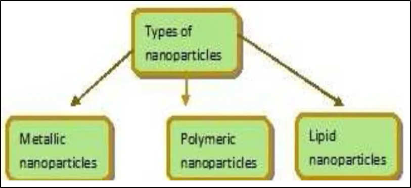
Fig. 1: - Types of Nanoparticles
Metallic nanoparticles
Silver
Due to their strong antibacterial efficiency against bacteria, viruses, and other eukaryotic microorganisms, silver nanoparticles have proven to be the most effective. They are without a doubt the most frequently utilised nanomaterials, being utilised as antimicrobial agents in the textile industry, for water treatment, sunscreen lotions, and other applications. Research have already shown that plants including Azadirachta indica20, Capsicum annuum21, and Carica papaya22 successfully biosynthesize silver nanoparticles [8].
Gold
In immunochemical research, gold nanoparticles (AuNPs) are utilised to identify protein interactions. In order to identify the presence of DNA in a sample, they are utilised as lab tracers in DNA fingerprinting. They are also employed in the detection of antibiotics known as aminoglycosides, such as streptomycin, gentamycin, and neomycin. Gold nanorods are being utilised to identify various bacterial groups, to diagnose cancer, and to find cancer stem cells.
Polymeric nanoparticles
Nano capsule: -
Drugs that can diffuse out under the right circumstances in response to environmental, chemical, thermal, or biological stimuli are contained in the oil-filled core of nanocapsules, which are made of polymeric membranes. Colloidal premade polymer interfacial deposition is used to create nanocapsules (PLA, PLGA, PCL, and PEG). It is appropriate for hydrophobic medication delivery. Lipid nanocapsules have been used to treat rat tumour instances that were multi-drug resistant. Although there hasn't been much research on using nanocapsules for drug delivery to RCC, Hureaux et alsuggestion .'s of a novel hybrid protein-lipid polymer nanocapsule of 180 nm as a nontoxic drug for codelivery of transcription factor p53 and lipophilic drug paclitaxel to induce HeLa cell apoptosis provides motivation to try it[9].
Nanospheres: -
The division of polymeric nanoparticles results in nanospheres. Nanospheres, which are matrix-type structures with spherical particle systems that range in size from 10 to 200 nm, are frequently utilised as carriers in clinical drug delivery systems. [7] In essence, the drug was dissolved, encapsulated, trapped, and connected to the polymer matrix. The medication formed a homogenous structure after being evenly distributed. Nanospheres can be crystalline or amorphous in form, and they have helped to prevent the drug's enzymatic and chemical breakdown. [10]. Nanospheres may or may not be biodegradable. Modified starch nanospheres, albumin nanospheres, gelatin nanospheres, polypropylene dextran nanospheres, and polylactic acid nanospheres are a few examples of biodegradable nanospheres.
Lipid Nanoparticles: -
Lipid nanoparticles exhibit exceptional qualities that are crucial to their therapeutic effect. The unique characteristics of nanoparticles (NP), such as their surface to mass ratio, extra colloidal particles, and their capacity to bind and convey substances, make NPs more intelligent to employ as pharmaceutical products [11]. Lipid nanoformulations create dispersions of moderately water-soluble drugs and can ease the formation of solubilized phases from which drug absorption is simple.
NLC
Prior to particle production, a liquid lipid (carrier oil) was blended with the solid lipid to create the second generation of lipid nanoparticles, also known as nanostructured lipid carriers (NLC) [12]. By altering the crystal packing structure, carrier oil is incorporated into the solid lipid matrix to increase loading capacity, physical and chemical stability, and allow for controlled release [16]. The impact of the solid lipid/carrier oil combination on drug loading and release properties has been the subject of numerous investigations [13]. Also, it has been demonstrated that the dispersion's stability against aggregation has been enhanced. Since NLCs were first created as drug delivery vehicles, there have been a lot more papers based on NLC formulations. This success can be explained by two key factors: Imaging and stability challenges in the production of lipid-based nanoparticles have been overcome, and there are novel uses for NLC formulations (e.g. colitis, P-gp efflux inhibition, theranostics) [14]. The newest type of lipid nanoparticles are called nanostructured lipid carriers (NLC), which are aqueous colloidal dispersions made of a mixture of solid and liquid lipids (oils). Its improved skin penetration capabilities led to recognition of it as a viable drug carrier system for topical application. The use of NLC in the treatment of skin conditions such psoriasis, dermatitis, and inflammations has been documented in the literature. With TDDS, the active compound's molecular characteristics are essential for transdermal potential. When the drug's molecular weight, for instance, is greater than 500 Da, it is thought that it cannot pass through skin. The mechanism behind NLC's enhanced skin penetration is still unclear at this time. NLCs have advantages over other delivery systems like creams, tinctures, and emulsions like controlled drug release, protection of active ingredients, and minimal skin irritation [15]. Furthermore, the nanoparticles' small particle sizes ensure that they are in close contact with the stratum corneum (SC), which increases the amount of active ingredient that permeates the skin.
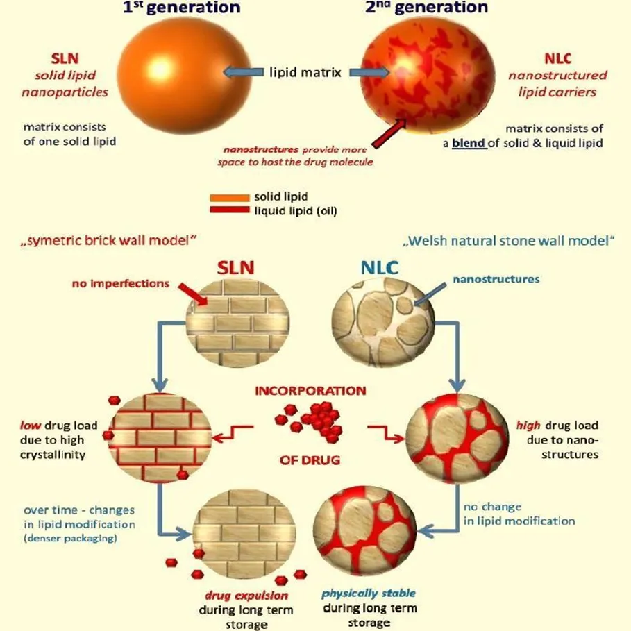
Fig. 2: - Difference between SLN and NLC
SLN
Solid lipid nanoparticles (SLNs) have great potential for delivering drugs and other bioactive substances. For the encapsulation of bioactive components, SLN offer a number of benefits over alternative colloidal delivery systems, including enhanced physical stability, protection against chemical degradation, no biotoxicity, precise control over release rates, and simpler manufacturing scale-up [16]. The main barrier to the use of SLNs in manufacturing is their propensity for uncontrolled aggregation when stored at room temperature, as well as the additional rejection of the bioactive chemical from the solid lipid matrix. According to a number of studies, the formation of hydrophobic surfaces as a result of the lipid's polymorphic transformation from unstable -form crystals into solid form crystals is what causes particle aggregation. Solid lipid nanoparticles (SLNs) have great potential for delivering drugs and other bioactive substances. For the encapsulation of bioactive components, SLN offer a number of benefits over alternative colloidal delivery systems, including enhanced physical stability, protection against chemical degradation, no biotoxicity, precise control over release rates, and simpler manufacturing scale-up [17]. The main barrier to the use of SLNs in manufacturing is their propensity for uncontrolled aggregation when stored at room temperature, as well as the additional rejection of the bioactive chemical from the solid lipid matrix. According to a number of studies, the formation of hydrophobic surfaces as a result of the lipid's polymorphic transformation from unstable -form crystals into solid form crystals is what causes particle aggregation. Due to surfactant arrest and induced interparticle interaction, hydrophobic patches cannot be covered, which ultimately leads to uncontrolled buildup. The polymorphic modification results in more ordered crystal packing, which causes a partial rejection of the encapsulated ingredient from the solid lipid matrix. Early in the 1990s, Professors R.H. Müller (Germany) and M. Gasco (Italy) began exploring the possibility of a novel formulation based on nanoparticles known as solid lipid nanoparticles (SLNs). 1,2 In contrast to other organic nanoparticles (such PLGA nanoparticles), their formulation, which was based on lipids, had the benefit of not requiring an organic solvent during the manufacturing process. They also demonstrated remarkable in vivo stability because they remained solid at body temperature. They were an option to lipid-based formulations in the past as well as organic nanoparticles because of the latter quality (e.g. liposomes). The modest drug loading, however, seemed to jeopardise the formulation's potential for use in the future. The development of the SLNs was aided by additional formulation research. SLNs incorporate the solid lipid, but NLCs entrap the drug in a mixture of solid and liquid lipids, thus resulting in a formulation with sustained release and eliminating the drawback of SLNs. With the profound understanding obtained in several sectors of biotechnology, biomedical engineering, and nanotechnology, the field of novel drug delivery systems is growing exponentially [18]. Some of the most modern formulation techniques make use of nanotechnology, which is the process of creating nanoscale structures that hold the API [19]. The National Nanotechnology Initiative (NNI) defines nanotechnology as the study and application of structures that are generally between one and one hundred nanometers in size. The overarching objective of nanotechnology and medicine is the same: to use targeted and controlled medication delivery to treat conditions as effectively and side-effect-free as feasible, and to identify conditions as precisely and early as possible [20]. Nanoparticles, solid lipid nanoparticles, nanosuspension, nanoemulsion, and nanocrystals are a few of the significant drug delivery systems created with the help of nanotechnology [21]. Solid Lipid Nanoparticles (SLNs) are the primary subject of this article. Introduced in 1991, solid-state nanoparticles (SLNs) offer a superior and substitute for conventional colloidal carriers like polymeric micro and nanoparticles, liposomes, and emulsions.
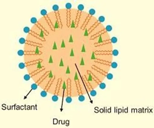
Fig. 3: - Solid lipid nanoparticles
Advantages of SLN
Disadvantages of SLN
Applications of SLN
SLN as a possible novel vaccine adjuvant
During immunization, adjuvants are utilized to boost the immunological response. Since the new, safer subunit vaccinations are less successful at immunizing, effective adjuvants are needed. The emulsion systems are new advancements in the adjuvant field. These are water-in-oil emulsions that break down quickly within the body. Since SLNs are solid, their lipid components will break down more gradually, exposing the immune system to them for a longer period of time.
Chemotherapy for Cancer Using Solid Lipid Nanoparticles
Over the past twenty years, a number of chemotherapeutic drugs have been investigated for their invitro and in vivo efficacy by encapsulating them in SLN. The results of these investigations have demonstrated to increase the effectiveness of chemotherapeutic medications while also lowering their negative effects. SLN is a good vehicle for delivering chemotherapeutic medications because of its many essential qualities, including higher drug efficacy, improved pharmacokinetics, decreased in-vitro toxicity, and the capacity to encapsulate chemotherapeutic chemicals with a variety of physicochemical properties.
Solid Lipid Nanoparticles for Focused Drug Delivery to the Brain
Given that solid lipid nanoparticles have a particle size of less than 50 nm, they may be advantageous for drug targeting. Minimal carrier size typically promotes decreased absorption by the endothelium. Modification of solid lipid nanoparticles' surfaces may potentially enable drug targeting. When it comes to treating illnesses of the central nervous system, SLNs are a promising drug targeting mechanism that can enhance a drug's ability to cross the blood-brain barrier. A study was conducted to address the restricted brain penetration of 5-fluoro-2'-deoxyuridine (FUdR) by synthesizing and encapsulating 3-,5'-dioctanoyl-5-fluoro-2'deoxyuridine (DO-FUdR) in solid lipid nanoparticles.
Targeted delivery of solid lipid nanoparticles for the treatment of lung diseases
One of the most difficult areas of pharmaceutical science study is targeted delivery of therapeutic molecules to specific organs or places. A new avenue for enhancing medication delivery was unlocked by the development of colloidal delivery systems, including liposomes, micelles, and nanoparticles. When comparing nanoparticles to other delivery systems, there are many benefits due to their unique features, which include small particle size, vast surface area, and the capacity to change their surface properties. Delivering targeted nanoparticles to the lungs is a growing field of study.
Solid Lipid Nanoparticles in the Disease of Tuberculosis
SLN have superior encapsulation efficiency and longer stability compared to liposomes. Furthermore, its synthesis procedure requires less organic solvents than that of polymeric nanoparticles. SLN has demonstrated efficacy in encapsulating anti-tubercular drugs (ATD) in experimental tuberculosis. Anti-tubercular medications, like pyrazinamide SLN systems, isoniazid, and rifampicin, were able to increase patient compliance and reduce dosage frequency.
METHODS OF PREPARATION OF SLN
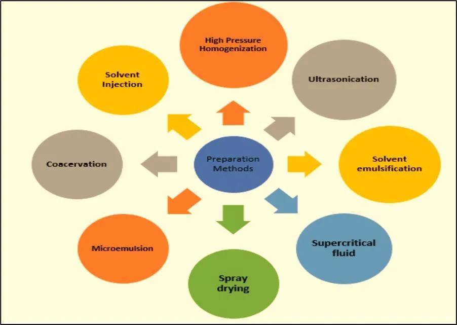
Fig. 4: - Methods of preraration of SLN
Cold Homoginization
On the other hand, the solid lipid is used during the cold homogenization process, which amounts to high pressure grinding of a solution. Because of the rise in temperature during homogenization, the lipid must be kept in its unmelted condition, which requires effective temperature control and regulation. The following three issues with the hot homogenization method have led to the development of cold homogenization. As with heat homogenization, the first stage entails dispersing or solubilizing the medication in the bulk lipid melt. The quick cooling of the drug-containing melt promotes the uniform dispersion of the drug within the solid matrix. Particle comminution is facilitated by low temperatures because they make the lipid more fragile. In a cold emulsifier solution, the solid lipid microparticles are distributed. At room temperature or lower, the pre-suspension undergoes high pressure homogenization.
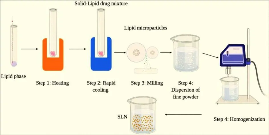
Fig. 5: - Cold homoginization
Hot Homoginization: -
Hot homogenization, which is performed at temperatures higher than the lipid's melting point, is sometimes referred to as emulsion homogenization. Using an Ultra-Turrax high-shear mixing apparatus, a pre-emulsion of the drug-loaded lipid melt and the aqueous emulsifier phase (at the same temperature) is produced. The quality of the pre-emulsion has a significant impact on the end product's quality, and droplets in the few micrometer size range are ideal [23]. It is important to remember that high pressure homogenization raises the sample's temperature (by about 10°C for 500 bar). Three to five homogenization cycles at 500 to 1500 bar are usually adequate. Particle coalescence, which happens as a result of the high kinetic energy of the particles, frequently causes an increase in particle size when the homogenization pressure or number of cycles is increased. Due to the lipid's liquid condition, which solidifies into particles when cooled to room temperature, the main product is a nanoemulsion. Lipid crystallization may be severely delayed in this case due to the small particle size and the presence of emulsifiers; the sample may stay as a supercooled melt for several monthods[24].
Evaporation Methods
The lipid-soluble material dissolves in the aqueous phase of a water-impermeable organic solvent (like cyclohexane). Lipid precipitates as 25 nm-sized nanoparticles after the solvent has evaporated, creating a dispersion of nanoparticles. The solution has been emulsified in a water phase and homogenized under high pressure. The organic solvent evaporated out of the emulsion at lower pressures.
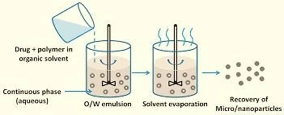
Fig. 6: - Solvent Evaporation Techniques
Double emulsion methods
Double w/o/w microemulsions can be made in two phases. First, to create a transparent system, a drug-containing aqueous solution is added to a mixture of melted fat, detergent, and co-surfactant at a temperature that is almost equal to the melting point of fat. In the second step, a clear w/o/w system is produced by mixing the produced w/o microemulsion with water, surfactant, and cosurfactant. To produce SLNs, warm micro double emulsions can be dispersed in cold water after being cleaned with dispersion medium using an ultrafiltration machine. Multiple emulsions show intrinsic instability when the internal aqueous droplets inside the oil phase coalesce due to the coalescing oil droplets and the layer rupturing on top of the internal droplets. Between their production and quenching in cold aqueous solutions, the transparent double microemulsions can be stabilized for a brief amount of time, which is necessary to make SLNs.
Supercritical fluid methods
This is a relatively novel way of generating SLN with the advantage of solvent-free processing. There are numerous variations of this platform technology that can be used to create powder and nanoparticles. SLN can be produced using the fast expansion of the supercritical carbon dioxide solutions (RESS) technology. It made sense to carry out this method with carbon dioxide (99.99%) as the solvent.
Characterization OF SLN
Particle size analysis: -
Particle size analysis (PSA) and Polydispersibility index (PDI) of SLN performed by using Malvern Zetasizer Nano ZS (Malvern Instruments, UK. Distilled water used as solvent for dispersed the formulation and maintained temperature at 25°C.
Zeta potential measurement: -
Zeta potential analysis of loaded SLN by using Malvern Zetasizer Nano ZS (Malvern Instruments, UK). Formulation diluted properly by using distilled water and zeta potential determined.
DSC: -
Differential scanning calorometry of SLN, drugs and its excipients analyzed by using DSC 7020 made in Hitachi High Tech Science Corporation, Japan.
TGA: -
Thermo-gravimetric analysis of SLN, drugs and its excipients analyzed by using TGA55 Instruments made in TA Instruments USA in range of 0-200°C. Take 10 mg power sample placed in platinum pan and heated under atmosphere (50ml/min) at heating rate of 10°C/min. After analysis of TGA weight loss verses temperature curve obtained.
PXRD: -
Utilising a Brucker D8 advanced diffractometer and Diffrac plus V1.01 software, the PXRD analysis of capecitabine, imvitor, and pemulen tr-1 was carried out. At a 2 value of 2-80°, a voltage of 40 kV, and a current of 40 mA, the diffraction pattern was recorded. By applying 2-3 drops of the formulation on an adsorbent Light Kaolin, a nanostructured lipid carrier was made solid in order to perform solid state characterization. The solid was then levigated to create a free-flowing powder. The primary aim of the present research work is to formulate and evaluate an optimized Curcumin-loaded Solid Lipid Nanoparticle (SLN) system to enhance the solubility, stability, bioavailability, and therapeutic efficacy of curcumin for effective cancer management.
1. To develop an optimized SLN formulation for the encapsulation of curcumin through ultrasound emulsification.
This objective focuses on the preparation of curcumin-loaded solid lipid nanoparticles using the ultrasound emulsification technique. Ultrasound emulsification is an efficient method for producing nanoparticles with a small particle size and uniform distribution. The study aims to select suitable lipids, surfactants, and processing conditions to achieve maximum drug entrapment, high stability, and efficient encapsulation of curcumin within the lipid matrix.
2. To investigate the influence of formulation variables on the performance and characteristics of Curcumin-SLNs using a suitable experimental design technique.
The formulation and process parameters, such as lipid concentration, surfactant concentration, sonication time, and homogenization conditions, can significantly affect the quality attributes of SLNs. Therefore, this objective aims to systematically evaluate the effect of these variables using an appropriate statistical optimization tool, such as Design of Experiments (DoE) or Quality by Design (QbD). The influence of these factors on particle size, polydispersity index (PDI), zeta potential, encapsulation efficiency, drug loading, and drug release behavior will be analyzed to obtain an optimized formulation.
3. To investigate the microstructure of Curcumin-loaded Solid Lipid Nanoparticles.
The microstructural characterization of the developed nanoparticles is essential for understanding their morphology, surface characteristics, and internal structure. This objective involves the use of advanced analytical techniques such as Scanning Electron Microscopy (SEM), Transmission Electron Microscopy (TEM), Differential Scanning Calorimetry (DSC), Fourier Transform Infrared Spectroscopy (FTIR), and X-ray Diffraction (XRD). These studies will help confirm the successful incorporation of curcumin into the lipid matrix, determine the physical state of the drug, and evaluate the structural integrity of the nanoparticle system. Curcumin is an ideal candidate for incorporation into solid lipid nanoparticles because of its hydrophobic nature and poor bioavailability. Encapsulation of curcumin in SLNs enhances its solubility, protects it from degradation, improves drug absorption, provides sustained drug release, increases cellular uptake, and enhances its anticancer efficacy. These advantages make Curcumin-SLNs a promising nanocarrier system for cancer management and targeted drug delivery applications.
EXPERIMENTAL WORK
Preparation of Curcumin-Loaded Solid Lipid Nanoparticles (SLNs)
Curcumin-loaded solid lipid nanoparticles (SLNs) were prepared using the hot homogenization followed by ultrasonication method. Initially, 10 mg of curcumin was accurately weighed and added to the melted solid lipid, Glyceryl Monostearate (GMS), in the oil phase. The surfactant Span 80 was then added to the lipid phase and mixed thoroughly until a clear solution was obtained. Simultaneously, 20 mL of distilled water was taken in a separate beaker as the aqueous phase and heated to the same temperature as the oil phase. The surfactant Pemulen TR-1 was added to the aqueous phase and stirred until completely dissolved. The hot aqueous phase was gradually added to the oil phase while maintaining the temperature above the melting point of Glyceryl Monostearate. A magnetic stirrer was placed in the mixture, and stirring was continued for 5 minutes to obtain a coarse emulsion. The resulting emulsion was then subjected to high-speed homogenization using an Ultra-Turrax homogenizer operated at 9400 rpm for 5 minutes to reduce the droplet size and form a fine emulsion. Following homogenization, the emulsion was transferred into a clean beaker and further processed using a probe sonicator at 60% amplitude for 5 minutes to obtain nanosized particles. Upon completion of sonication, solid lipid nanoparticles were formed. The nanoparticle dispersion was allowed to cool gradually to room temperature, leading to the solidification of the lipid matrix and stabilization of the nanoparticles. Finally, the prepared Curcumin-loaded SLN dispersion was transferred into a suitable container and stored in a cool place until further characterization and evaluation.
RESULT AND DISCUSSION
Particle size
According to Table, the Batch had the lowest size (Z-Average) and PDI, which were 104.0 r.nm and 0.438, respectively. Figures which demonstrate the Z-average (nm) of CS-1-SLN in terms of number percentage and intensity, respectively.
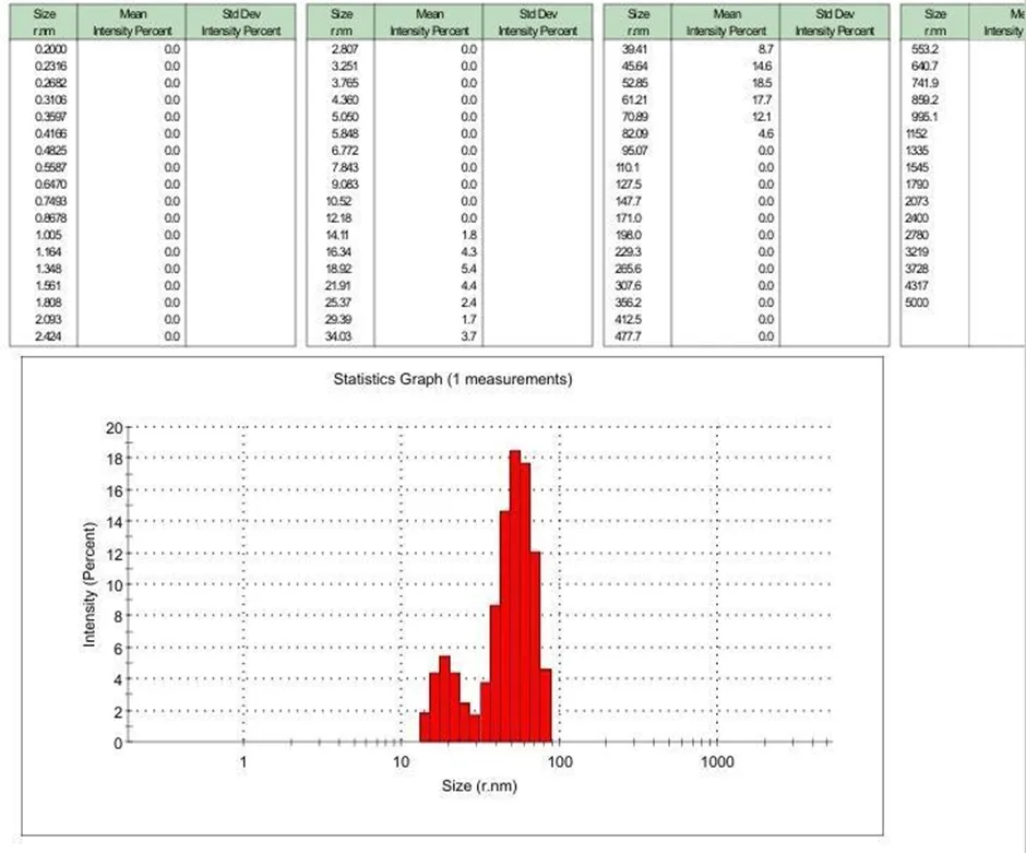
Fig. 7: - Particle size distribution
Figure. shows that 7 (% Mean number) particle had size 52.85 nm, followed by 17.7(% Mean number) corresponding to 61.21 nm, 14.6 (%Man number) particles of 45.64 nm, 12.1 (% Mean number) particles of 70.89, 8.7 (% Mean number) particles of 39.31 nm. As shown, the Z-average was 104.0 (r.nm). The Polydispersity Index, a constant resulting from a cumulants analysis of the intensity autocorrelation function picked up by the DLS, is a measure of how much light scatters. The Polydispersity Index is adjusted to exclude values below 0.05 and has no dimensions. When utilising severely monodisperse criteria, the Polydispersity Index is adjusted so that values below 0.05 are rarely occasionally seen. It has no dimensions. Values above 0.7 frequently imply that the sample has an extremely wide size distribution.
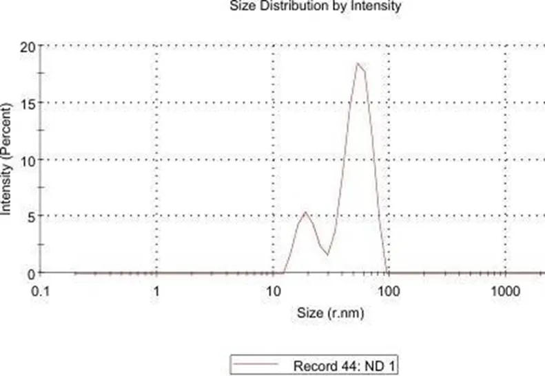
Fig. 8 Particle size and PDI of Batch-ND
As noted Fig. 8. the designed Curcumin-SLN had narrow size distribution with PDI value of 0.369.
Zeta potential
For monitoring the behaviour of dispersive systems in liquids, colloid chemists employ the zeta potential. Furthermore, the zeta-potential characterises the electrical double layer on the solid/liquid interface, which is crucial for flotation and flocculation processes. The stability of colloidal dispersions can be predicted using ZP. For electric repulsion to be effective, the zeta potential must be strong (>30 mv).
ND
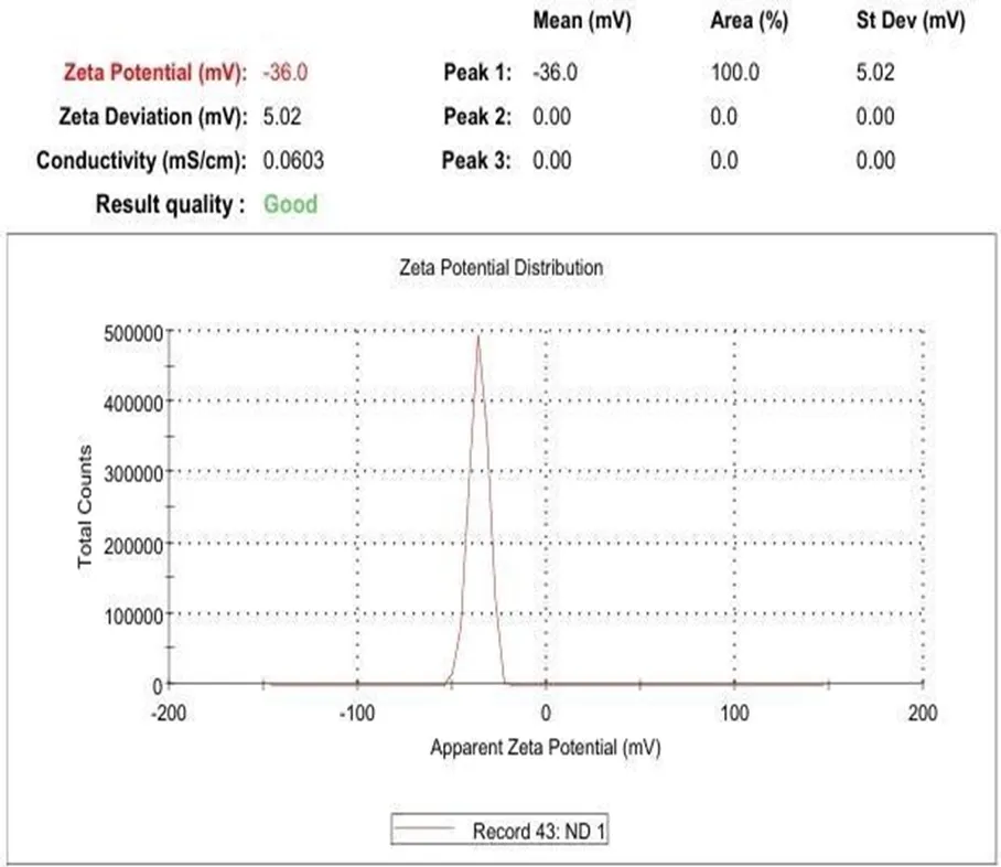
Fig. 9 Zeta potential of Batch-CS-1
As portrayed in (Figure. 9), Curcumin-SLN had a zeta-potential of -36.0 mV suggesting substantial physical stability.
Differential scanning calorimetry
Thermal analysis used in differential scanning calorimetry to ascertain the physical and chemical characteristics of medicines and polymers. Heat released or absorbed during such a shift can be measured using DSC. The methods by which the quantity of heat needed to raise the temperatures of a sample and a reference object is measured in relation to temperature. The CS-1-SLN and Drug lipid melt (CS-1 DLM) and structural components' thermal behaviour.
Batch
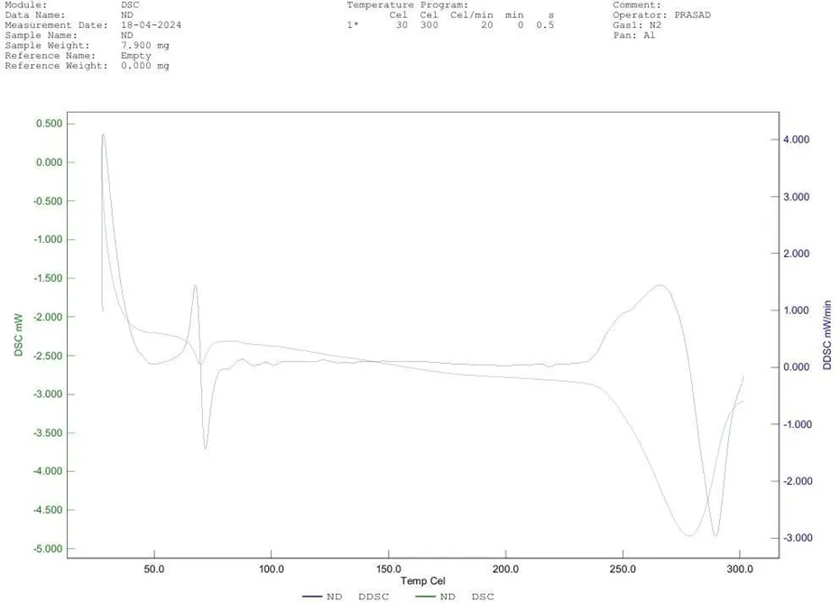
Fig. 10 DSC of Batch
DLM
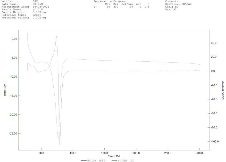
Fig. 11 DSC of DLM
CONCLUSION
Curcumin-loaded solid lipid nanoparticles (Curcumin-SLNs) were successfully prepared using the ultrasound emulsification method. Curcumin, a bioactive polyphenolic compound derived from Curcuma longa, exhibits significant anticancer activity through multiple mechanisms, including antioxidant, anti-inflammatory, and pro-apoptotic effects. However, its clinical application is hindered by poor aqueous solubility, low bioavailability, rapid metabolism, and fast systemic elimination. To address these limitations, solid lipid nanoparticles (SLNs) have been developed as an advanced drug delivery system. SLNs enhance the physicochemical stability of curcumin, improve its solubility, and provide controlled and sustained drug release. Furthermore, these nanoparticles promote enhanced cellular uptake and targeted delivery to tumor tissues, thereby improving therapeutic efficacy. The prepared Curcumin-SLNs exhibited a spherical morphology with a mean particle size (Z-average) of 104.4 nm, a polydispersity index (PDI) of 0.438, and a zeta potential of −36.0 mV. These results indicate the successful formation of a moderately uniform and physically stable nanoparticle formulation suitable for anticancer drug delivery applications.
For a Results and Discussion section, you could additionally interpret the characterization data:
The particle size of 104.4 nm falls within the optimal nanometric range for enhanced cellular internalization and passive tumor targeting via the enhanced permeability and retention (EPR) effect. The PDI value of 0.438 suggests a moderately broad particle size distribution, indicating acceptable but not highly uniform nanoparticle dispersion. The zeta potential of −36.0 mV reflects good colloidal stability due to strong electrostatic repulsion between particles, which reduces the likelihood of aggregation during storage and application. Overall, the physicochemical characteristics confirm the successful preparation of stable curcumin-loaded SLNs with potential for improved anticancer drug delivery.
REFERENCES







Bhuvaneshwari Nehete, Mrudula Pachpande, Vishakha Pardeshi, Netrali Narkhede, Sagar Sarode*, Yogesh Sonawane, Dipak Kumbhar, Formulation and Evaluation of Curcumin Solid Lipid Nanoparticles for Management of Cancer, Int. J. Med. Pharm. Sci., 2026, 2 (7), 179-193. https://doi.org/10.5281/zenodo.21139130
 10.5281/zenodo.21139130