

We use cookies to ensure our website works properly and to personalise your experience. Cookies policy
Malla Reddy Pharmacy College, Maisammaguda, Dhulapally, Medchal District, Hyderabad-500100
Nanotechnology is an emerging field that uses very small particles called nanoparticles for different pharmaceutical and biomedical applications. In this study, lotus petal-derived nanoparticles were prepared using a simple and eco-friendly ultrasound-assisted acid hydrolysis method. Fresh lotus petals were dried, powdered, and extracted using an ethanol-water mixture.. The extract was treated with acid and ultrasound to produce nanoparticles. The prepared nanoparticles were characterized using UV-Visible spectroscopy, FTIR, and SEM. The results confirmed the successful formation of nanoparticles. UV–Visible spectroscopy was performed to confirm the presence of lotus nanoparticles based on their optical characteristics. A distinct absorbance peak was observed at approximately 248 nm (λmax), indicating the presence of lotus-derived nanoparticles. FTIR spectroscopy was employed to identify the functional groups present in the synthesized lotus nanoparticles. The analysis was carried out using the ATR-FTIR technique by placing the dried nanoparticle sample on the ATR crystal and scanning over the range of 4000–400 cm⁻¹. The SEM micrograph revealed that the synthesized lotus nanoparticles were predominantly spherical with slight agglomeration, exhibiting a relatively smooth surface morphology. Most particles were observed between 50 and 100 nm, indicating a fairly uniform size distribution. Phytochemical screening showed the presence of important plant compounds such as flavonoids, phenolics, alkaloids, saponins, proteins, carbohydrates, and glycosides. The nanoparticles also showed good antioxidant activity. Overall, this study suggests that ultrasound-assisted acid hydrolysis is a simple, effective, and environmentally friendly method for producing lotus petal-derived nanoparticles with potential pharmaceutical applications.
Nanotechnology is a rapidly emerging field of science and technology that focuses on the manipulation and control of matter at the nanoscale, typically between 1 and 100 nanometers (nm). According to the National Nanotechnology Initiative (NNI), nanotechnology involves the understanding and application of materials and devices whose dimensions fall within this size range. At the nanoscale, materials exhibit unique physical, chemical, optical, electrical, and magnetic properties due to their high surface area, surface energy, and quantum effects, which differ significantly from those of their bulk counterparts. The term "nano" is derived from the Greek word meaning "dwarf" and represents one-billionth (10⁻⁹) of a meter. Nanotechnology encompasses various scientific disciplines, including physics, chemistry, biology, materials science, engineering, molecular biology, and semiconductor technology. It involves the design, synthesis, characterization, and application of nanomaterials and nanostructures to develop innovative products and technologies. Nanoparticles are among the most important components of nanotechnology. They can be classified into two major categories: organic and inorganic nanoparticles. Organic nanoparticles primarily consist of carbon-based materials, whereas inorganic nanoparticles include metallic nanoparticles (such as gold, silver, copper, and zinc), metal oxide nanoparticles (such as titanium dioxide and zinc oxide), magnetic nanoparticles (such as iron oxide), and semiconductor nanoparticles. Nanoparticles can be synthesized in a variety of shapes and morphologies, including spheres, rods, tubes, cylinders, and sheets. Their physicochemical properties are influenced by factors such as size, shape, composition, synthesis method, and the surrounding medium. This diversity in structure and composition enables nanoparticles to exhibit unique functionalities and broad applicability. Due to their remarkable properties, nanoparticles have found extensive applications in numerous fields, including medicine, drug delivery, biosensing, diagnostics, electronics, cosmetics, food technology, environmental remediation, catalysis, and energy storage. In particular, nanomedicine has gained significant attention because nanoparticles can enhance drug delivery efficiency, improve therapeutic outcomes, and facilitate disease diagnosis. Ultrasonic-assisted acid hydrolysis is an advanced and sustainable technique that combines acid treatment with ultrasonic waves to improve extraction and nanoparticle synthesis. Ultrasound waves produce a process known as acoustic cavitation, where microscopic bubbles form and collapse rapidly inside the liquid medium. Ultrasonic-assisted acid hydrolysis (UAAH) combines chemical acid catalysis with high-frequency sound waves. It uses the physical phenomenon of acoustic cavitation to significantly accelerate reaction kinetics. As ultrasonic waves pass through a liquid, they create and violently collapse microscopic bubbles. This generates extreme local temperatures, pressures, and intense micro-jets. Lotus is an aquatic medicinal plant that has been used in traditional medicine for centuries. Different parts of the lotus plant, including flowers, leaves, seeds, and roots, possess therapeutic properties. Lotus (Nelumbo nucifera), commonly referred to as Asian lotus, is grown as a perennial aquatic plant, belonging to the member of the family Nelumbonaceae, with widely being distributed across Asia and Australia. Lotus contains flavonoids, alkaloids, polysaccharides, and other bioactive substances, which have immunomodulatory, antioxidant, memory-improving, and nervous system-regulating effects. The lotus flower is rich in proanthocyanidins, polyphenols, flavonoids, alkaloids, sterols, quercetin, and dietary fiber, which have pharmacological activities such as antioxidant effects, memory and cognitive improvement, antibacterial properties, blood sugar and lipid regulation, anti-tumor effects, and improvement of the nervous system.
MATERIALS AND METHODOLOGY:
1. MATERIALS:
Fresh lotus flowers procured from local market. The remaining all the chemicals were purchased from SD fine chemicals private limited, Mumbai India. All the chemicals and media were used without any pretreatment.
2. METHODS:
2.1. Collection and Pre-treatment of Lotus Petals
Fresh lotus petals are collected and washed thoroughly with distilled water to remove dust and impurities. The petals are then dried under shade for about 7 days to preserve their bioactive compounds. Once dried, the petals are ground into a fine powder to increase the surface area for extraction.
2.2. Preparation of Lotus Petal Extract
10 grams of powdered lotus petals were weighed and mixed with 200 ml of distilled ethanol and water in a ratio of 1:2. The mixture was heated at 60–70°C for 30 minutes with continuous stirring to facilitate extraction of bioactive compounds. After heating, the mixture was cooled and filtered using Whatman filter paper to obtain the crude extract.
2.3. Acid Hydrolysis
The lotus petal powder extract was carefully measured and treated with 25 ml of hydrochloric acid (HCl), allowing the acid to react thoroughly with the extract under controlled conditions. Here, HCl acts as a catalyst and speeds up the reaction.
2.4. Ultrasonic-Assisted Nanoparticle Formation
The hydrolysed extract is transferred into a sonication vessel and subjected to ultrasonic treatment at a frequency of 20–40 kHz for 30 minutes. An ice bath was used to maintain temperature and prevent overheating. This step reduces particle size and promotes the formation of uniform nanoparticles.
2.5. Neutralization
The solution was neutralized by adjusting the pH to approximately 7 using sodium hydroxide (NaOH) solution. Continuous stirring is maintained during the process to ensure uniform pH throughout the solution and stabilize the nanoparticles.
2.6. Separation and Purification
The neutralized mixture was centrifuged at 10,000–15,000 rpm for 15–20 minutes to separate nanoparticles. The resulting particles were collected and washed with distilled water or ethanol to remove impurities. This washing process is repeated 2–3 times to ensure purity.
2.7. Drying
Finally, the purified nanoparticles were dried, preferably at 37°C, to maintain structural integrity. This process results in dry and stable nanoparticles derived from lotus petals.
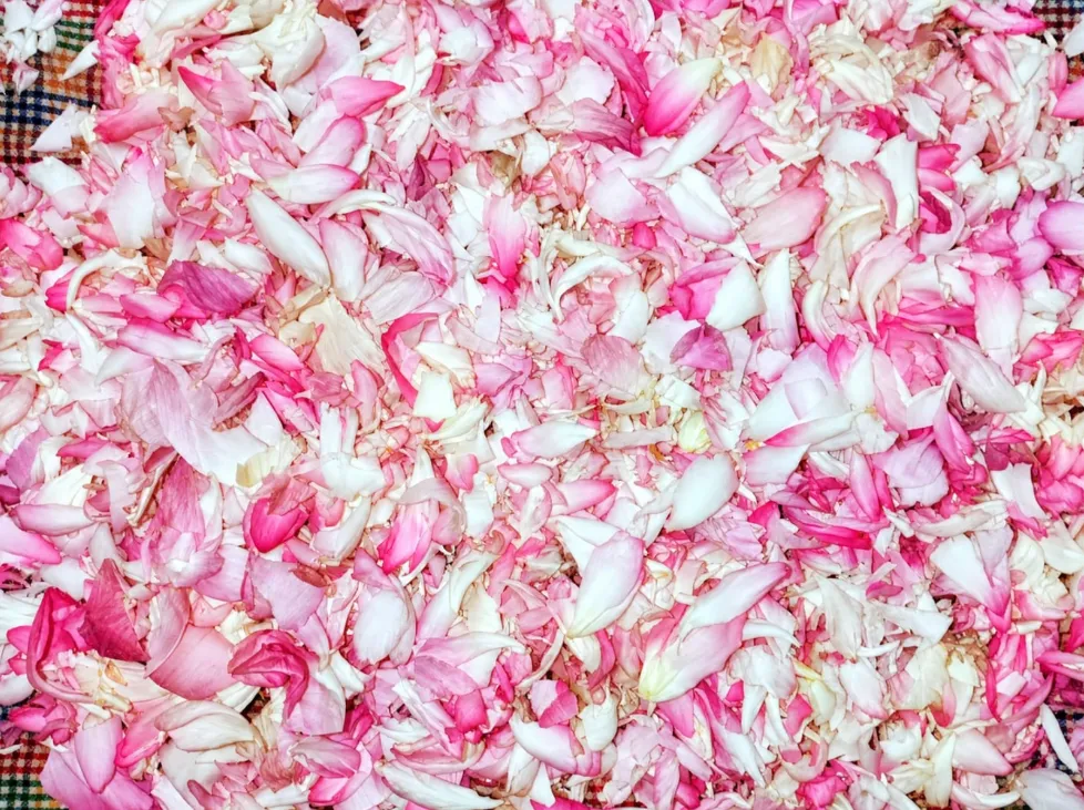
Fig no: 1 Fresh lotus petals
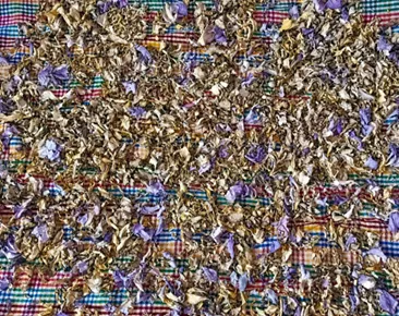
Fig no :2 Dried lotus petals
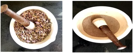
Fig no: 3 Grinding of dried lotus petals into coarse powder
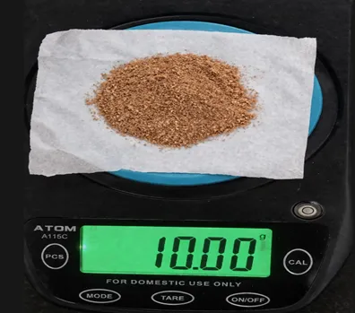
Fig no :4 weighing of lotus powder
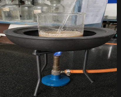
Fig no :5 Heating of powder with ethanol water (1:2)
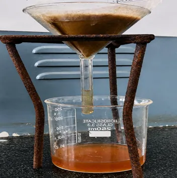
Fig no: 6 Extraction of lotus powder
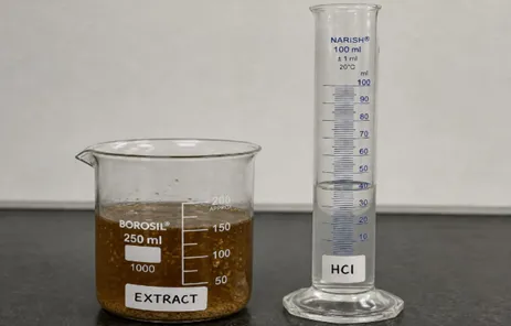
Fig no: 7 Acid Hydrolysis of lotus petals Extract
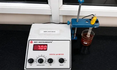
Fig no: 8 Neutalization of the Extract
3. Phytochemical screening tests:
Phytochemical analysis were performed for phenolics, glycosides, saponins, proteins, and carbohydrates flavonoids, alkaloids, amino acids and carbohydrates by performing shinoda alkaline reagent, lead acetate, dragendroffs test, mayers, ferric chloride, gelatin test, keller–killiani Test, borntrager's test, Foam Test, Hemolysis Test, Biuret, Ninhydrin test, Molisch, benedicts test. Phytochemical screening confirmed the presence of these compounds, which play an important role in nanoparticle formation and stabilization.
Table no 1. Phytochemical screening
|
Test Method |
Observation |
Results |
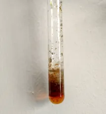
1.shinoda test: the extract is treated with magnesium turnings& conc. HCL, & appeared of red Colour confirmed the presence of flavonoids |
|
+ |
|
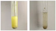
2. Alkaline Reagent Test : The extract was treated with sodium hydroxide solution. A yellow colour was formed, which disappeared on the addition of dilute acid, confirming the presence of flavonoids. |
|
+
|
|
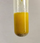
3. Lead Acetate Test: Lead acetate solution was added to the extract. Formation of a yellow precipitate confirmed the presence of flavonoids. |
|
+ |
|
B. ALKALOIDS
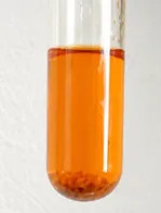
4. Dragendroff's Test The extract was treated with Dragendroff's reagent. Formation of an orange or reddish precipitate confirmed the presence of alkaloids. |
|
+ |
|
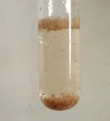
5. Mayer's Test The extract was treated with Mayer's reagent. Formation of a cream-coloured precipitate indicated the presence of alkaloids. |
|
+ |
|
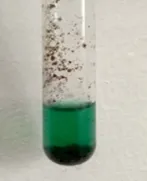
6. Ferric Chloride Test The extract was treated with ferric chloride solution. Development of a green colour confirmed the presence of phenolic compounds and tannins. |
|
+ |
|
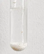
7. Gelatin Test The extract was treated with gelatin solution. Formation of a white precipitate indicated the presence of tannins.
|
|
+ |
|
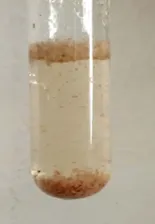
8. Keller–Killiani Test The extract was treated with glacial acetic acid, ferric chloride, and concentrated sulfuric acid. Formation of a brown ring at the interface confirmed the presence of cardiac glycosi des. |
|
+ |
|
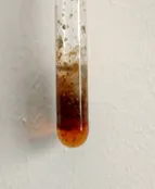
Borntrager's Test The extract was treated with benzene and ammonia solution. Formation of a pink/red colour in the ammoniacal layer indicated the presence of glycosides. |
|
+ |
|
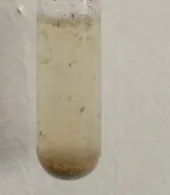
Foam Test The extract was shaken vigorously with water. Formation of stable and persistent froth confirmed the presence of saponins.
|
|
+ |
|
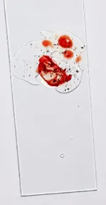
Haemolysis Test A drop of blood was mixed with a few drops of aqueous saponin solution. Rupture of red blood cells (RBCs) confirmed the presence of saponins.
|
|
+ |
|
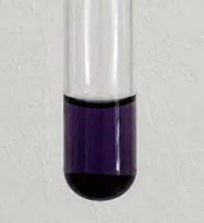
Biuret Test The extract was treated with sodium hydroxide and copper sulfate. Appearance of a violet colour indicated the presence of proteins.
|
|
+ |
|
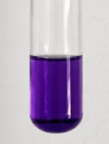
Ninhydrin Test The extract was treated with ninhydrin reagent. Development of a purple colour confirmed the presence of amino acids. |
|
+ |
|
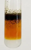
Molisch Test The extract was treated with Molisch reagent and concentrated sulfuric acid. Formation of a violet ring at the junction confirmed the presence of carbohydrates. |
|
+ |
|
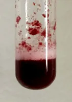
Benedict's Test The extract was heated with Benedict's reagent. Formation of a brick-red precipitate indicated the presence of reducing sugars. |
|
+ |
RESULTS AND DISCUSSIONS:
I. Analytical Studies:
UV–Visible spectroscopy was performed to confirm the presence of lotus nanoparticles based on their optical characteristics. The technique operates on the principle of electronic transitions, particularly π–π* transitions associated with aromatic and conjugated biomolecules present in plant-derived materials. Lotus extract contains various phytochemicals, including flavonoids and phenolic compounds, which typically exhibit strong absorption in the UV region. The sample was carefully loaded into a clean cuvette and scanned over a wavelength range of 200–800 nm. A distinct absorbance peak was observed at approximately 248 nm (λmax), indicating the presence of lotus-derived nanoparticles. The cuvette was handled with care to avoid contamination and to minimize ambient light interference during measurement. Multiple scans were performed to ensure accuracy and reproducibility of the results. A calibration curve was prepared using standard concentrations to enable quantitative analysis. The obtained results confirm the successful synthesis and identification of lotus nanoparticles through UV–Visible spectroscopic analysis.
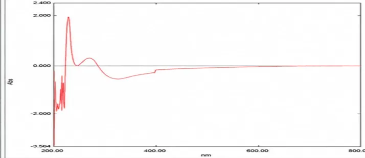
Fig no 10: UV-Visible Spectrophotometer output showing absorbance spectrum
FTIR spectroscopy was employed to identify the functional groups present in the synthesized lotus nanoparticles. The analysis was carried out using the ATR-FTIR technique by placing the dried nanoparticle sample on the ATR crystal and scanning over the range of 4000–400 cm⁻¹. A broad absorption band observed at 3342 cm⁻¹ corresponds to O–H stretching vibrations, indicating the presence of hydroxyl groups of phenols and alcohols. The peaks at 2919 cm⁻¹ and 2819 cm⁻¹ are assigned to C–H stretching vibrations of aliphatic CH₂/CH₃ groups, confirming the presence of aliphatic compounds and lipid constituents. A distinct absorption band at 1713 cm⁻¹ is attributed to C=O stretching, indicating the presence of carbonyl groups such as esters or aldehydes. The peak at 1634 cm⁻¹ corresponds to C=C stretching or amide/O–H bending, suggesting the presence of proteins, phenolic compounds, and absorbed water molecules. The bands at 1455 cm⁻¹ and 1394 cm⁻¹ are assigned to CH₂ and C–H bending vibrations, representing aliphatic and methyl groups, while the peak at 1429 cm⁻¹ is due to O–H bending/C–H deformation, indicating phenolic compounds. The absorption bands at 1148 cm⁻¹, 1101 cm⁻¹, 1036 cm⁻¹, and 1016 cm⁻¹ are attributed to C–O and C–O–C stretching vibrations, confirming the presence of alcohols, ethers, polysaccharides, carbohydrates, glycosidic linkages, and flavonoids. These characteristic FTIR peaks confirm that the lotus nanoparticles contain abundant hydroxyl, carbonyl, phenolic, aliphatic, carbohydrate, polysaccharide, and flavonoid functional groups, which are responsible for their stability and biological activity.
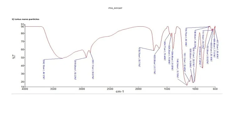
Fig No.11 FTIR peak assessment for lotus nano particles
Table no:2 FTIR Peak Assignment & Functional group Interpretation of Lotus Nanoparticle
|
Peak |
Assignment |
Interpretation |
|
3342 |
O-H stretching |
Hydroxy groups of phenols/alcohols |
|
2919 |
C-H stretching |
Aliphatic -CH2/-CH3 groups |
|
2851 |
Symmetric C-H stretching |
Alkanes/lipids present in phytochemicals |
|
1713 |
C=O stretching |
Carbonyl groups of esters, aldehydes. |
|
1634 |
C=Cor amide/O-H bending |
Proteins, phenolics and absorbed water molecules |
|
1455 |
CH2bending |
Aliphatic compounds |
|
1429 |
O-H bending/ C-H deformation |
Phenolic compounds |
|
1394 |
C-H bending |
Methyl groups of organic constituents |
|
1148 |
C-O stretching |
Alcohols, ethers and polysaccharides |
|
1101 |
C-O-C stretching |
Carbohydrates and glycosidic linkages |
|
1016 |
C-O stretching |
Alcohol and carbohydrate groups |
|
1036 |
C-O stretching |
Polysaccharides and flavonoids. |
|
873 |
Aromatic C-H bending |
Aromatic ring vibrations |
1.3 Scanning Electron Microscopy (SEM):
Scanning Electron Microscopy (SEM) was employed to examine the surface morphology and particle size of the synthesized lotus nanoparticles. Prior to imaging, the dried nanoparticle powder was uniformly mounted onto an aluminum SEM stub using double-sided conductive carbon tape. The sample was sputter-coated with a thin layer of gold to enhance electrical conductivity and minimize charging effects during imaging. The stub was then placed inside the SEM chamber, and micrographs were recorded under appropriate operating conditions (magnification: 50.00 KX, accelerating voltage: 15.00 kV, working distance: 4.6 mm). The SEM micrograph revealed that the synthesized lotus nanoparticles were predominantly spherical with slight agglomeration, exhibiting a relatively smooth surface morphology. The nanoparticles were well distributed, although a few particles formed small aggregates, which is commonly observed due to intermolecular interactions during drying. The measured particle sizes ranged from 42.35 nm to 119.42 nm, confirming the successful synthesis of nanoparticles within the nanoscale range. Most particles were observed between 50 and 100 nm, indicating a fairly uniform size distribution. The SEM analysis confirmed the formation of nanosized lotus particles with spherical morphology and acceptable size uniformity. The observed morphology and particle size distribution demonstrate the successful synthesis of lotus nanoparticles and suggest that the prepared nanoparticles possess suitable structural characteristics for potential biomedical, pharmaceutical, and antioxidant applications.
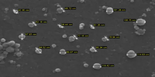
Fig no: 12 SEM image showing size distribution of Nanoparticles
2. Anti-Oxidant Activity
2.1. Hydrogen Peroxide (H₂O₂) Scavenging Assay
Hydrogen peroxide is a reactive oxygen species that can generate highly reactive hydroxyl radicals, leading to oxidative stress and cellular damage. Therefore, compounds capable of scavenging hydrogen peroxide are considered effective antioxidants. The present study demonstrated that lotus nanoparticles possess appreciable hydrogen peroxide scavenging activity. The antioxidant activity increased progressively from 20% inhibition at 10 µg/mL to 65% inhibition at 50 µg/mL, indicating that higher concentrations of lotus nanoparticles exhibit greater free radical scavenging potential. The antioxidant activity of lotus nanoparticles may be attributed to the presence of phytochemicals such as flavonoids, phenolic compounds, tannins, and alkaloids present in lotus extract, which act as reducing and hydrogen-donating agents. These bioactive compounds remain associated with the nanoparticles during green synthesis and contribute to neutralizing hydrogen peroxide. Although ascorbic acid exhibited higher antioxidant activity (80% inhibition at 50 µg/mL), lotus nanoparticles showed considerable scavenging efficiency, suggesting their potential as a natural antioxidant. The results indicate that biosynthesized lotus nanoparticles could be useful in pharmaceutical, biomedical, and nutraceutical applications where antioxidant properties are desirable. Overall, the study confirms that lotus nanoparticles exhibit moderate to strong hydrogen peroxide scavenging activity in a concentration-dependent manner, supporting their potential role as effective natural antioxidant agents.
Table no: 3 Hydrogen Peroxide (H₂O₂) Scavenging activity
|
Concentration (µg/ml) |
Absorbance (Ascorbic acid) |
% inhibition (Ascorbic acid) |
% Absorbance (lotus NPs) |
%inhibition (Lotus NPs) |
|
0(Blank) |
0.800 |
0.0% |
0.800 |
0.0% |
|
10 |
0.560 |
30.0% |
0.640 |
20.0% |
|
20 |
0.400 |
50.0% |
0.520 |
35.0% |
|
30 |
0.320 |
60.0% |
0.440 |
45.0% |
|
40 |
0.240 |
70.0% |
0.360 |
55.0% |
|
50 |
0.160 |
80.0% |
0280 |
65.0% |
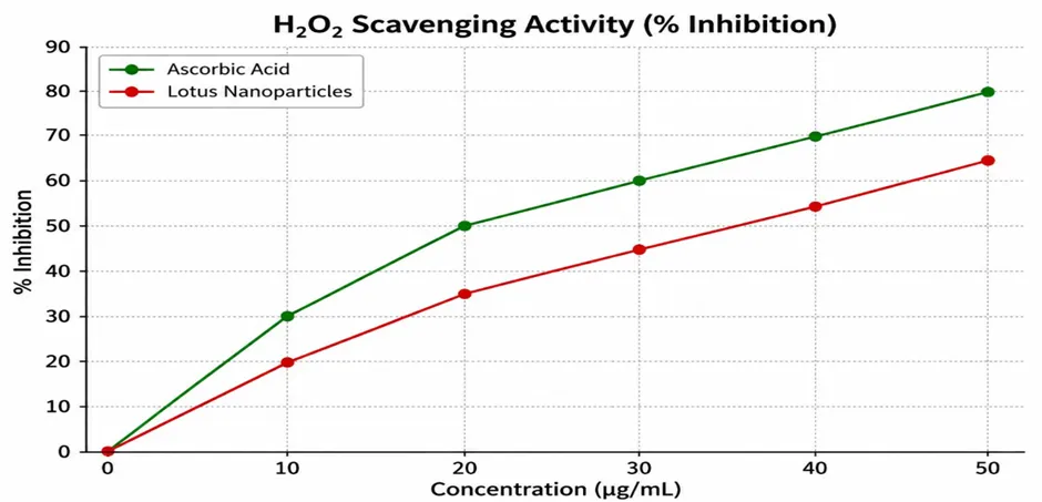
Fig no: 13 Hydrogen Peroxide (H₂O₂) Scavenging activity
2.2. Total phenolic content
The results indicate that lotus nanoparticles possess significant antioxidant activity due to their phenolic compounds. As the concentration increased from 200 to 1000 µg/mL, the antioxidant activity increased from 20% to 85%, demonstrating a clear dose-dependent effect. The reduction in absorbance with increasing concentration confirms the presence of phenolic constituents capable of reducing the Folin–Ciocalteu reagent. The high antioxidant activity of lotus nanoparticles may be attributed to bioactive phytochemicals such as phenolic compounds, flavonoids, and other reducing agents present on the nanoparticle surface. These compounds effectively donate electrons or hydrogen atoms to neutralize free radicals, thereby enhancing antioxidant capacity. Overall, the study demonstrates that lotus nanoparticles exhibit strong antioxidant potential with increasing concentration. The maximum antioxidant activity of 85% at 1000 µg/mL suggests that lotus nanoparticles are a promising natural source of antioxidants and may have potential applications in pharmaceutical, biomedical, and nutraceutical fields. The results indicate that lotus nanoparticles possess significant antioxidant activity due to their phenolic compounds. As the concentration increased from 200 to 1000 µg/mL, the antioxidant activity increased from 20% to 85%, demonstrating a clear dose-dependent effect. The reduction in absorbance with increasing concentration confirms the presence of phenolic constituents capable of reducing the Folin–Ciocalteu reagent. The high antioxidant activity of lotus nanoparticles may be attributed to bioactive phytochemicals such as phenolic compounds, flavonoids, and other reducing agents present on the nanoparticle surface. These compounds effectively donate electrons or hydrogen atoms to neutralize free radicals, thereby enhancing antioxidant capacity.
Table no: 4 Total Phenolic content of Anti-oxidant Activity
|
Concentration (µg/ml) |
Absorbance (control) |
Absorbance (lotus Nanoparticles) |
%Antioxidant (% inhibition) |
|
200 |
0.700 |
0.560 |
20.0 |
|
400 |
0.700 |
0.420 |
40.0 |
|
600 |
0.700 |
0.280 |
60.0 |
|
800 |
0.700 |
0.175 |
75.0 |
|
1000 |
0.700 |
0.105 |
85.0 |
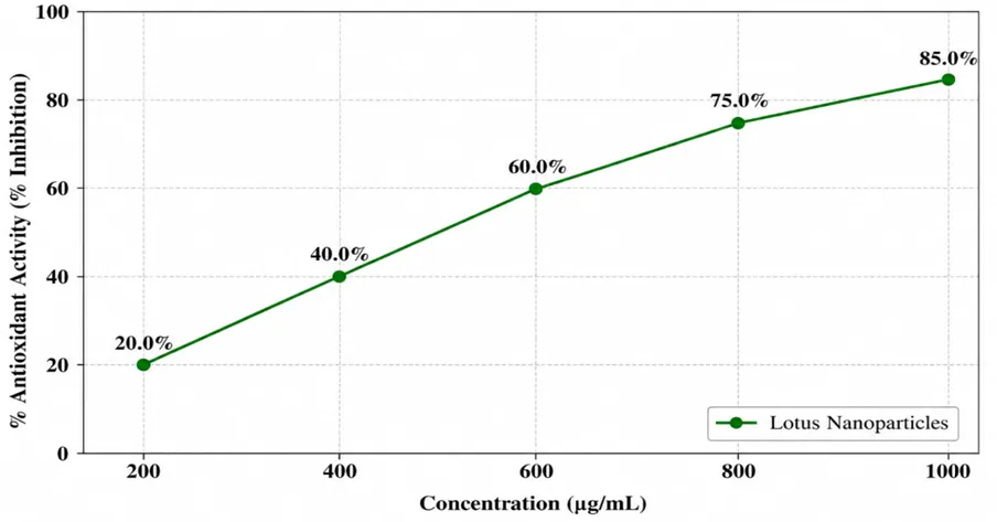
Fig No14: Total phenolic content of Anti-oxidant Activity
SUMMARY AND CONCLUSION
The present study successfully demonstrated the sustainable synthesis of lotus petal-derived nanoparticles using the ultrasound-assisted acid hydrolysis (UAAH) method. Lotus petals, rich in bioactive phytochemicals such as flavonoids, phenolics, alkaloids, glycosides, saponins, proteins, and carbohydrates, were utilized as a natural source for nanoparticle synthesis. Phytochemical screening confirmed the presence of these compounds, which play an important role in nanoparticle formation and stabilization. The synthesized nanoparticles were characterized using UV–Visible spectroscopy, FTIR, and SEM. UV–Visible spectroscopy showed a characteristic absorption peak at 230 nm, confirming nanoparticle formation. FTIR analysis identified various functional groups responsible for the stabilization of the nanoparticles, while SEM analysis revealed that the nanoparticles were spherical, well-dispersed, and ranged from 42.35 to 119.42 nm in size. Antioxidant studies, including the Hydrogen Peroxide Scavenging Assay and Total Phenolic Content assay, demonstrated a concentration-dependent increase in antioxidant activity, reaching 85% inhibition at the highest concentration tested. Overall, the study confirms that ultrasound-assisted acid hydrolysis is an efficient, eco-friendly, and cost-effective method for synthesizing lotus petal-derived nanoparticles with excellent antioxidant potential. These nanoparticles have promising applications in pharmaceutical, biomedical, nutraceutical, and antioxidant-based formulations, and further investigations on their therapeutic efficacy and large-scale production are recommended.
REFERENCES

Asha Ranjani V.*, Divya B., Sathwika B., Sai Pravallika K., Sustainable Ultrasound -Assisted Acid Hydrolysis for Lotus Nano Particles: Synthesis, Characterization and Antioxidant Studies, Int. J. Med. Pharm. Sci., 2026, 2 (9), 308-320. https://doi.org/10.5281/zenodo.22788270
 10.5281/zenodo.22788270