

We use cookies to ensure our website works properly and to personalise your experience. Cookies policy
Department of Pharmaceutics, The Erode college of pharmacy, Erode, Tamil Nadu, India
Cancer continues to be a leading global morbidity and mortality and given the disadvantages of chemo- therapies such as low tumor specificity, toxicity to normal tissues, resistance to drugs, low bioavailability, there are efforts for developing novel approaches for designing drugs. Nano-technology has revolutionised all the dimensions of therapeutic treatments particularly that of cancers by facilitating targeting, directed & controlled & personalized use of therapeutic compounds. In this article, a review of nanoparticle based drug delivery approach to combat canceris presented. The rationale of nanoparticles including the principle of passivetargeting through Enhanced Permeability and Retention Effect(EPR) and that of active targeting via receptor directed ligands (e.g. RGD, folic acid, &anti-EpCAM antibodies etc.) in achieving improved specificity to cancer- cells and thereby targeting effectiveness have been emphasized. Various Nano- carriers used are broadly classified into 5 categories as lioposomes ,polymeric micelles, dendrimers, polymeric nano- particlesand various others multifunctional Nano systems are thoroughly review. The design aspect, mechanism of drug delivery, the current clinical and preclinical reports of different Nano systems are analyzed. The paper highlights the new development of bio-mimetic approaches including red blood cells, cancer cells, macro- phages, platelets, neutrons & NK-cells derived Nano carriers & there by improving the stealth behaviour of Nanoparticles. In particular, new approaches combining Nano-technology and immnotheropy, immnogen iccell death, photo- thermal therapy, photo- dynamictherapy, nanovaccines, combo immune-phototherapym is presented. It also reviews, personalised nano-medicine & identifies potential bottleneck for future development, such as reproducibillity in manuf acturing; regulatory guidelines; & patient specifity to EPReffect, the barrier crossing potential of Nanopaticles, longtime safe & scalability & commercial manufacturing.
Cancer is a one of the leading cause of death globally. This disease poses a formidable public health challenge. Global Cancer Statistics 2022 reported that worldwide, approximately 20 million new cases of cancer and 9.7 million cancer-related deaths occurred in 2022 alone. Breast cancer accounted for 2.3 million new cases and almost 670,000 deaths, making it the most commonly diagnosed type of cancer globally. As populations grow, age, and lifestyle changes, the cancer burden is expected to significantly grow in coming decades with an estimated 35 million new cases per year by 2050 [1]. Traditionally used cancer treatment methods involve surgery, radiotherapy and chemotherapy. The effective improvement of survival outcomes is noticed in many patient populations undergoing conventional treatments, however; drawbacks are associated with each of treatments such as limitations in clinical effectiveness with the surgery approach with widespread diseases or when the malignant tissues were metastatic with conventional radiotherapy, damage to peripheral normal cells by their poorly selective manner and systematical side effects with conventional cytotoxic anticancer agents (like bone marrow, GI epithelium and skin epithelium) and relatively poor pharmacokinetic profiles and reduced accumulation at the target region [2, 3]. Among these, anthracyclines, such as doxorubicin (DOX), represent potent chemotherapy agents but clinical usage is hampered by dose-limited cardiotoxicity, and myelosuppression associated with a very narrow therapeutic index. Current evidence suggests that continuous, local, and sustained administration of drugs can enhance therapeutic outcomes in comparison to intermittent high-dose regimens [4]. Nano technology can present a significant potential to enhance the effectiveness of chemotherapy. In nanoparticle- based approach, the therapeutic agents have been incorporated into a Nanotechnology material, ranging from 10–500 nm, which functions as a Nano carrier to protected the sensitive biological active compounds, releasing the drugs in sustained controlled mode, enhancing permeability and retention effects, controlling the solubility of hydrophobic agents, prolong circulation time, shielding vulnerable biomolecules, improving intracellular uptake and targeted drug delivery into cancer tissues. The Nano science can be further tailored for various targeting modes, i.e., passive targeting, active targeting [5]. First approved FDA nanoparticle was Doxil, DOX with a Polyethelene glycol (PEG) labeled liposome system, presented beneficial Pharmacological parameters in compared to DOX including reduction of cardiotoxicity and enhancing the efficacy. Nonetheless, many issues associated with the delivery system of the first generation nano drug system such as limited accumulation at the tumor, insufficient of intra-cellular release, and the rapid uptake by the reticuloendothelial system (RES) were arisen. Repeated administrations of PEG-conjugated nanocarriers have been described in clinical settings lead to acceleration of blood clearance (ABC phenomenon, the PEG dilemma) due to the development of anti- PEG antibodies and the reduced efficacy in cellular uptake and intra cellular drug release [6,7]. Biomimetic nanotechnology has created the concept that can successfully achieve better targeting. Instead of utilizing surface functionalization, a variety of biomimetic nano systems have been developed by using natural derived membranes for encapsulating synthetic nanocarriers (cell membrane camouflaged nanoparticles) such as:red blood cells, platelets, stem cells, macrophages, neutrophil, natural killer (NK) cells, and even cancer cells. Using these naturally- derived membrane components, not only can maintain biological characteristics of cells, such as immune evading, prolonged circulation, tumor-homing ability and enhanced the therapeutic efficacy, but can still retain the versatility of man-made Nano systems [8]. Beyond drug delivery, the therapeutic applications of nano technology have continuously expanded into advanced field such as:cancer therapy with immunotherapy, immune checkpoint blockers, inducing immunogenic cell death, photothermal and photodynamic therapies, gene therapy, nano-cancer vaccines, theranostic diagnosis/therapy andpersonalized therapy. The development of personalized medicine through various nanotechnological approaches, depending on individual tumor biological characters, holds great future [9,10]. In the current review, we provide an exhaustive assessment of the contemporary nanotherapy against cancer, highlight principles of both passive and active tumor targeting strategies, summarize recent developments on major classes of nano carriers, including liposomes, polymeric micelles, dendrimers, polymeric Nanoparticles and biomimetic Nanoparticles system, and give insights into promising application strategies in immunotherapy, phototherapy, nanovaccines and theranostics. Finally, we address important clinical limitations with regard to passive tumor targeting (i.e., variability of EPR effects), biological barriers, large-scale fabrication, regulatory issues, safety aspects and prospect of individualized Nanomedicine.
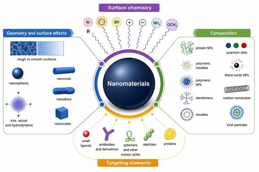
FIG: 1 Targeting Elements
Mechanistic Foundations of Tumor Targeting
EPR Effect of Passive Targeting
Over the last few decades, the enhanced permeability and retention (epr) effect, described in 1986, has served as the primary mechanistic explanation for tumor drug delivery via nanoparticles. Because tumors constantly create new blood vessels in order to feed their rapid growth (neovascularization) there can be tumor-associated angiogenesis where abnormal blood vessels with 100 to 780 nanometer large pores form between the endothelial cells enabling transit of blood-born nanoparticles into the interstitial space of the tumor due to the absence of the highly organized microvascular architecture seen in healthy tissue [9]. Moreover, the absence of functional lymphatics in some solid tumors prevent clearance of extravasated macromolecules into the lymphatic system. while decades of extensive study have been conducted based upon this premise, the translation alability of epr has come into much dispute. It’s clinical utility has been found to be highly dependent on a variety of factors including vasculature architecture, tumor pressure and patient specific physiological differences [13]. Furthermore,passive targeting possesses it’s own inherent problems, most notably an inability to effectively differentiate tumor tissue from surrounding normal tissues [10]. Most importantly, this passive strategy cannot consistently deliver therapeutic agent concentrations to areas within bulky solid tumors that are spatially distant from functional tumor vessels. These limitations thus have led to the generation of an active targeting strategy as a necessary adjunct, rather than a substitute, to passive targeting.
Active Targeting Strategies
Active targeting involves targeting specific cell-surface receptors over-expressed on cancer cells or on angiogenic tumor-associated endothelial cells through functionalization of nanoparticle surfaces with specific ligands. Such receptor mediated interactions lead to enhanced targeting of nanoparticles to tumors, and also receptor mediated internalization of tumor targeted particles with their encapsulated drugpayload. Several types of targeting ligands have been investigated including: - fibronectin has an rgd (arginylglycylaspartic acid) binding motif. The motif, an rgd sequence present in fibronectin is specifically recognized by members of the integrin family of proteins found on most surfaces in and surrounding the matrix and over-expressed in angiogenic tumor endothelia cells [19]. Rgd-targeted nps showed five times greater accumulation in tumors compared to non-targeted nps and a five-fold increase in overall tumor-specific drug release in studies in tumor bearing animals. Survival in mice treated with rgd-functionalized particles (13 days) was higher than in those injected with nontargeted nps (21 days) [12] - folate is a vitamin-like substance that serves as a precursor for coenzyme f4. It is well known that several human tumors up-regulate the expression of cell-surface folate receptors (fr). Over-expression of fr is reported in epithelial, neuronal, renal cell carcinoma, as well as some leukemias and brain tumors. It has become widely accepted that fr represent a viable target for tumor therapy and have been extensively used for targeting nanoparticles [13]. The targeted delivery can potentially enhance drug retention in tumor tissue and increase intercellular drug delivery to receptor-positive tumor cells. - the cdx peptide is part of the snake neurotoxin candoxin. It was employed to guide therapies for cns tumors through the blood-brain barrier [14]. - carbohydrate-based targeting for cancer vaccines and immunotherapies exploit mansose lectin binding capability to identify dendritic cells or macrophages and deliver these targeted particles to the site of antigen presentation or the area of action [15]
Nanocarrier Platforms In Cancer Therapy
Liposomes
Liposomes are the only nano delivery system that mimic natural biological cell membrane composed of lipid bilayers and can provide a stable encapsulating environment within the hydrophobic lipid domain and the hydrophilic inneraqueous space [6]. The unique structural property allow liposomes encapsulate both lipophillic drugs in the lipid bilayer and hydrophillic drugs in the center. Liposomes have merits such as good biocompatibilty, biodedradablity and low inherent toxicity [6]. Lipoosomes can be classified to unilamellar vesicle (uvs) with only one lipid bilayer and multilamellar vesicles (mlvs) consist of multiple lipid bilayers, depend on the number of the bilayers. The use of complex lipid compositions in the multilayer core and shell design of mlvs allows their drug cargo capacity to be increased and also control the drug-release rate. There have been a series of liposome designs with enhanced anti-tumor activity in-vitro and in- vivo demonstrated in the literature. Emtansine-loaded macrophage membrane-coated liposomes inhibited lung metastasis in animal models and enhances epr-based drug internalization of breast cancer cells [16]. A multistage dual system based on multivesicular liposomes simultaneously entrapping paclitaxel(ptx)- hyaluronic acid (ha)- pei/dna complexes is designed using ha coating on multivesicular liposomes encapsulating the two types of therapy to co deliver paclitaxel and dna to inhibit genes (e.g. egfr) and cancer proliferation [16]. Other examples include the biomimetic liposomal nanozymes, a combination of mesoporous manganese dioxide, doxorubicin, and collagenase to enhance tumor penetration capability, reduction of hypoxia, and protection against doxorubicin induced cardiotoxicity [17]. The nano-pt/vp@mlipo system, which encapsulate ultra-small platinum nanoparticles and verteporfin in the liposomes camouflaged by macrophage membranes, can efficiently kill cancer cells in orthotopic murine tumor models, exhibit remarkable in vivo therapeutic effects in term of deep spheroid penetration and lung metastasis suppression and extend mouse survival [18].
Polymeric micelles
Polymeric micelles are spherical nanoparticles assembled by the self-assembly of amphiphilic block copolymers in aqueous media. The micelle is characterized by a hydrophobic core, in which hydrophobic drugs can be embedded, and a hydrophilic shell, which provides stability and prevents micelle agglomeration. Moreover, the small size of micelles, ranging from 10 to 100 nm, facilitates their accumulation in and penetration into tumor tissues [20, 21, 22, 23]. Chol-peo micelles loaded with dox were shown to have a slow and sustained drug release because of the encapsulation of dox in cholesterol-modified lipophilic core that mimics biological membrane [21]. The rgd/tat dual peptide functionalization of peo-pcl block copolymer, combined with the spermine-modified pcl core for sirna delivery, enabled concurrent tumor oncogene inhibition and chemotherapy [20]. Deoxycholic acid (da) coupled with pei was applied to prepare dual functional ptx/sirna micelles to inhibit tumor genes and concurrently delivery paclitaxel into the cells [21]. Inspired from the segmentation design of naturally occurring cell membranes, advanced biomimetic multicompartment micelles were developed to carry multiple different hydrophobic cancer drugs simultaneously [22].
Dendrimers
Dendrimers are 3d, dense, well-defined, highly branched macromolecular structures that are characterized by a central core, progressive generations of branches and reactive functional groups on the periphery. Dendrimers provide a means for precise control oversize and molecular weight in addition to molecular monodispersity [23]. Loading withdrugs is achieved by a multitude of methodologies, from physical encapsulation in internal cavities to covenent attachment of drug to the surface functional groups. Comparing pamam dendrimers loaded with dox and targeted towards il-6 or arg, the larger il-6 ligand contributed to the enhanced cellular internalization and loading efficiency [24]. Surface modification using phosphorylcholine moiety reduced toxicity and improved drug release [25]. The cationic nature of dendrimers leads to membrane ruptures, causing apoptotic death, particularly as dendrimer generation increases [26]. As such, various modifications of zeta charge or incorporation anionic or zwitterionic groups on the surface have been proposed to address these limitations [26].
Polymeric carriers
A broad class of drug delivery vehicles are biodegradable nanoparticles and nanogels that offer tunable size and other physicochemical characteristics [27, 28, 29]. The pp-75 endosomolytic polymer, designed froml-phenylalanine grafted to an polyamide backbone, is effective in nanoparticle-targeted endocytosis, facilitates endosomal release via proton-sponge mechanism, and enables the selective intracellular release of doxorubicin without targeting normal cells [27]. Hpma copolymer drug-conjugates were among the earliest biodegradable polymer based cancer drugs that entered phase i/ii clinical trials [28]. Studies using negatively charged zwitterionic poly(amidoamine) nanogels have shown less toxicity and greater biocompatibility compared with positively charged counterparts, providing excellent drug carriers [29].
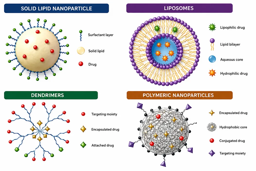
FIG: 2 Different Polymeric Carriers
Biomimetic Cell Membrane Coating Technology
The RBC Membrane Coating
RBC membrane coated particles are some of the first and most well-studied nanoparticle applications. The major paradigm shift in nanoparticle-based cancer therapy over the last decade has been cell membrane-coating nanoparticles, which take a top down rather than trying to mimic complex cellular biological functions one ligand at a time (that in practice would still be fundamentally insufficient). Instead, they isolate full plasma membranes from live cells, which are then transferred onto the surfaces of pre-formulated nanoparticle cores [8]. The biomimetic particle maintains the ability of the synthetic core biomimetic platforms to deliver and release drugs, while displaying the biological identity of the cell it came from, all surface proteins, glycoproteins, receptors and signaling molecules (in natural orientation). Human blood cells, namely red blood cells, are the most abundant circulating cells, and cancirculat for up to 120 days (which is a function of their “do not eat me signal”, CD47) [30]. MHC proteins are absent on the rbcs, significantly decreasing their immunogenicity. RBC membrane-coated particles exhibit both, achieving circulation half-lives of about 39.6h (compared with15.8h forpegylated equivalents) and a >2-fold enhancement in the probability of accumulation in tumors [31].
An elegant demonstration of cell membrane coated particles for cancer is a new TT-RBC-NP (Targeted Theranostic RBC membrane-coated Nanoparticle), developed to facilitate the treatment of breast cancer. These particles are made by coating them with human red blood cell membrane afterfunctionalizing withanti-epcam antibodies via DSPE-PEG-biotin lipid insertionand then encapsulating into aplga nanoparticle co-loaded with doxorubicin(DOX)and a fluorescent imaging agent(FITC). The rationale for selecting epcam (CD326) was that it is an appealing active targeting moiety for being significantly overexpressed in several carcinomas includingbreast cancer. The nps were stable in suspensionfor> 1 month at 4 °C and size ~159.4, zeta potential 33 mv, and core–shell construction as confirmed by TEM [32]. DOX release at 48h was approximately 50% and increased to about80% over5days following ahiguchikinetics model. This pattern of release could support therapeutic benefits over a bolus form. The targeting performance was excellent; fluorescence microscopic imaging revealed poor interaction with epcam-negative fibroblasts and a 23-fold enhanced interaction with the epcam positive MCF-7breastcancer cells compared with targeted RBC-coated nano particles, and this result was further confirmed by flow cytometry with a48-fold increase in fluorescence intensity that was specific for cancer cells. The toxicity experiments demonstrated potent activity of thett-RBC-nps compared with Free DOX (45% kill) and the untargeted version of the coated Nanoparticles (40% kill), yielding about 30% viable cancer cells at 72h(2-D culture) and only 22% cell viability at 48h(3-D spheroid tumor models[32]. Similar to pegylated controls, no significant macro phage up take occurred and test showedapproximately5% hemolysis (vs>60% with Free DOX), which is excellent [32].
Cancer cell membrane coating
This innovative biomimetic approach exploits the “homotypic cell-cell adhesion phenomena”, the inherent tendency for cancer cells to preferably adhere to cancer cells of the same histocompatibility type. In this way, cancer cell membrane-coated nanoparticles (CCNP s) display a range of tumor-associated antigens on their outer surface and achieve tumor-targeted accumulation. Due to these two characteristics, CCNP s are suitable as both drug carriers and tailored nanovaccinees [33]. In fact, prophylacticb16f10 melanoma mice pretreated with Cp gloadedccnps were protected at a high rate of 86%from tumor development for as long as 150 days [33]. Macrophage, Platelet, and Other cell Membrane Coatings Macrophage membranes possess integrins, such as 4, which naturally bind to Vascular Cell Adhesion molecule-1 (VCAM-1), found on both cancer cells and tumor vasculature, thus providing both active tumor targeting and immune evasion [34]. Anti-PD-L1 antibodies linked to platelets microparticles also demonstrated significant delay of postsurgical tumor regrowth inthesetting of severemurinedamage by local immunotherapy [35]. Platelets membrane coatings can target damaged vasculature and post surgery wound site via naturally mediated integrin interactions. They also inherently present a surface for binding to circulating tumor cells via Neutrophils membrane so it possesses the intrinsicanti-metastasic property. Thenk cell membranes can even stimulate(polarized)M1 macrophage and possess inherent lethality towards tumor cells, thereby eliciting a systemic anticancer immune response and suppresding both local and distant growth viaabscopal effect when coupled with PTT [36].
Integration with Phototherapy
Photothermal therapy(ptt)
The principles of PTT rely on the use of phototransduction agents (PTA) which arecertain nanostructures that absorb light from NIR region and convert heat to raise temperature locally to cause cell death. NIR window (700–1350 nm) is especially applicable for deep tissues as biological tissues absorb in this regime of optical energy absorption [37]. Various ptas such as gold nanorods and nanoshells with tunable Plasmon resonances, ags nanodots with Size dependent photothermal effect and pegylatedcu Se nanoparticles with prominent plasmon resonances in NIR are explored. Cell membrane-shelled semiconducting polymer Nanoparticles were designed with combination of immunity and cell affinity with tumor accumulation and long circulations time and excellent photo acoustic signal. Cell membrane modified ironoxide nano clusters were embedded in Poly pyrrole members(CM-LFPP) and they had greatnir-II absorption and photothermal conversion efficiency and exhibited good photoacoustic and mrinimaging-guided theranostics in prostate cancer models [39].
Photodynamic therapy(pdt)
PDT operates by a much different mechanism: Photosensitizer molecules get excited to the higher energy state after receiving irradiation with visible wavelength and transfer the energy to molecular oxygen and generate photoactivity ROS which attacks cells(lipid andprotein and DNA) in multiple ways. The water insolvency and low selectivity issues can be addressed through encapsulating photosensitizer intonasoparticles [40]. AIE fluorogens-based photosensitizers could efficiently generate ROS, to get an effective ROS production efficiency in the aggregates state which in turn enhance the efficiency for image guided PDT. The ucnps which absorb the NIR light to emit bright visible signals to activate co encapsulated photosensitizers, significantly improved effective tissue penetration of PTT [41]. The graphenes oxide nanocarriers coupled to photosensitizers and Tumor-specific peptides produced tumor specific targetin gand significantly reduced metastatic outgrowth and tumor regrowthin vivo[42]. It has been demonstrated that, the synergistic combining of PTT and PDT of cell membrane camouflaged nanocarrier results in significant synergy as heat enhances photosensitizing, ROS leads to decreased celluar heat shock response.
Nanoparticle-Based Immunotherapy and Immunogenic Cell Death
Immune Checkpoint Blockade
Tumors also evasion of immunological destruction via different immunossupresive mecanisms, as increased expression of immune checkpoint ligands like PD-L1 and CTLA-4, which functionally blocked T cell cytotoxic function. To address this issue, in contrast to systemic antibody therapy, delivery of checkpoint blockers to the tumor microenvironment by nanoparticle-based systems enabled better PK and fewer systemic autoimmune side effects [43]. For example, PD-1-displaying nanovesicles derived from engineered HEK293T cells effectively antagonize tumor PD-L1 resulting in a survival increased from 10% of conventional mAb treatment to 20% with addition of combinatorial payload [44]. A typical example of combining local photothermal therapy and systemic immunotherapy was given when PLoS ONE 9(3): e91274. DOI: 10.1371/journal.pone.0091274 (2014). PLGA nanoparticles encapsulating indocyanine green (ICG) and the TLR7 agonist, imiquimod (R837), were utilized along with systemic anti-CTLA-4 immunotherapy to perform NIR-activated photothermal killing of the primary tumor, while activating distal antitumor immune responses capable of eliminating metastases [45]. Immunogenic Cell Death (ICD) ICD is a type of apoptosis characterized by shedding or exposure of damage associated molecular patterns (DAMPs) from dying cells, such as ATP, HMGB1 and calreticulin, to which activates APC recruitment and adaptive immune responses towards tumors, turned dying tumor cells into endogenous vaccines [46]. A good illustration where ICD induced ICD and reduced the overall tumor growth, locally anddistally, was given by Photodynamic therapy and NK cell-based nanoparticles that mediated the abscopal effect [47]. More recently, iron oxide nanoparticles conjugated with myeloid-derived suppressor cells membranes revealed increased tumor progression and ICD induction during the photothermal ablation therapy [47]. In one of most dramatic survival improvements reported in the preclinical literature, multilamellar lipid-polymeric nanoparticles encapsulating CpG and coated and directed toward dying tumor cells increased survival of B16F10-OVA animals from 20 to 100% [48]. CANCER NANOVACCINES AND ARTIFICIAL ANTIGEN-PRESENTING CELLS Cancer Cell Membrane-Coated Nanovaccines The core idea in cancer nanovaccines is to take advantage of the body’s adaptive immune response, with its remarkably specific memory, to identify and kill cancerous cells. One technical challenge in this field remains: While many efforts have focused on personalizingcancer vaccinations based on tumor antigens, picking out the most critical antigens for each patient is extremely difficult. Cancer cell membrane-coated nanoparticles circumvent this issue by essentially displaying all the antigens from a patient’s own cancer [49]. One potent example demonstrating cell-mediated immune responses and overcoming the need to pre-select tumor antigens was provided by calcium phosphate nanocarriers containing CpG and the DAMP molecule HSP70p, that were co-coated with cell membranes from cancer cell models. They induced both TRP2- and OVA-specific immune responses at levels up to 7.2 times higher than those of the DAMP signal alone and almost completely cured tumors when it was combined withanti-PD-1 treatment [50]. Furthermore, mannose-decorated CCNPs loaded with the TLR7 agonist R837 not only triggered cell-mediated immune response but their uptake by dendritic cells was increased 2 times compared to non-modified particles, facilitating more effective antigen uptake and amplified anti-tumor immune response [51]. Artificial Antigen-Presenting Cells and Virus-Like Particles aAPC bypass the normal role of endogenous immune cells in antigen processing and directly deliver the second signal for immune cell activation, in the form of costimulatory molecules (signal 2, such as CD80/CD86) as well as the first signal, represented by peptide loaded MCH molecules (signal 1). Using an approach that mimicked the immune synapse, aAPC based on iron oxide nanoparticles promoted the proliferation of antigen specific T cells more than 15-fold in vitro [52]. Biomimetic aAPC made from magnetic nanoclusters coated with leukocyte membranes modified with azide group successfully activate and redirect CTLs to the tumor and inhibit growth of murine tumors [53], Physical manipulation of aAPC's geometrical shape (from spherical to elliptical morphology) and size further enhances T cell responses by tuning the interfacial antigen/MHC complex and CD80/86/CD28 geometry [53]. Recently, plant-derived virus-like particles (VLPs) based on cowpea mosaic virus showed extremely efficient activate local innate immunity leading to marked tumor regression in many models such as B16F10 lung metastasis, 4T1 breast cancer, CT26 colon cancer and ID8 ovarian cancer. Four weeks after treatment commencement, more than 75% of the treated mice completely rejected tumor challenge at second time, indicating long-term immune protection. In addition, combined with radiotherapy in clinical trials for canine patients, plant VLPs eliminated primary tumors totally [54]. THERANOSTIC NANOPLATFORMS Ther anostics, a new strategy that takes advantage of both diagnostic and therapeutic functions carried on a unique nanoscale platform, represents another exciting clinical perspective of nanomedicine. Treatment schemes are plagued by non-synchronised, and thus frequently ineffective, sequential cycles of treatment followed by imaging for assessment of outcome [55]. The possibility of coadministrating diagnosis and therapy, along with real-time follow of biological behavior in vivo, make a more efficient approach by utilizing theragnostic nanoparticles that offer real-time imaging of tumor response, bio distribution, and even the distribution of the therapy, within a single patient procedure [55]. The advantages of utilizing theranostic platforms, in terms of being able to visualize, with no invassive techniques, the biodistribution of injected nanoparticles, to assure targeting of the tumor or affected location, to monitor the longitudinal evolution of the therapy, and to assess therapeutic response early, lead to new solutions in oncology. Examples of such platforms consist of (a) gold nanoparticles as MRI contrast agent, X-ray computed tomography (CT) agent and capable of photothermal therapy, (b) Ironoxide nanoparticles acting as MRI contrast agent and delivery Vehicle for drugs, (c) up conversion nano particles (UCNPs) capable of emitting light to excite photosensitizers for photodynamic therapy (PDT) after they convert the infrared irradiation to visual wavelengths, and (d) high quantum–yield luminescent quantum dots (QDs) used for fluorescence imaging [56]. Ironoxide nanoclusters embedded in polypyrrole are one exemplary cell membrane coated nanotherapeutic that offers both imaging and photothermal functions for treating prostate cancer using dual-modal photoacoustic and MRI visualization and photothermal eradication of the cancer [39].
CURRENT CHALLENGES AND FUTURE PERSPECTIVES
Despite great scientific achievements using active targeting strategies, biomimetic surfaces, combination therapy approaches and nanocarrier systems, it is still essential to overcome substantial roadblocks to clinicall use of these approaches. The manufacturing challenges with many nanomaterials, especially biological-derived materials, remain a significant obstacle in translating these to clinic [57]. Creating cell membranes would necessitate an arduous selection/ harvest/ fragmentation/ extrusion/ filtration process and would be difficult to scale or guarantee batch-to-batch uniformity, in particular for engineered cell membranes with controlled protein expression [57]. Biological barriers to delivery represent a substantial challengeat many points in the pipeline. Even the most complex nanostructure must navigate from the bloodstream, traverse dense tumor extracellular matrix, extravasate through tumor vasculature, get to the cancer cell by the endocytosis route, evade breakdown in lysosomes, and release the cargo at the desired intracellular site. Any step failure could negate the efficacy of the therapy [58]. Regulatory challenges also present a major obstacle. Biomimetic nanomedicine fits nowhere comfortably; existing regulations address either biologically based medicines or inorganic drug delivery devices, so unique regulations are necessary [59]. Due to the heterogeneity of the EPR effect with regards to persons and tumors (differing sizes, compositions, and pathological states), the ability to predictably target through EPR cannot be guaranteed and represents an important patient group specific problem [60]. Personalized nanomedicines which take benefit from specific tumor antigen and cell membrane antigens; combinatorial approaches targeting multiple therapeutic strategies that harness synergies to improve outcomes, like photoimmunotherapy or photothermal combined with cancer immunotherapy and the use of stimuli-responsive nanomaterials which are released triggered by local tumor stimuli such as low pH, high ROS levels, or enzymes [17].
CONCLUSION
Nanotechnology has opened a completely new dimension for cancer treatment with smart and intelligent drugs delivered in targeted and controlled manner, providing opportunities to tackle the limitations of conventional chemotherapy. This review was concluded by outlining the significant developments in cancer nanotechnology with improved drug solubility, pharmacokinetics, tumor selectivity, and controlled drug delivery as well as reducing the systemic toxicity with various nanocarrier platforms (liposomes, micelles, dendrimers, biodegradable polymeric nanoparticles) starting with basic mechanistic concepts (passive EP R - and active ligand-directed targeted approach). In this review, we analyzed the unique advances made with biomimetic cells membrane-coated nanoparticles, a new generation of tumor-specific nanomedicines, capitalizing on their ability to mimic cells (RBC, MCs, platelets, neutrophils and NK cells) leading to inherent tumor cell capture, effective evasiveness of host immune responses, prolonged systemic circulation, precise targeted targeting and therapeutic responses in a multifunctional manner. Biomimetic cell membrane-coated nanoparticles ( TT-RBC-NPs) have overcome all the problems that faced up to today. In addition to that, synergistic combination of nanotechnolgy with photothermal or photodynamic therapy, inmunomodulators, cancer vaccination or immunogenic cell death therapies with real-time diagnostics/imaging has made that the current state-of-art be very promising for targeting primary tumors and for developing new therapies to fight metastasis. Although this technology holds tremendous potential for development into new and better therapies against cancer, the challenges of scale-up manufacturing under GMP conditions, long-term biosafety, delivery through physical and biological barriers and interpatient variability of EPR effect must be addressed. Biomimetic cell membrane-based nanotherapy will surely be a part of a targeted, effective and safer cancer therapeutic strategies of the future combining diagnostic tools, drug delivery and immune therapy for treatment individual patients.
CONFLICT OF INTEREST
The authors declare that there are no conflicts of interest associated with this manuscript. ChatGPT (OpenAI) was used solely as an assistive tool for creating and designing illustrative figures/images included in the manuscript. The authors reviewed, edited, and approved all generated visual content and take full responsibility for the accuracy, originality, and scientific integrity of the manuscript and its figures.
REFERENCES

P. R. Radhika*, Aishwarya P. A., Jeyaraman R., Kathiresan N., Priyadharshini M., Rajeshkannan R., R. Sambath Kumar, Recent Advances in Biomimetic Nanoparticle - Based Drug Delivery for Precision Cancer Therapy, Int. J. Med. Pharm. Sci., 2026, 2 (9), 55-67. https://doi.org/10.5281/zenodo.22269935
 10.5281/zenodo.22269935