

We use cookies to ensure our website works properly and to personalise your experience. Cookies policy
Faculty of Pharmacy, Mansarovar Global University, Sehore (M.P)
Cancer is a fatal disease that can affect people of any age or gender. Humanity is greatly concerned about it since it is one of the main causes of death worldwide. In the upcoming years, it is anticipated that millions of cases of skin cancer would arise. It was predicted that melanoma will result in new cases and vast number of deaths overall. Skin cancer is among the most deadly forms of cancer, and both its mortality and morbidity rates continue to rise steadily. Chemotherapy is currently one of the most promising options, but it has a number of disadvantages. Skin cancer has become a significant worldwide health problem due to its increasing prevalence among Caucasian populations. Three primary types of skin cancer have been identified: melanoma, basal cell carcinoma (BCC), and squamous cell carcinoma (SCC). Thanks to nanotechnology, which has benefits including more accurate drug delivery, enhanced imaging, and better diagnostic methods, there are now additional treatment choices for skin cancer. Its primary role in this field lies in developing nanocarriers that enable the targeted and efficient transport of therapeutic agents. The primary use of nanotechnology in the treatment of skin cancer is the development of nanocarriers that enable accurate drug delivery. Liposomes, polymeric nanoparticles, dendrimers, gold nanoparticles, magnetic nanoparticles, quantum dots, and others are examples of nanocarriers. Nanomedicine is crucial in the treatment of skin cancer because of its strong anti- carcinogenic qualities and ability to deliver drugs straight to the sites of tumors, improving therapeutic results, reducing toxicity, and slowing tumor growth. Although nanotechnology shows great promise, many of its treatments remain under research and development. Before being used widely in clinical settings, more research is required to maximize safety and effectiveness.
One of the main causes of death globally, cancer continues to be a serious health issue. In 2018, an estimated 9.6 million people died from cancer, while 18 million new cases were diagnosed Aim to lower death rates and stop the spread of cancer As skin cancer spreads worldwide, more and more individuals are becoming victims of its frequently lethal effects. Globally, the number of skin cancer cases has surged, with the United States leading the way. Skin tumors are characterized according to their source cell and clinical aspects Squamous cell carcinoma (SCC), basal cell carcinoma (BCC), and cutaneous malignant melanoma, often known as melanoma are the two primary forms of non-melanoma skin cancer (NMSC). Through the introduction of novel techniques for both diagnosis and therapy, nanotechnology has recently transformed the field of cancer research. Developments in nanomaterials, including quantum dots and gold nanoparticles, have greatly improved molecular diagnostics. These nanotechnology-based instruments aid in the creation of biomarkers, which facilitate quicker and more accurate cancer diagnosis and bolster early detection initiatives. Moreover, researchers have shown strong interest in applying nanosystems for drug delivery. Nanosystems that can precisely deliver chemotherapeutic drugs into the tumor microenvironment and transcend biological barriers have been made possible by advancements in this field
Nanocarriers In Drug Delivery for Skin Cancer
Numerous research have looked at using nanoparticles to co-deliver medicinal drugs for the treatment of skin cancer11.The aggregation of nanocarriers in malignant growing tissue with a defective vascular enhances their maintenance impact and porosity. Patients could receive chemotherapy with this. Liposomes, polymeric nanoparticles, dendrimers, gold nanoparticles, magnetic nanoparticles, and quantum dots are only a few of the nanocarriers that can be used to treat skin malignant tumors Because of their distinct physical and chemical characteristics, nanocarrier-based delivery systems can act as artificial platforms for diagnostic agents, offering cancer patients greater hope by aiding in the detection and monitoring of tumors [15]. Nanotechnology also makes it possible to deliver proteins, DNA, and medications precisely to tumors, reducing the possibility that these materials would inadvertently build up in nearby tissues. By facilitating the targeted transport of medications to malignant tissues, the increased permeability and retention (EPR) effect of nanomedicine offers enormous potential for more precise tumor targeting. Certain medications' ability to attach selectively to components of cancer cells or the tumor microenvironment enhances this targeted precision even further, safeguarding healthy tissues and enhancing treatment outcomes. By enhancing the solubility of weakly water-soluble medications, reducing immunological reactions, prolonging circulation time, changing pharmacokinetics, and boosting bioavailability, nanoparticles also improve medicinal efficacy. Additionally, they facilitate the co- administration of many medicines in combination therapy and permit regulated drug release
Nanoparticle-Based Systems for Drug Delivery Applications:
While there are several additional types of nanoparticles used in drug delivery, the most often used ones include liposomes, polymeric, gold, dendrimers, and quantum dots. The advantages and disadvantages of various nanoparticles are listed in Table 01.
Table 01: Advantages and disadvantages of various nanoparticles51
|
S. No. |
Nanoparticles |
Advantages |
Limitations |
|
1 |
Gold |
Excellent biocompatibility, high stability, and strong photothermal treatment potential |
Possible harm and accumulation in organs during extended use |
|
2 |
Dendrimers |
Options for surface functionalization, precise size control, and high drug-loading capacity |
Some types may be toxic, and synthesis can be complex |
|
3 |
Liposomes |
Can encapsulate both hydrophilic and lipophilic drugs; low toxicity; highly biocompatible |
Susceptible to instability and rapid elimination by the immune system |
|
4 |
Quantum Dots |
High optical efficiency and stability for imaging applications |
Potential toxicity due to the presence of heavy metals |
|
5 |
Polymeric Nanoparticles |
Adjustable drug-release characteristics for more targeted delivery |
Potential toxicity depends on polymer type; production process can be complex |
Gold Nanoparticles in Drug Delivery
Titanium dioxide (TiO₂), gold (AuNP), silver (Ag), nickel (Ni), and platinum (Pt) are examples of metallic nanoparticles. Gold nanoparticles may be made into a variety of forms, such as nanospheres, nanoshells, nanorods, and nanocages. They are usually between 1 and 150 nm in size. For biochemical uses including gene transfer, imaging agents, and medicinal medications, these structures provide a flexible platform. In contrast to previous biomedical nanotechnologies, they combine optical, electrical, chemical, and physical characteristics The blood-brain barrier (BBB) may be crossed by gold nanoparticles smaller than 50 nm, according to studies. They are useful for monitoring and directing surgical procedures as well as for enhancing the receptivity of tissues and cells to treatment The combination of antibody-conjugated gold nanoparticles with confocal reflectance microscopy has enabled the development of extremely sensitive cancer imaging techniques. Additionally, they don't cause any allergic or immunological reactions and are biocompatible and non-toxic Fig.1 shows the usage of gold nanoparticles in drug delivery for skin cancer. For instance, photothermal treatment is another application for gold nanoparticles. By selectively injecting AuNP into tumor cells, photothermal therapy is a typical technical therapeutic strategy that has been evolving as a cancer therapy A significant benefit of using gold nanoparticles (AuNPs) in photothermal treatment is their high affinity for thiol groups, which makes it simple for them to connect to therapeutic chemicals and target molecules. The ability of AuNPs to absorb near-infrared (NIR) light further makes them perfect for applications involving photothermal therapy. One drawback of employing AuNPs is that they become coated with serum proteins after delivery, which alters their biological characteristics. The mononuclear phagocytic system consequently detects and removes them from the circulation in a timely manner To overcome this issue, AuNPs are frequently coated with substances like polyethylene glycol (PEG), which reduce unwanted protein adsorption, prolong circulation time, and improve their biocompatibility In order to increase the effectiveness of the nanoparticles, anticancer medications have recently been administered via polyelectrolyte capsules attached to AuNPs or contained within polymer nanogels that break down when exposed to laser light. Enhancing these conjugated nanoparticles' stability and biocompatibility by adding substances like PEG, enzymes, hormones, or colors is essential to maximizing their targeting effectiveness toward bacteria or tumors. Targeted drug delivery is still one of AuNPs' most useful therapeutic uses. Currently, the most popular method for targeting these medications to certain tissues is to attach AuNPs to anticancer treatments for tumor-specific targeting or to antibiotics for infections As previously noted, AuNPs can be delivered into tumor cells using two primary approaches, they are passive delivery, which involves PEG conjugation, and active delivery, where AuNPs are attached to antibodies or hormones that target tumor-specific proteins. PEG’s unique properties, such as reduced lymphatic clearance and minimal recognition by the immune system, contribute to stabilizing AuNPs in the bloodstream [43,44 ]. Gold nanoparticles (AuNPs) and epigallocatechin-3-gallate (EGCG), the main active ingredient in green tea, were tested for their anticancer properties in an animal research. The findings showed that EGCG efficiently inhibited tumor development and that, in comparison to EGCG alone, it was more potent in preventing melanoma cell proliferation when combined with AuNPs. Figure 4.2 provides more information on the uses of gold nanoparticles.
To produce nanoparticles, a variety of physiochemical methods have been employed, many of which have led to significant environmental requirements. Of all the metal nanoparticles, gold nanoparticles are the most important because of their long history of medical use. Table 02 describes particles used to extract gold nanoparticles are Table 2 Sources that are used to extract gold nanoparticles
|
S. No. |
Type of Source |
Source |
|
1 |
Biological |
Proteins, fatty acids, sugars, enzymes, and phenolic compounds |
|
2 |
Plant Extract |
Root part of Euphorbia fischeriana; pulp of Punica granatum |
|
3 |
Synthesis from Bacteria |
Bacillus subtilis, marine bacteria, Marinobacter algicola, Cupriavidus, and Shewanella |
|
4 |
Synthesis from Fungi |
Penicillium citrinum, Macrophomina phaseolina, etc. |
The intracellular concentration of therapeutic medicines can be raised by using nanoparticles to deliver medications to target cells more precisely. Following chemotherapy, this accuracy is particularly crucial since cancer cells may become resistant to repeated medication treatments. This technique uses a variety of agents and stabilizers. Important roles are played by biomolecules found in plant extracts, including proteins, polyphenols, flavonoids, reducing sugars, polysaccharides, alkaloids, amino acids, vitamins, and ketones. A plant must possess at least one chemical that can reduce metal ions to their elemental form in order to be chosen for biosynthesis. After Au³⁺ ions are first reduced to Au⁰, the gold surface is coated to stabilize the nanoparticles and stop them from aggregating. Fungal autolysate, intracellular synthesis, or extracellular synthesis are the three methods by which fungi can produce gold nanoparticles. Both the specific fungal strain and the experimental setup affect the final nanoparticles' size and dispersion
The application of GNP conjugates for drug administration has seen some notable developments over the past ten to fifteen years, however there are still challenges. Because of their improved bioavailability, longer treatment duration and circulation, and less side effects from their specialized impact on cancer cells, medicines conjugated with GNPs, whether antibacterial or anticancer, often exhibit superior effectiveness than their free equivalents.
Dendrimers in Drug Delivery
The use of gnp conjugates for drug delivery has seen several important developments over the past ten to fifteen years although there are still challenges drugs conjugated with gnps whether antibacterial or anticancer often exhibit greater effectiveness than their free counterparts due to their improved bioavailability longer therapeutic duration and circulation and less side effects from their targeted action on cancer cells known as dendrimers these highly homogeneous extensively branching macromolecules consist of a central core many internal layers called generations and an exterior terminal functional group their exact control over size shape and surface properties is made possible by their well-defined structure which makes them ideal candidates for drug delivery systems they can bind to their surface or encase medications to release therapeutic molecules in a specific and regulated way figure 03 displays the dendrimer structure.
By using the enhanced penetration and retention epr effect in tumor tissues dendrimers make passive targeting possible by altering their surfaces with ligands like folic acid or antibodies dendrimers can actively target cancer cells controlled release surface groups and internal cavities can produce ph-sensitive or enzyme-responsive drug release to minimize harm to healthy tissues
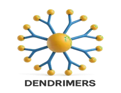
Fig.01: D structure of dendrimers
Because of their special capacity to deliver medications precisely and precisely, dendrimers are becoming more potent instruments in nanomedicine. Enhancing anticancer medications' internal behavior is one of the main issues facing cancer treatment today. Attaching polymers to these medications has shown promise in making them more stable, soluble in water, and less likely to cause unintended immune reactions. In solid tumors, drug–dendrimer conjugates tend to accumulate at the target site, which not only improves solubility but also limits harmful side effects elsewhere in the body. Researchers have explored different ways to load therapeutic agents, genetic material, targeting ligands, and imaging dyes into dendrimers through encapsulation, complexation, or chemical bonding taking advantage of their highly organized structures. Because of these features, dendrimers are opening new possibilities in both cancer treatment and diagnostic imaging. Recent studies, for example, highlight how poly(amidoamine) (PAMAM) dendrimers can carry drugs like 5-fluorouracil and methotrexate directly into skin cancer cells, enhancing drug penetration and retention in tumors while reducing toxicity to the rest of the body
Advantages
when administered with controlled release devices targeted administration minimizes damage to healthy cells by concentrating medication treatments on diseased areas at lesser dosages drugs can have long- lasting therapeutic effects when drugs that arent very soluble in water become more soluble this is known as bioavailability these devices versatility allows for simultaneous imaging and therapy which makes it possible to employ cutting-edge techniques like theranostics.
concerns such potential cytotoxicity immunogenicity and manufacturing scalability persist despite promising results future research should focus on long-term safety studies regulatory approvals and the development of cost-effective production methods for the targeted administration of drugs in the treatment of skin cancer dendrimer nanoparticles provide a flexible and efficient platform because of their distinct structural characteristics drugs may be administered precisely which may enhance patient outcomes and lessen adverse effects more studies and clinical trials are required to fully comprehend their potential in cancer
Liposomes In Drug Delivery
Nanotechnology's particular benefits in drug transport, imaging, and diagnostics have led to a growing awareness of its potential in cancer treatment. Preclinical and clinical studies have examined a variety of nanocarriers; those that combine many advantageous characteristics have significant therapeutic potential. Some examples that have been employed to improve the administration of therapies including immunotherapy, radiation therapy, gene therapy, and chemotherapy are liposomes, polymeric micelles, inorganic nanoparticles, drug conjugates, and virus-like nanoparticles. The limitations of cancer vaccines, tyrosine kinase inhibitors, and therapeutic monoclonal antibodies have been steadily addressed by nanotechnology-based approaches, in addition to notable advancements in immunotherapy. Liposomes are a promising nanotechnology-based approach in cancer immunotherapy. These lipid-based nanoparticles have been applied in numerous clinical trials for purposes such as vaccination and targeted cancer treatments. One notable liposomal formulation, Doxil, has received approval for cancer therapy. Because liposomes can encapsulate or associate with a variety of therapeutic agents including peptides, proteins, antibodies, and small-molecule chemotherapeutics. They offer significant potential to enhance the effectiveness of cancer immunotherapies
A variety of drugs have been incorporated into liposomes to improve the efficacy of skin cancer treatment for example encapsulating doxorubicin enhances tissue penetration while minimizing cardiotoxicity similarly liposomal delivery of 5- fluorouracil 5-fu increases retention in the skin and reduces the risk of local irritation natural compounds such as curcumin and resveratrol which possess anticancer properties also experience enhanced solubility and stability when administered via liposomal carriers Liposome-based therapies enable site-specific treatment of breast cancer, enhancing the therapeutic efficacy of the associated drug by improving its pharmacokinetics and pharmacodynamics and ensuring delivery to the targeted site. Despite being powerful anticancer medications, anthracyclines harm the body's rapidly dividing cells, such as the blood, hair follicles, and the gastrointestinal tract's mucosa. To assist prevent this off-target damage, these medications are encapsulated in liposomes. Because liposomal delivery concentrates the drug at the tumor site, it improves therapeutic efficacy and reduces negative side effects.
Liposome nanocarriers have a great deal of potential, but in order to increase their therapeutic value, a number of issues need to be resolved. Two significant challenges still remain: scaling up manufacturing and maintaining stability during production. Effective medication penetration for topical treatments is restricted by the skin barrier, and the sophisticated nature of these nanocarrier systems complicates regulatory clearance procedures. To assure long-term safety and efficacy, future research is thus concentrating on creating multifunctional liposomes that may integrate therapeutic administration with diagnostic imaging, investigating patient-specific customized nanomedicine techniques, and carrying out extensive clinical studies
Quantum Dots
There are several reasons why liposome nanocarriers are better than conventional cancer treatments. They reduce systemic toxicity and protect healthy tissues by facilitating localized drug delivery. Because the encapsulated drugs may concentrate at tumor sites, their stability and bioavailability are enhanced, and therapy accuracy is raised. These characteristics often allow effective treatment with lower doses by decreasing the likelihood of side effects. Additionally, liposomes may be modified for combination therapies, such as combining chemotherapy with photodynamic therapy, which provides a more comprehensive and effective way to treat cancer. Skin cancer is still one of the most prevalent malignancies in the world, including melanoma and non-melanoma types. Although traditional therapies like chemotherapy, radiation, and surgery are often used, they frequently have drawbacks such drug resistance, systemic side effects, and inaccurate targeting. To improve the accuracy and efficacy of drug administration in this environment, nanotechnology- specifically, quantum dots-offers a unique platform, especially when paired with imaging tools for more precise and customized skin cancer therapy
Quantum dots combine real-time imaging and tailored medication delivery to create efficient theranostic agents. They can more precisely deliver drugs by preferentially binding to receptors that are overexpressed on cancer cells when coupled with certain ligands, such folic acid. Furthermore, it is possible to create quantum dots that will release therapeutic chemicals in response to enzyme activity, temperature changes, or pH variations, allowing for targeted and regulated therapy. Additionally, their inherent fluorescence makes it possible to track the location of tumors and the distribution of drugs in real time, which offers useful feedback throughout treatment
Diagnostic Imaging:
Because they make tumor-specific biomarkers more visible quantum dots qds are highly effective imaging techniques for the early diagnosis of melanoma64 the precision of tumor localization is enhanced by their strong and consistent fluorescence
Drug Delivery
Molanoma cells have been effectively treated with doxorubicin and other chemotherapeutic drugs using quantum dots (QDs). Research demonstrates enhanced cytotoxic effects on cancer cells while minimizing damage to nearby healthy tissues
Photodynamic and Photothermal Therapy
As energy donors in photodynamic treatment (PDT), quantum dots (QDs) trigger photosensitizers that produce reactive oxygen species, which destroy cancer cells. QDs use photothermal treatment to locally ablate tumor tissues by converting near-infrared light into heat
Quantum dots provide several key advantages in biomedical applications, especially in cancer therapy. Their high specificity and selectivity enable precise targeting of diseased cells, greatly improving therapeutic accuracy. Owing to their multifunctional capabilities, they can simultaneously facilitate both imaging and drug delivery, making them well-suited for integrated diagnostic and therapeutic strategies. Efficient targeting also allows for lower drug doses, minimizing potential side effects. Additionally, the surface of quantum dots can be readily modified with various functional groups, offering flexibility for tailored applications.
There are some disadvantages to quantum dots despite their great potential. Since many quantum dots include potentially hazardous heavy metals like cadmium, toxicity is a major issue. Because of their inadequate body clearance and low biodegradability, they also raise long-term safety concerns. These concerns, in addition to regulatory barriers and a lack of comprehensive long-term safety data, have hindered the widespread adoption of quantum dot-based medications in clinical settings.
Current research is focusing on alternatives such as carbon- and silicon-based quantum dots, which are non-toxic and biodegradable. Additionally, developments in biosensors, microneedle patches, and combination therapies (for example, QDs paired with siRNA) are being explored to enable more personalized and effective treatments
Polymeric Nanoparticles:
For targeted drug delivery, polymeric nanoparticles (PNPs) have become a flexible and exciting platform in nanomedicine. Because of their adaptable size, safe degradability, biocompatibility, and capacity to transport a variety of therapeutic substances, they are ideal for the treatment of skin cancer. This study focuses on melanoma and discusses current developments in the design and production of polymeric nanoparticles, as well as their processes and uses in the treatment of skin cancer. It also discusses the difficulties facing this subject now and possible future paths. For targeted drug delivery in nanomedicine, polymeric nanoparticles (PNPs) have shown great promise and versatility. Skin cancer can be effectively treated with them because of their customizable size, biocompatibility, biodegradability, and capacity to carry a broad range of therapeutic chemicals. Recent developments in the synthesis and engineering of polymeric nanoparticles are reviewed in this article, along with an investigation into their modes of action and potential uses in the treatment of skin malignancies, especially melanoma. It also talks about the difficulties that exist now and outlines possible paths forward in this field
Natural or manufactured polymers, including polylactic acid (PLA), chitosan, poly(lactic-co-glycolicacid) (PLGA), and polyethylene glycol (PEG), combine to form polymeric nanoparticles, which are colloidal systems. In general, they are between 10 and 1000 nmin size and may be designed to encapsulate both hydrophilic and hydrophobic medications
PLGA: Biodegradable polymer approved by the FDA.
Chitosan: Exhibits mucoadhesive properties and improves skin permeability.
PEG: Extends circulation time and lowers immunogenicity.
Because of their nanoscale size and surface characteristics, polymeric nanoparticles can pass through the stratum corneum and settle in the deeper layers of the skin. Multiple pathways can lead to the release of drugs from these nanoparticles: stimuli- responsive release, which is triggered by particular factors like pH shifts, temperature changes, or enzymatic activity at the tumor site; diffusion- controlled release, where the drug slowly moves through the polymer matrix; and erosion-controlled release, which involves the degradation of the polymer to free the drug
Topical Delivery: Polymeric nanoparticles applied topically enhance drug penetration and retention in the skin, making them well-suited for treating superficial skin cancers. Chitosan-based PNPs, in particular, have demonstrated effective delivery across the skin barrier. Gene Therapy and Immunotherapy: Polymeric nanoparticles can be utilized to carry siRNA/miRNA and immunomodulatory drugs to alter cancer cells or strengthen the immune system. For instance, PLGA nanoparticles containing anti-PD-1 antibodies have shown encouraging results in models of melanoma.
Photodynamic Therapy: Polymeric nanoparticles can encapsulate photosensitizing agents for localized, light- triggered cytotoxic effects. Controlled release upon irradiation helps minimize damage to surrounding healthy skin
The use of polymeric nanoparticles in skin cancer therapy offers several advantages, including the ability to deliver medications to tumor cells precisely, control and sustain drug release, improve drug stability and solubility, lower systemic toxicity, and integrate therapeutic agents with imaging molecules for multifunctional treatment strategies
Notwithstanding their potential, polymeric nanoparticles face a number of difficulties and restrictions. Their ability to penetrate the skin can be restricted by the stratum corneum's ability to function as a barrier. These nanoparticles' stability and integrity may also be impacted by environmental influences. Because of safety and long-term toxicity concerns, only a small number of formulations have advanced to clinical trials, making it difficult to scale up manufacturing while maintaining consistent quality and regulatory obstacles
FUTURE PERSPECTIVES
The development of intelligent, stimuli-responsive systems that react to certain triggers is the main goal of future polymeric nanoparticle research. The integration of these nanoparticles with microneedle patches to improve medication delivery and epidermal penetration is gaining traction. Additionally, biodegradable natural polymers like alginate and hyaluronic acid are being used more often. Furthermore, a promising avenue for more potent treatments for skin cancer is provided by individualized nanomedicine strategies catered to specific tumor indicators
CONCLUSION
For targeted treatment of skin malignancies such as melanoma, squamous cell carcinoma, and basal cell carcinoma, nanocarriers are a potential development. Liposomes, polymeric nanoparticles, dendrimers, gold nanoparticles, and quantum dots are examples of nanocarriers that carry medications straight to the tumor location, overcoming the drawbacks of traditional chemotherapy. Therapeutic efficacy is increased and systemic toxicity is decreased by this focused administration. There are unique benefits and drawbacks associated with each form of nanoparticle. Their shown promise in diagnosis and therapy need more study to assure their safety and enable practical usage.
REFERENCES

Quadri Mohammed Soheb*, Rajeev Kumar Malviya, A Review on Nanocarrier for Combination Delivery for Skin Cancer, Int. J. Med. Pharm. Sci., 2026, 2 (8), 726-736. https://doi.org/10.5281/zenodo.22094733
 10.5281/zenodo.22094733