

We use cookies to ensure our website works properly and to personalise your experience. Cookies policy
1Bachelor of Pharmacy, Pallavan Pharmacy College, Kanchipuram, India.
2Assistant professor, Department of Pharmacology, Pallavan Pharmacy College, Kanchipuram, India.
3Principal, Head of Department of Pharmacognosy, Pallavan Pharmacy College, Kanchipuram, India.
4Vice Principal, Head of Department of Pharmacology, Pallavan Pharmacy College, Kanchipuram, India
Nowadays, Nanotechnology has emerged as a powerful platform for antimicrobial therapy due to the unique physicochemical properties of nanomaterials rapid development of multidrug-resistant (MDR) microorganisms, along with the reduced effectiveness of conventional antimicrobial agents, necessitating the development of alternative, safe, and efficient antibacterial agents. In this context, the synthesis of nanoparticles from plant derived compounds and biodegradable polymers is being increasingly recognized as a promising approach due to their environmentally friendly properties as well as improved biological performance. In the current study, a green synthesis method was employed to synthesize quercetin-functionalized silver nanoparticles (Q-AgNPs) using oxidized amylose as a biopolymeric matrix, stabilizer, and dispersing agent. Oxidized amylose is a naturally occurring polysaccharide that contains many aldehyde and hydroxyl groups, which play a significant role in the synthesis of nanoparticles. In addition, silver nanoparticles with quercetin functionalization adds antioxidant power, antimicrobial synergy, and anti-inflammatory effects. It enables green synthesis with quercetin acting as a natural reducing/capping agent for better stability. It enhances free-radical scavenging beyond free quercetin's capability. It strengthens antibacterial action through dual ROS and cytokine-based mechanisms. It also lowers toxicity to healthy cells while preserving antimicrobial efficacy. The successful synthesis of nanoparticles was confirmed through a series of comprehensive analytical tests, including UV-Vis spectroscopy, XRD, FTIR, NTA, and FEG-TEM analysis, which indicated the synthesis of highly crystalline, spherical, and uniformly distributed nanoparticles of 32 nm size. The oxidized amylose-stabilized quercetin-functionalized silver nanoparticles were found to possess improved antioxidant and broad-spectrum antibacterial properties compared to quercetin alone, which demonstrated improved antibacterial activity against pathogenic bacteria responsible for wound and systemic infections. Thus, the synergistic effect of phytoconstituents and oxidized amylose-functionalized silver nanoparticles act as hybrid agent, eco-friendly approach to overcome bacterial resistance for the development of novel antibacterial agents.
Quercetin is a naturally occurring flavonoid widely distributed in fruits, vegetables, and medicinal plants such as onions, apples, berries, and tea. It is a prominent phytoconstituent and a foundational component of flavonoids [2] Chemically defined as 3,3′,4′,5,7-pentahyroxyflavone, it consists of a molecular structure featuring three interconnected rings: two aromatic rings (A and B) and a heterocyclic ring (C) enriched with five hydroxyl groups, which are pivotal for its biological activities. It has attracted significant scientific attention due to its broad spectrum of biological activities, including antioxidant, anti-inflammatory, anticancer, and antimicrobial properties (Fig1). The antimicrobial activity of quercetin is particularly important in the current era of increasing multidrug-resistant (MDR) microorganisms. Quercetin exerts its antimicrobial effects through multiple mechanisms, including disruption of microbial cell membranes, inhibition of nucleic acid and protein synthesis, interference with energy metabolism, and suppression of virulence factors. It has demonstrated effective antibacterial activity against both Gram-positive and Gram-negative bacteria, as well as antifungal and antiviral properties. Moreover, quercetin is considered a safe, biodegradable, and ecofriendly compound, making it a promising candidate for green antimicrobial applications. Its ability to act as a reducing and stabilizing agent further enhances its potential use in nanoparticle synthesis for improved antimicrobial efficacy.
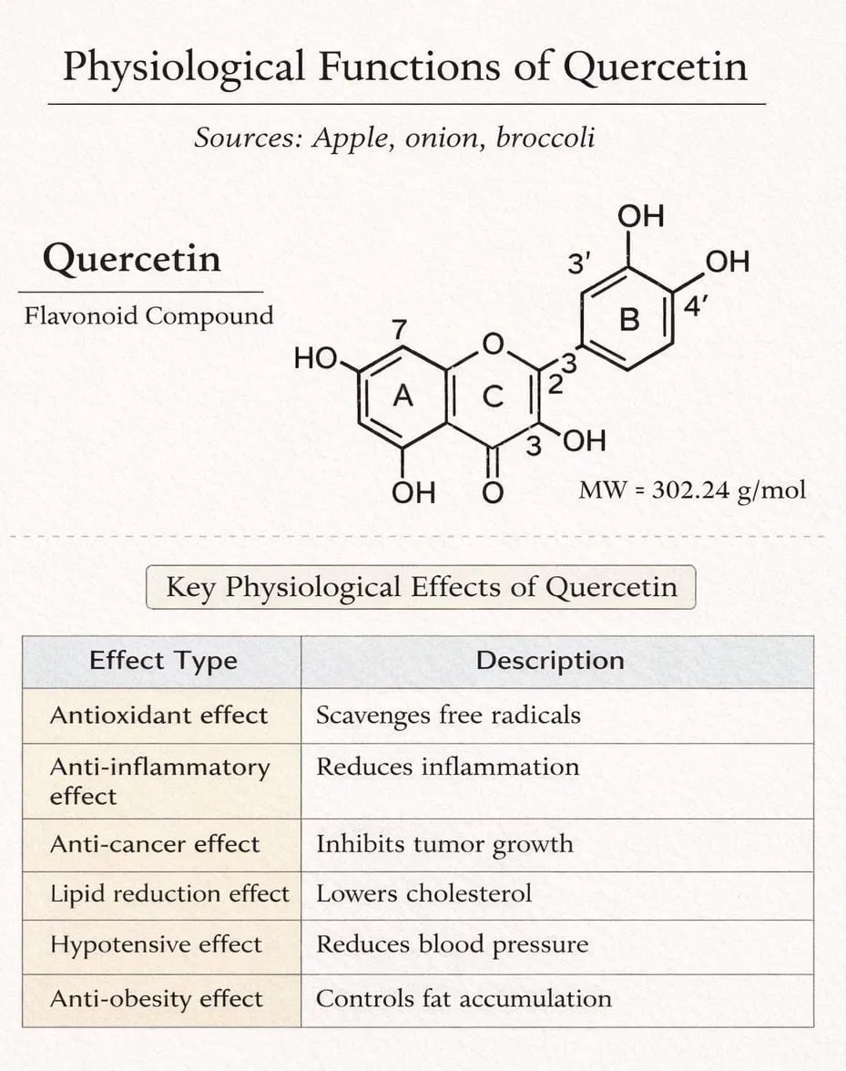
Fig1: Sources, Structure & Functions of Quercetin
Despite its wide range of therapeutic benefits, quercetin flavonoid suffer from poor intestinal absorption, resulting in limited systemic bioavailability and restricting its pharmacological applications. However, its therapeutic efficacy is challenged by its limited aqueous solubility, rapid metabolism, and the action of bacterial efflux pumps, which reduce its intracellular concentration. These limitations hinder its ability to exert sustained antibacterial effects and restrict its efficacy in combating systemic infections. To improve stability of a quercetin, nanoparticles like gold, silver, and copper were probably used. In this work, the silver nanoparticles were taken. The AgNp’s synthesized using Quercetin especially to increase its anti-microbial activity and synergistic effect. This combination improves microbial membrane disruption, reactive oxygen species (ROS) generation, and inhibition of cellular enzymes and DNA replication. This study developed a hybrid antibacterial agent by using silver nanoparticles and Quercetin Oxidized amylose. Silver nanoparticles (AgNPs), possess unique physico-chemical properties, including size-dependent behavior, high surface-area-to-volume ratio, and excellent biocompatibility, making them ideal candidates for drug delivery and therapeutic applications. Functionalizing nanoparticles with quercetin enhances its bioavailability, enables sustained release, and facilitates targeted delivery to the site of action. Biogenic synthesis approaches offer a sustainable and eco-friendly strategy to fabricate nanoparticles using natural compounds as reducing and stabilizing agents. Oxidized amylose, a modified biopolymer derived from starch, has garnered attention for its enhanced antimicrobial properties. The oxidation process introduces functional groups that confer potent activity against microbial pathogens, including bacteria and fungi. As a result, oxidized amylose is being explored as a potential agent for developing novel antimicrobial materials, such as wound dressings, food packaging, and therapeutic agents. Its biocompatibility, biodegradability, and potential for synergistic effects with other antimicrobials make it an attractive candidate for addressing antimicrobial resistance and promoting public health.
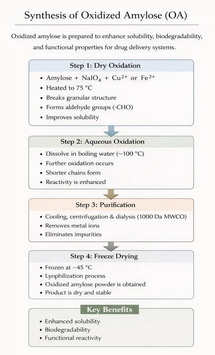
Fig 2: Synthesis of Oxidized Amylose
Instead Amylose, oxidized amylose possesses greater solution stability than native amylose primarily because the introduction of carboxyl and carbonyl groups during oxidation reduces the tendency of the molecules to rearrange into insoluble crystalline structures, a process known as retrogradation. While native amylose has low water solubility and tends to precipitate (retrograde) in water, oxidation breaks down the hydrogen bonds, increasing water solubility, and introducing electrostatic repulsive forces between the chains. This functionalized hybrid agent hold promises for overcoming the limitations of free quercetin, enabling enhanced therapeutic efficacy in both antibacterial and anticancer applications
MATERIALS AND METHODOLOGY
MATERIALS
The Quercetin (99%pure) was procured from Sigma–Aldrich, silver nitrate (AgNO3 99.9%), Amylose (99%) [The average degree of polymerization was 1230 according to gel permeation chromatography (GPC) analysis Hydrogen peroxide (H2O2, 30.0%), hydrochloric acid (HCl, 37.0%), and α-amylase, agar– agar, nutrient broth, antibacterial discs, DPPH, experiments were conducted using deionized water as the solvent medium.
METHODOLOGY
Method for the preparation of quercetin -silver Nanoparticles with oxidized amylose (Fig3):
Quercetin Oxidized amylose was first used as environmentally friendly reducing agent and stabilizer to synthesize silver nanoparticles. The preparation of (Q-AgNPs) involves dissolving 3 mg of quercetin in 100 mL of deionized water, followed by sonication for 5 min to ensure complete dissolution. Then, 1 mL of a 1 mM aqueous AgNO₃ solution is added to the quercetin solution, and the mixture is exposed to sunlight for approximately 5 min. The solution turns brown, indicating the successful formation of Q-AgNPs. Then Q-AgNP’s was added to the mixture of Oxidized amylose during the cooling process. The quercetin would be encapsulated into the cavity of helices of oxidized amylose to obtain the hybrid antibacterial agent. The optimal concentration of quercetin 3 mg was achieved by using UV–visible spectrophotometry, as no further increase in the absorption peak was observed, indicating that the nanoparticles remained stable at this concentration. Quercetin served as both a capping and reducing agent, facilitating the reduction of metal ions and stabilizing the formed nanoparticles. Statistical analysis was conducted to evaluate antibacterial activity, by ensuring clarity and accuracy.
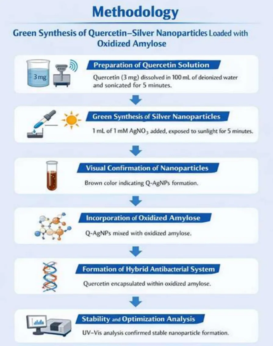
Fig 3: Synthesis of Q-Ag NP’s with Oxidized Amylose
CHARACTERIZATION
UV–VIS spectroscopy
It was confirmed that quercetin was successfully attached to the surface of silver nanoparticles using a special tool called UV-vis spectroscopy. The light absorbed by the quercetin and the quercetin-silver nanoparticles over a range of wavelengths, from 190 to 1100 nanometers, using a Shimadzu UV 1900i spectrophotometer. The baseline was carefully corrected using double distilled water, so we could get accurate results. This was an important step to ensure our measurements were reliable.
Fourier transform infrared (FTIR)
To understand how quercetin interacts with the nanoparticles, a technique FTIR spectroscopy was used. A spectra was obtained using a PerkinElmer instrument, looking at a range of 400-4000 cm⁻1 and scanning 16 times. By comparing the spectra of quercetin and quercetin nanoparticles, the parts of the molecule were involved in forming and stabilizing the nanoparticles. This helps to figure out how the nanoparticles work. This shows that certain groups of atoms were playing a key role in making the nanoparticles stable.
X‑ray powder diffraction (XRD) analysis:
To look at the structure of quercetin and Q-AgNPs, a special XRD tool called a Rigaku Miniflex600 was used which helps to understand what the tiny particles are made of. The readings were taken over a range of 20 to 80°, moving in small steps of 0.02°, and doing it quickly at a rate of 4° per minute. The readings were figured out by comparing them to standard patterns from a big database called ICDD, which has a special card number, 01– 0893722, used as a reference. This helps to identify the different parts that make up the samples.
Field emission gun‑transmission electron microscope 200 kV (FEG‑TEM 200kV)
The morphological, structural, and size distribution characteristics of the synthesized Q-AgNPs determined using FEG-TEM on a FEI Tecnai G2, F30 instrument operated at an accelerating voltage of 200kv to take a closer look at the tiny particles. The images were taken at different zoom levels, from 58 times bigger to 1 million times bigger, to find all the details. This helps to understand the structure of the particles well. To figure out the size of the particles, a technique called NTA, which was done with equipment from Malvern Instruments Ltd in the UK.
Assessment of antibacterial activity by Disk diffusion method:
The disk diffusion method is a phenotypic technique where a paper disk impregnated with a known concentration of an antimicrobial agent is placed on an agar plate inoculated with a test microorganism. The antimicrobial agent diffuses into the agar, creating a concentration gradient. After incubation, the diameter of the zone of inhibition (clear area where microbial growth is inhibited) around the disk is measured to assess the susceptibility of the microorganism.
RESULT AND DISCUSSION:
Preparation and characterization of AgNPs by UV–visible spectroscopy:
Successfully quercetin-mediated silver nanoparticles (Q-AgNP) were synthesized. Quercetin acted as both a reducing and stabilizing agent. The formation of nanoparticles was confirmed through UV– visible spectroscopy by the appearance of a characteristic localized surface plasmon resonance (LSPR) band at 430 nm. This peak is indicative of nanoscale silver formation with defined size distribution and morphology. A visible color transition from a clear solution to pale brown further supported nanoparticle formation. The presence of a single, sharp absorption peak suggests the formation of a relatively uniform and stable nanoparticle dispersion. (Fig4)
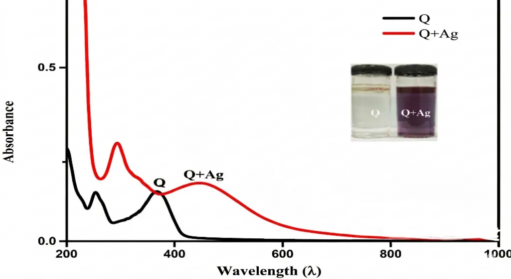
Fig 4: UV -Vis spectra showing the characteristic LSPR Peaks of Q-AgNPs at 430nm, Confirming the formation of silver nano particles in solution.
Fourier Transform Infrared Spectroscopy (FTIR):
FTIR analysis revealed notable differences between pure quercetin and QAgNPs, confirming the involvement of functional groups in nanoparticle synthesis. The hydroxyl (O–H), carbonyl (C=O), and ether (C–O) groups observed in quercetin showed reduced intensity or disappearance in Q-AgNPs, indicating their participation in the reduction of silver ions and stabilization of nanoparticles. Additionally, aromatic C=C stretching and C–H bending vibrations were partially retained, suggesting their contribution to surface capping. These findings support the role of quercetin as an electron donor, facilitating the reduction of Ag⁺ ions into metallic silver nanoparticles while simultaneously preventing aggregation (Fig 5).
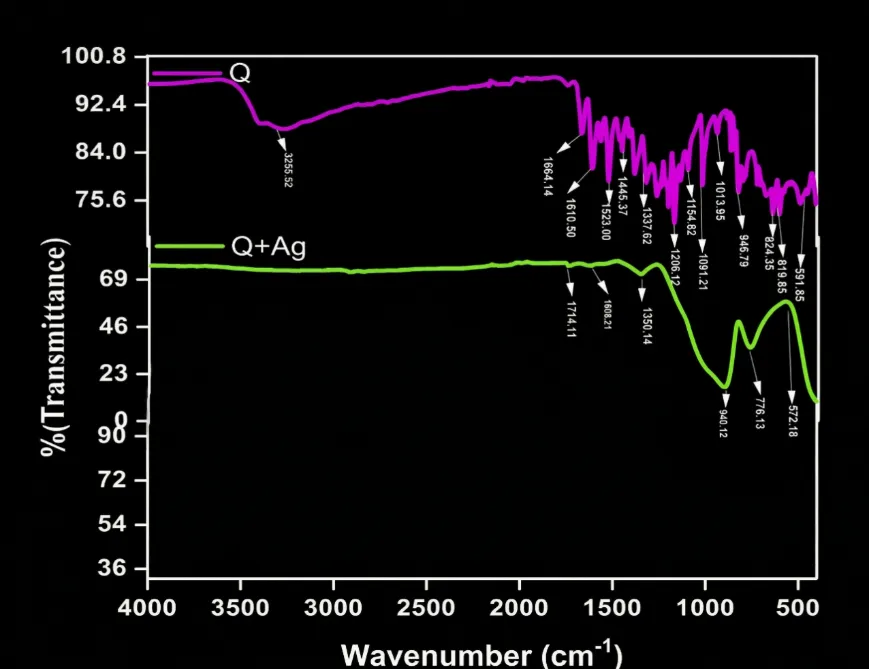
Fig 5: FT-IR spectra represent the functional groups of Q and Q-AgNPs.
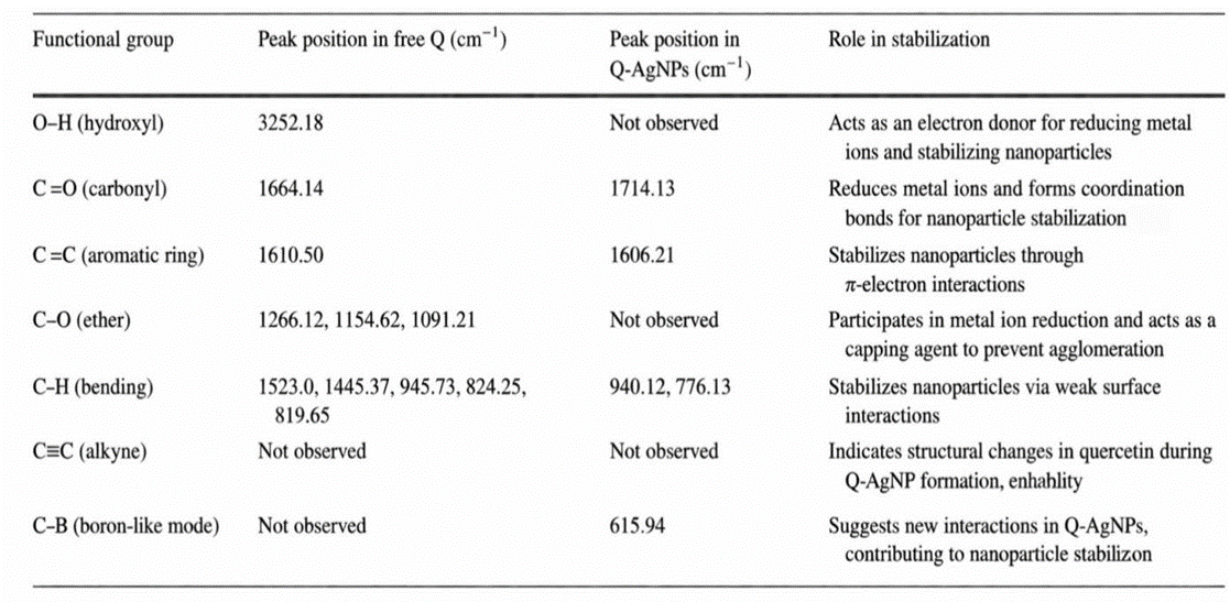
Fig 6: Comparative table summarizing the functional groups identifed in free quercetin and Q-AgNPs based on the FTIR analysis.
X-ray Diffraction (XRD):
The crystalline nature of the synthesized Q-AgNPs was examined using X-ray diffraction analysis following purification through repeated centrifugation and washing. The XRD pattern confirmed the presence of well-defined crystalline structures, providing insight into lattice arrangement and particle phase purity.
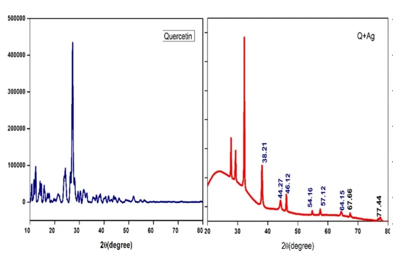
Fig.7: X-ray difraction of Qu and Q-AgNPs at 2θ values.
Transmission electron microscopy:
The morphology and size distribution of Q-AgNPs were analyzed using transmission electron microscopy. The samples were prepared by placing nanoparticle suspensions onto carbon-coated copper grids and allowing them to dry before imaging. TEM images revealed predominantly spherical nanoparticles with smooth surfaces and an average diameter of approximately 32 nm. The particles were closely spaced but maintained a consistent interparticle distance, indicating good dispersion and stability. These structural characteristics are crucial, as the biological activity of QAgNPs is strongly influenced by their physicochemical properties such as size, shape, and surface functionality. (Fig:8)
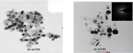
Fig:8 TEM images of Quercetin silver nanoparticles with average size of 32nm at 200kv.
Antibacterial Activity of Quercetin-Mediated AgNPs:
The antibacterial activity of quercetin, Q-AgNPs, and chloramphenicol (positive control) was evaluated using the disk diffusion method against both Gram-positive bacteria (Staphylococcus aureus and Bacillus subtilis) and Gram-negative bacteria (Salmonella typhi and Escherichia coli). (Fig:9) Q-AgNPs exhibited enhanced antibacterial activity compared to free quercetin, with inhibition zones measuring 15 ± 1 mm (S. aureus), 13 ± 2 mm (S. typhi), 9 ± 2 mm (E. coli), and 6 ± 1 mm (B. subtilis). Table: 1 The improved antimicrobial performance can be attributed to the synergistic effect of quercetin and silver nanoparticles. These nanoparticles enhance cellular interaction, disrupt bacterial membranes, and increase permeability, leading to leakage of intracellular components and eventual cell death. Overall, the results highlight the potential of quercetin-functionalized silver nanoparticles as effective antimicrobial agent.
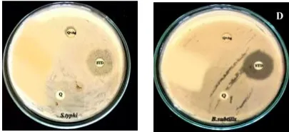
Fig9: Comparative efficacy of Q-AgNPs inhibiting the growth of bacterial species S. typhi & B. subtilis (Standard: Chloramphenicol)
Table1: Zone of inhibition diameter (mm)
|
Microbial strain |
Chloramphenicol (mm) |
Q (mm) |
Q-AgNPs (mm) |
|
Staphylococcus aurens |
31 ± 02 |
01 ± 02 |
15 ± 02 |
|
Salmonella typhi |
34 ± 01 |
06 ± 01 |
13 ± 01 |
|
Escherichia coli |
29 ± 01 |
03 ± 01 |
08 ± 02 |
|
Bacillus subtilis |
46 ± 01 |
01 ± 02 |
07 ± 01 |
CONCLUSION:
In conclusion, the present work establishes a sustainable and biologically relevant strategy for the green synthesis of silver nanoparticles using quercetin, with oxidized amylose serving as an effective biopolymeric stabilizer and carrier system. The integration of a flavonoid-based reducing agent with a structurally modified polysaccharide enabled precise control over nanoparticle nucleation, growth, and long-term colloidal stability, while maintaining environmental compatibility. Comprehensive characterization confirmed the formation of well-dispersed, nanoscale silver particles with favorable surface functionality. The enhanced antimicrobial performance of the synthesized nanocomposite can be attributed to the synergistic interplay between silver ions, quercetin’s redox and membrane-disruptive properties, and the sustained release behavior imparted by oxidized amylose. This multifunctional architecture promotes improved interaction with microbial cell walls, increased reactive oxygen species generation, and effective inhibition of microbial proliferation. Importantly, the use of oxidized amylose minimizes nanoparticle aggregation and potentially reduces silver-associated cytotoxicity, thereby improving biocompatibility. Overall, this study highlights the potential of phytochemical–biopolymer engineered silver nanoparticles as advanced antimicrobial systems. The findings provide a foundation for the rational design of green nanomaterials with enhanced therapeutic efficacy and safety, paving the way for future investigations into mechanistic pathways, in vivo performance, and broader biomedical and pharmaceutical applications.
REFERENCES


A. Faizun Nisha*, G. Hemalatha, M. Vignesh, K. Kaviya, J. Karthi, S. Swarnalatha, Biogenic Synthesis and Characterization of Silver Nanoparticles Using Quercetin and Oxidised Amylose: Assessment of Antimicrobial Activity, Int. J. Med. Pharm. Sci., 2026, 2 (7), 457-467. https://doi.org/10.5281/zenodo.21273363
 10.5281/zenodo.21273363