View Article

  • A Review on Happy Hormone: Dopamine

  • 1Assistant Professor, Department of Pharmaceutical Quality assurance, Faculty of Pharmacy, SSSRGI, Vadasma, Mehsana, Gujarat, India- 382705.
    2Professor, Department of Pharmaceutical Chemistry, Faculty of Pharmacy, SSSRGI, Vadasma, Mehsana, Gujarat, India- 382705
     

Abstract

Dopamine is a vital neurotransmitter that plays a central role in the regulation of motivation, reward, mood, cognition, learning, movement, and several physiological processes. Although commonly referred to as a “happy hormone,” dopamine is technically a neurotransmitter and functions as an important chemical messenger within the central and peripheral nervous systems. It is synthesized primarily from the amino acid tyrosine through the sequential action of tyrosine hydroxylase and aromatic L-amino acid decarboxylase. Dopaminergic pathways, particularly the mesolimbic, mesocortical, nigrostriatal, and tuberoinfundibular pathways, are associated with reward and motivation, cognitive functions, motor control, and endocrine regulation, respectively. Alterations in dopamine signaling have been implicated in several neurological and psychiatric disorders, including Parkinson’s disease, schizophrenia, attention-deficit/hyperactivity disorder, depression, and substance-use disorders. Dopamine also influences physiological functions such as prolactin secretion, cardiovascular activity, and renal function. This review summarizes the biosynthesis, metabolism, receptors, signaling pathways, physiological functions, and clinical significance of dopamine. It also highlights the relationship between dopamine, reward mechanisms, lifestyle factors, and mental well-being. A better understanding of dopamine homeostasis may contribute to improved approaches for maintaining neurological health and managing dopamine-related disorders.

Keywords

Dopamine; Hormone; Neutological disorders; Mental health

Introduction

× Popup Image

What is Dopamine? [1-9]

Dopamine was first described by George Barger, James Ewens, and Henry Dale in 1910 as an epinephrine-like monoamine compound. However, in the 1950s Kathleen Montagu showed that dopamine occurred in the brain by itself, and a series of studies by Arvid Carlsson and collaborators demonstrated that dopamine is a bona fide neurotransmitter, a finding that would earn Carlsson the 2000 Nobel Prize in Physiology and Medicine. In a landmark experiment, he pharmacologically blocked all dopamine neurotransmission in rabbits, which rendered them completely paralyzed, and then fully recovered their behavior with an injection of the dopamine precursor L-DOPA, demonstrating that dopamine was essential for self-initiated movement. A similar effect was quickly reproduced by Oleg Hornykiewicz and collaborators in human Parkinsonian patients. Within a few years, dopamine jumped from relative obscurity to being critical for life as we know it. [1-2]

“Explore dopamine as a key neurotransmitter involved in reward, motivation, mood, cognition, and motor control, highlighting its biosynthesis, receptor-mediated signaling, physiological functions, and role in neurological and psychiatric disorders.”

Dopamine is an important neurotransmitter that plays a key role in the brain’s reward, motivation, pleasure, learning, and emotional regulation systems. It is commonly known as the “happy hormone,” although it is technically a neurotransmitter rather than a hormone. Dopamine is produced from the amino acid tyrosine and acts through different dopamine receptors in the brain and other parts of the body. Balanced dopamine levels are essential for normal mood, movement, attention, and cognitive function. Abnormal dopamine activity is associated with conditions such as Parkinson’s disease, schizophrenia, depression, and addiction. Thus, dopamine is crucial for maintaining both brain function and overall well-being. [3-5]

General Pharmacology of Dopamine

Dopamine is an endogenous catecholamine neurotransmitter and neurohormone that produces its effects by activating dopamine D₁–D₅ receptors and, at higher concentrations, β₁- and α₁-adrenergic receptors. Its pharmacological effects are dose-dependent: lower concentrations predominantly cause D₁-mediated renal and mesenteric vasodilation, moderate concentrations stimulate β₁ receptors and increase cardiac contractility and cardiac output, while higher concentrations activate α₁ receptors, producing vasoconstriction and increased blood pressure. Dopamine has a very short plasma half-life and is rapidly metabolized by monoamine oxidase (MAO) and catechol-O-methyltransferase (COMT). Clinically, intravenous dopamine has been used primarily for selected cases of acute circulatory failure, although its use is now more limited because alternative vasopressors and inotropes are often preferred. Adverse effects may include tachycardia, arrhythmias, hypertension, nausea, and peripheral vasoconstriction; extravasation can cause local tissue injury. [6,7]

Chemistry of Dopamine

Dopamine is a catecholamine neurotransmitter with the chemical name 3,4-dihydroxyphenethylamine and molecular formula C₈H₁₁NO₂. Structurally, it consists of a benzene ring containing two hydroxyl (–OH) groups at the 3rd and 4th positions (catechol group), attached to an ethylamine side chain. This catecholamine structure is responsible for its ability to participate in various biochemical and neuronal processes. [8,9]
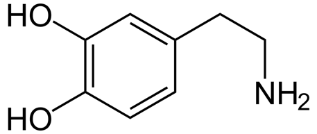
Figure 1. Structure of Dopamine

  • Chemical name: 3,4-Dihydroxyphenethylamine  
  • Molecular formula: C8H11NO2
  • Molecular weight: 153.18 g/mol
  • Functional groups: Two hydroxyl groups and one primary amine group
  • Chemical class: Catecholamine neurotransmitter

Synthesis, Storage, Release and Re-uptake of Dopamine [10-13]

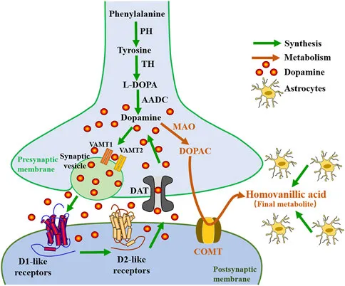
Dopamine is synthesized in presynaptic dopaminergic neurons and stored in synaptic vesicles before being released into the synaptic cleft. Its movement through the synapse occurs through three major steps:

  1. Synthesis:

Dopamine (DA) is produced in the neuronal cytoplasm via tyrosine hydroxylase (TH) acting on tyrosine to form 3,4-dihydroxyphenylalanine (DOPA) and then L-aromatic-amino-acid decarboxylase (LAAAD) acting on DOPA to form dopamine.
  1. Storage:
    Newly synthesized dopamine is transported into synaptic vesicles by the vesicular monoamine transporter-2 (VMAT2). Vesicular storage protects dopamine from degradation and prepares it for neurotransmission.
  2. Release:
    When an action potential reaches the presynaptic terminal, voltage-gated calcium channels open, causing calcium ions (Ca²⁺) to enter the neuron. This triggers vesicle fusion with the presynaptic membrane and releases dopamine into the synaptic cleft by exocytosis. Dopamine then binds to D1-like and D2-like receptors on target cells.
  3. Re-uptake:
    After its action, dopamine is primarily removed from the synaptic cleft by the dopamine transporter (DAT) located on presynaptic neurons. The transported dopamine can be repackaged into vesicles by VMAT2 or metabolized by enzymes such as monoamine oxidase (MAO) and catechol-O-methyltransferase (COMT).

Figure 2. Synthesis, release and re-uptake of Dopamine

Effect of Dopamine on the Nervous System [14, 15]

Dopamine is an important neurotransmitter that regulates several functions of the central and peripheral nervous systems. Its effects depend on the brain region, receptor type, and amount of dopamine released.

Major effects include:
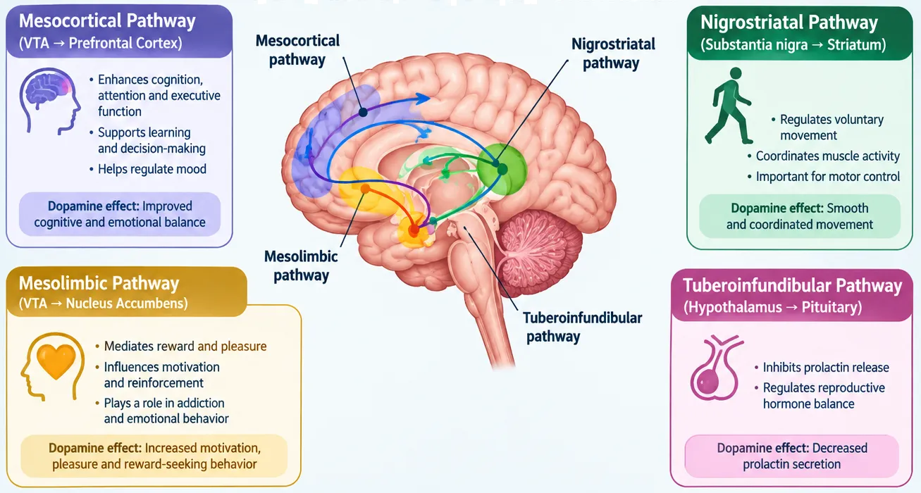
  • Reward & Pleasure: Dopamine contributes to the brain’s reward and reinforcement system, influencing feelings of satisfaction and motivation.
  • Motivation & Behavior: It promotes goal-directed behavior, attention, and the drive to perform rewarding activities.
  • Movement Control: Dopamine in the nigrostriatal pathway helps regulate voluntary movement. Reduced dopamine activity in this pathway is associated with Parkinson’s disease.
  • Learning & Memory: Dopamine supports learning, memory formation, attention, and cognitive flexibility.
  • Mood & Emotions: Dopaminergic signaling contributes to emotional regulation and overall mental well-being.
  • Hormonal Regulation: Dopamine released from the hypothalamus inhibits prolactin secretion from the pituitary gland.
  • Autonomic Functions: Dopamine also influences cardiovascular, renal, and other autonomic functions.

Figure 3. Effects of Dopamine on nervous system

Signs of Dopamine Imbalance [16, 17]

Dopamine imbalance refers to altered dopamine signaling or activity, rather than simply having a measurable “high” or “low” dopamine level. Depending on the affected brain pathway, it may influence mood, motivation, cognition, movement, and behavior.

  • Low dopamine activity: reduced motivation, fatigue, difficulty concentrating, low mood, reduced pleasure (anhedonia), and slowed movements.
  • Excessive or dysregulated dopamine activity: increased impulsivity, agitation, heightened reward-seeking, sleep disturbance, or psychotic symptoms such as hallucinations or delusions in certain disorders.
  • Movement-related changes: tremor, rigidity, or slowed movement can occur when dopamine signaling is significantly reduced in motor pathways, as seen in Parkinson’s disease.
  • Cognitive and behavioral changes: changes in attention, learning, decision-making, and reward-driven behavior may occur with disrupted dopamine signaling.

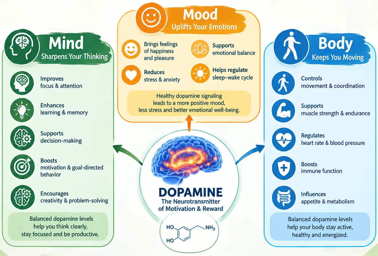
How Dopamine aeffects the Mind, Mood & Body

Dopamine is a neurotransmitter that helps coordinate communication between nerve cells. It influences the mind, mood, behavior, and several body functions through different neural pathways.

Table 1. Effect of Dopamine on different area

Area

Effect of Dopamine

Mind

Supports attention, learning, memory, focus, decision-making, and motivation.

Mood

Contributes to pleasure, reward, satisfaction, and emotional responses. Dopamine is more strongly linked to motivation and reward-seeking than to happiness itself.

Motivation

Encourages goal-directed behavior and reinforces activities that the brain considers rewarding.

Movement

Helps control voluntary movements, coordination, and motor function.

Body

Influences heart and blood-vessel function, kidney function, and sympathetic nervous activity.

Hormonal control

Dopamine from the hypothalamus helps regulate prolactin secretion by the pituitary gland.

Balance

Both too little and too much/disrupted dopamine signaling can contribute to neurological and psychiatric problems.

Figure 4. Effect of Dopamine on Mind, Mood & Body

Dopaminergic Reward System [20-22]

The dopaminergic reward system is a neural network that regulates reward, motivation, pleasure, learning, and goal-directed behavior. Dopamine-producing neurons in the ventral tegmental area (VTA) project mainly to the nucleus accumbens and prefrontal cortex, forming important pathways involved in reward and motivation.

Major components of reward system:

  • VTA: Major source of dopamine neurons.
  • Nucleus Accumbens: Processes reward, reinforcement, and motivation.
  • Prefrontal Cortex: Involved in decision-making, cognition, and behavioral control.
  • Amygdala & Hippocampus: Contribute to emotional and memory-related aspects of rewarding experiences.

Dopamine signaling is particularly important for reinforcement learning—helping the brain learn which behaviors or experiences are rewarding and increasing the likelihood of repeating them.

Dopamine and disease [20-24]

Dopamine-related diseases are generally caused by abnormal dopamine signaling in specific brain pathways, rather than simply having “high” or “low” dopamine throughout the body.

Table 2. Diseases associated with high or low level of dopamine

Dopamine status / signaling

Associated conditions

Low dopamine activity

Parkinson’s disease, depression, anhedonia (reduced ability to experience pleasure), and some cognitive/motivational disorders

High or excessive dopamine activity

Schizophrenia/psychosis (particularly increased D2 signaling in some pathways), mania, and certain forms of impulsive or addictive behavior

Dysregulated dopamine signaling

ADHD, substance-use disorders, restless legs syndrome, and other neurological/psychiatric conditions

Parkinson’s disease

Parkinson’s disease (PD) is a progressive neurodegenerative disorder characterized mainly by bradykinesia (slowness of movement), rigidity, resting tremor, and postural instability. A major pathological feature is the degeneration of dopamine-producing neurons in the substantia nigra pars compacta, resulting in reduced dopamine availability in the nigrostriatal pathway. This dopamine deficiency disrupts the basal ganglia circuits responsible for coordinated voluntary movement, leading to the characteristic motor symptoms of Parkinson’s disease.

Pharmacological significance: Since dopamine itself does not effectively cross the blood–brain barrier, levodopa (L-DOPA) is used as a dopamine precursor and is commonly combined with carbidopa to increase its availability in the brain and reduce peripheral adverse effects.

Schizophrenia/psychosis

Schizophrenia is a complex psychiatric disorder characterized by symptoms such as delusions, hallucinations, disorganized thinking, reduced motivation, and cognitive difficulties. Dopamine signaling is an important component of its neurobiology, although schizophrenia involves multiple neurotransmitter systems. Current models suggest increased presynaptic dopamine signaling, particularly in the striatum, is associated with psychotic symptoms, while reduced dopaminergic activity in the prefrontal cortex has been linked with some negative and cognitive symptoms.

Pharmacological significance: Most antipsychotic drugs reduce dopamine signaling by blocking D₂ receptors; some newer agents act as D2 partial agonists, providing a different way of modulating dopamine activity.

ADHD

Attention-Deficit/Hyperactivity Disorder (ADHD) is a neurodevelopmental disorder characterized by persistent difficulties with attention, impulsivity, and hyperactivity that can affect daily functioning. Dopamine plays an important rle in attention, motivation, reward processing, and executive functions. In ADHD, alterations in dopamine signaling and neurotransmitter regulation, particularly within frontostriatal brain networks, are thought to contribute to symptoms such as reduced sustained attention, impulsivity, and difficulty with motivation and reward processing.

Pharmacological significance: Stimulant medicines such as methylphenidate and amphetamine-based medications increase dopamine and norepinephrine signaling in relevant brain circuits and are commonly used to manage ADHD symptoms.

Dopamine Agonists and Antagonists [23,24]

Dopamine agonists and antagonists are drugs that modify dopamine signaling by acting on dopamine receptors.

Table 3. Agonists and Antagonists of Dopamine receptors


Dopamine Agonists

Dopamine Antagonists

Definition

Activate dopamine receptors or mimic dopamine

Block dopamine receptors and reduce dopamine signaling

Main action

↑es Dopaminergic activity

↓es Dopaminergic activity

Examples

Levodopa*, bromocriptine, pramipexole, ropinirole, apomorphine

Haloperidol, risperidone, olanzapine, metoclopramide

Common uses

Parkinson’s disease, restless legs syndrome, hyperprolactinemia

Schizophrenia, psychosis, nausea/vomiting

Possible effects

Nausea, dizziness, hallucinations, impulse-control problems

Movement disorders, increased prolactin, sedation, metabolic effects

*Levodopa is technically a dopamine precursor, not a direct dopamine receptor agonist. It is converted into dopamine in the brain.

Clinical Importance of Levodopa [25-30]

Levodopa (L-DOPA) is the most effective precursor used to increase dopamine levels in the brain. Because dopamine itself does not cross the blood–brain barrier effectively, levodopa is administered to enter the brain and is then converted into dopamine by aromatic L-amino acid decarboxylase (AADC).

Major Clinical Importance:
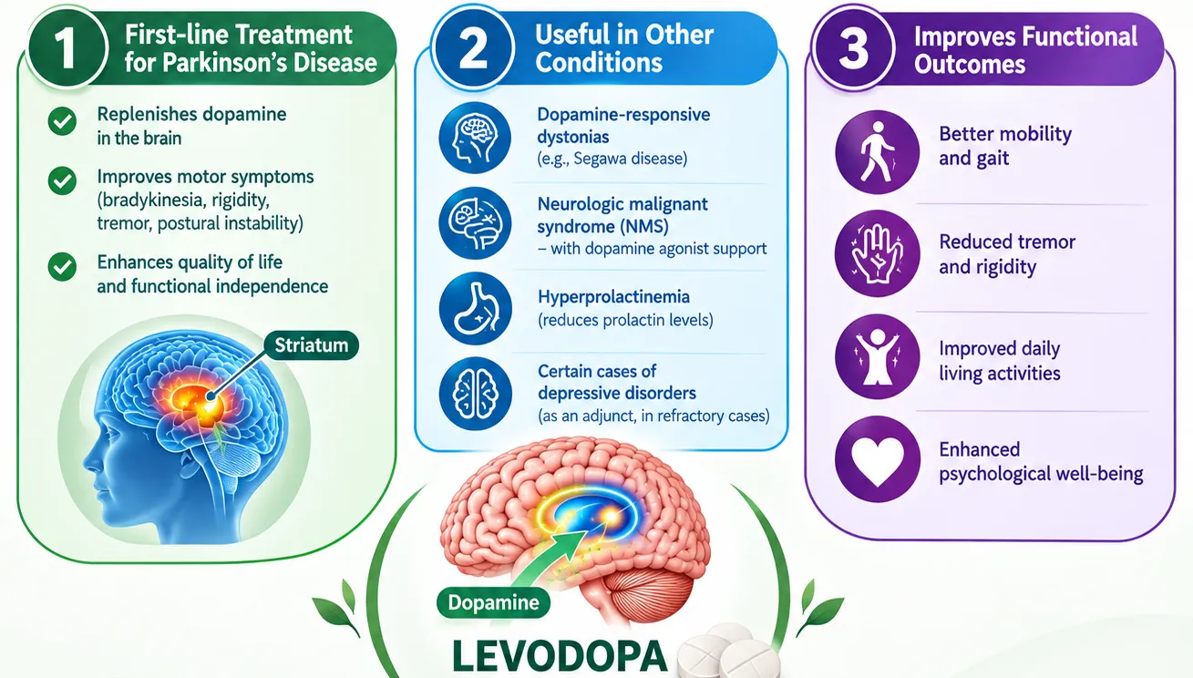
  • Parkinson’s disease: Mainstay treatment for the motor symptoms of Parkinson’s disease.
  • Improves motor symptoms: Helps reduce bradykinesia, rigidity, and tremor.
  • Usually combined with carbidopa/benserazide: These inhibit peripheral conversion of levodopa to dopamine, allowing more levodopa to reach the brain and reducing peripheral adverse effects.
  • Symptomatic treatment: Levodopa improves symptoms but does not cure Parkinson’s disease or stop its underlying neurodegeneration.
  • Long-term therapy: Chronic use may lead to motor fluctuations (“wearing-off”) and dyskinesias.

Figure 5. Clinical importance of Levodopa

Dopamine as a Precursor of Melanin [30]

Dopamine can contribute to melanin biosynthesis, particularly in certain specialized tissues and organisms. Dopamine is an aromatic catecholamine that can undergo oxidation to quinone intermediates, which subsequently participate in reactions leading to the formation of melanin pigments. In humans, however, the principal precursor of melanin in melanocytes is L-tyrosine, which is converted to L-DOPA and then dopaquinone through the action of tyrosinase. Dopamine-derived melanogenesis is therefore distinct from the classical tyrosine–L-DOPA pathway and is particularly relevant to the formation of neuromelanin in dopaminergic neurons. Neuromelanin accumulates mainly in the substantia nigra and locus coeruleus and is associated with dopamine metabolism and neuronal protection.

How to Support Dopamine Naturally

Rather than trying to “boost” dopamine as much as possible, the goal is to support healthy dopamine signaling and balance. Dopamine function can be supported naturally through a healthy lifestyle and balanced daily routine. Regular physical exercise, adequate sleep, a balanced diet rich in tyrosine-containing foods, healthy sunlight exposure, stress management, social interaction, and engaging in enjoyable or meaningful activities may help maintain normal dopamine signaling. Protein-rich foods such as eggs, dairy products, legumes, nuts, seeds, and fish provide tyrosine, an important precursor for dopamine synthesis. Maintaining these healthy habits may promote balanced dopamine activity, motivation, mood, cognitive function, and overall brain health.
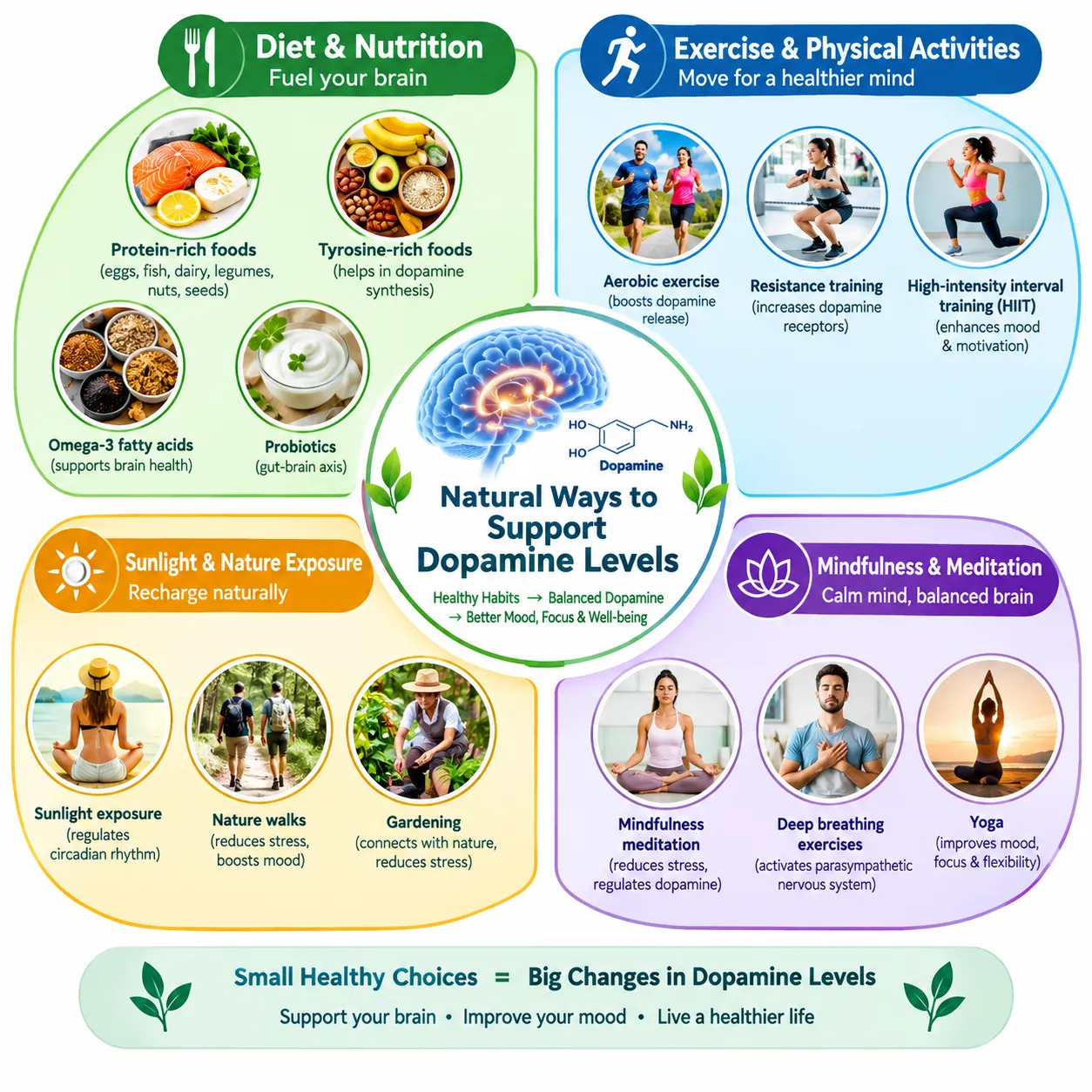
Figure 6. Natural way to support Dopamine

Future of Dopamine as the “Happy Hormone” [31,32]

The future of dopamine research is focused on developing a deeper understanding of its role in reward, motivation, mood, cognition, and neurological health. Advances in neuroscience, artificial intelligence, personalized medicine, and targeted drug delivery may help identify individual patterns of dopamine signaling and support more precise treatments for dopamine-related disorders. Research into dopamine receptors, neural pathways, biomarkers, and lifestyle-based interventions may further improve approaches to maintaining healthy dopamine function. Overall, future research is expected to move beyond the simple concept of dopamine as a “happiness hormone” toward a more comprehensive understanding of its role in brain function, behavior, and overall well-being.

CONCLUSION

Dopamine, popularly known as the “happy hormone,” is an essential neurotransmitter that plays a significant role in the nervous system, influencing reward, motivation, mood, cognition, learning, attention, and motor control. Balanced dopamine signaling is important for maintaining normal brain function and overall well-being, while its dysregulation may contribute to various neurological and psychiatric conditions. Dopamine function can be naturally supported through regular physical activity, adequate sleep, a balanced diet containing tyrosine-rich foods, healthy daylight exposure, stress management, social interaction, and meaningful activities. Thus, maintaining healthy dopamine signaling is important for a balanced mind, positive mood, proper nervous-system function, and overall health.

REFERENCES

  1. Benes FM: Carlsson and the discovery of dopamine, Trends Pharmacol. Sci. (2001), 22:46-47.
  2. Berry MS. Cottrell GA: Dopamine: excitatory and inhibitory transmission from a giant dopamine neurone. Nat. New Biol. (1973), 242, 250–253.
  3. Hornykiewicz O: A quarter century of brain dopamine research. In: Dopaminergic Systems and their Regulation. eds. G.N. Woodruff, J.A. Poat & P.J. Roberts pp. 3–18. London: Macmillan (1986).
  4. Schultz W: Predictive reward signal of dopamine neurons. J. Neurophysiol. (1998), 80, 1–27.
  5. Wise RA: Dopamine, learning and motivation. Nat. Rev. Neurosci. (2004), 5, 483–494.
  6. Soares S, Atallah BV & Paton JJ: Midbrain dopamine neurons control judgment of time. Science (2016), 354, 1273–1277.
  7. Watabe-Uchida M, Uchida N: Multiple dopamine systems: weal and woe of dopamine Cold Spring Harb. Symp. Quant. Biol. (2018), 83:83-95.
  8. Poulin JF, et al: Defining midbrain dopaminergic neuron diversity by single-cell gene expression profiling. Cell Rep. (2014), 9, 930–943.
  9. Hornykiewicz O: Dopamine miracle: from brain homogenate to dopamine replacement. Movement Disord (2002), 17, 501–508.
  10. Bertler Å & Rosengren E: Occurrence and distribution of dopamine in brain and other tissues. Experientia (1959), 15, 10–11.
  11. Sulzer D, Cragg SJ, Rice ME: Striatal dopamine neurotransmission: Regulation of release and uptake Basal Ganglia. (2016); 6:123-148.
  12. Blaschko H: Metabolism and storage of biogenic amines. Experientia (1957), 13, 9–12.
  13. Conti J, Strope E, Adams RN & Marsden CA: Voltammetry in brain tissues: chronic recording of stimulated dopamine and 5-hydroxy-tryptamine release. Life Sci. (1978), 23, 2705–2716.
  14. Alexander SPH, Mathie A and Peters JA: Dopamine receptors, in Guide to Receptors and Channels. Br. J. Pharmacol. (2004), 141, S2s.
  15. Matsumoto M & Hikosaka O: Two types of dopamine neuron distinctly convey positive and negative motivational signals. Nature (2009), 459, 837–841.
  16. Jaber M, Robinson SW, Missale C & Caron MG: Dopamine receptors & brain function. Neuropharmacology (1996), 35, 1503–1519.
  17. Kebabian JW & Calne DB: Multiple receptors for dopamine. Nature (1979), 277, 93–96.
  18. Salamone JD & Correa M: The mysterious motivational functions of mesolimbic dopamine. Neuron (2012), 76, 470–485.
  19. Mazzoni P, Hristova A, Krakauer J W: Why don’t we move faster? Parkinson’s disease, movement vigor, and implicit motivation, J. Neurosci. (2007), 27, 7105–7116.
  20. Eshel N, Tian J, Bukwich M & Uchida N: Dopamine neurons share common response function for reward prediction error. Nat. Neurosci. (2016), 19, 479–486.
  21. Berke JD & Hyman SE: Addiction, dopamine, and the molecular mechanisms of memory. Neuron (2000), 25, 515–532.
  22. Shiner T. et al: Dopamine and performance in a reinforcement learning task: evidence from Parkinson’s disease. Brain (2012), 135, 1871–1883.
  23. Schultz W: Multiple dopamine functions at different time courses. Annu. Rev. Neurosci. 30, 259–288 (2007).
  24. Howe MW & Dombeck DA: Rapid signalling in distinct dopaminergic axons during locomotion and reward. Nature (2016), 535, 505–510.
  25. Bromberg-Martin ES, Matsumoto M and Hikosaka O: Dopamine in motivational control: rewarding, aversive, and alerting. Neuron (2010), 68, 815–834.
  26. Pasquereau B & Turner RS: Dopamine neurons encode errors in predicting movement trigger occurrence. J. Neurophysiol. (2015), 113, 1110–1123.
  27. Anderson BA et al.: The Role of dopamine in value-based attentional orienting. Curr. Biol. (2016), 26, 550–555.
  28. Adel M, Griffith LC: The role of dopamine in associative learning in Drosophila: an updated unified model Neurosci. Bull. (2021), 37:831-852.
  29. Wise RA, Robble MA: "Dopamine and Addiction". Annual Review of Psychology (2020),71(1):79106.
  30. Watabe-Uchida M, Zhu L, Ogawa SK: Whole-brain mapping of direct inputs to midbrain dopamine neurons Neuron. (2012), 74:858-873.
  31. Birkmayer W & Hornykiewicz O: Der L-dioxyphenylalanin (=DOPA) – Effekt bei der Parkinson-Akinese. Wien. Klin. Wschr (1961), 73, 787–788.
  32. da Silva JA, Tecuapetla F, Paixão V & Costa RM: Dopamine neuron activity before action initiation gates and invigorates future movements. Nature (2018), 554, 244–248.
  33. Berry MS. Cottrell GA: Dopamine: excitatory and inhibitory transmission from a giant dopamine neurone. Nat. New Biol. (1973), 242, 250–253.
  34. Hornykiewicz O: A quarter century of brain dopamine research. In: Dopaminergic Systems and their Regulation. eds. G.N. Woodruff, J.A. Poat & P.J. Roberts pp. 3–18. London: Macmillan (1986).
  35. Schultz W: Predictive reward signal of dopamine neurons. J. Neurophysiol. (1998), 80, 1–27.
  36. Wise RA: Dopamine, learning and motivation. Nat. Rev. Neurosci. (2004), 5, 483–494.
  37. Soares S, Atallah BV & Paton JJ: Midbrain dopamine neurons control judgment of time. Science (2016), 354, 1273–1277.
  38. Watabe-Uchida M, Uchida N: Multiple dopamine systems: weal and woe of dopamine Cold Spring Harb. Symp. Quant. Biol. (2018), 83:83-95.
  39. Poulin JF, et al: Defining midbrain dopaminergic neuron diversity by single-cell gene expression profiling. Cell Rep. (2014), 9, 930–943.
  40. Hornykiewicz O: Dopamine miracle: from brain homogenate to dopamine replacement. Movement Disord (2002), 17, 501–508.
  41. Bertler Å & Rosengren E: Occurrence and distribution of dopamine in brain and other tissues. Experientia (1959), 15, 10–11.
  42. Sulzer D, Cragg SJ, Rice ME: Striatal dopamine neurotransmission: Regulation of release and uptake Basal Ganglia. (2016); 6:123-148.
  43. Blaschko H: Metabolism and storage of biogenic amines. Experientia (1957), 13, 9–12.
  44. Conti J, Strope E, Adams RN & Marsden CA: Voltammetry in brain tissues: chronic recording of stimulated dopamine and 5-hydroxy-tryptamine release. Life Sci. (1978), 23, 2705–2716.
  45. Alexander SPH, Mathie A and Peters JA: Dopamine receptors, in Guide to Receptors and Channels. Br. J. Pharmacol. (2004), 141, S2s.
  46. Matsumoto M & Hikosaka O: Two types of dopamine neuron distinctly convey positive and negative motivational signals. Nature (2009), 459, 837–841.
  47. Jaber M, Robinson SW, Missale C & Caron MG: Dopamine receptors & brain function. Neuropharmacology (1996), 35, 1503–1519.
  48. Kebabian JW & Calne DB: Multiple receptors for dopamine. Nature (1979), 277, 93–96.
  49. Salamone JD & Correa M: The mysterious motivational functions of mesolimbic dopamine. Neuron (2012), 76, 470–485.
  50. Mazzoni P, Hristova A, Krakauer J W: Why don’t we move faster? Parkinson’s disease, movement vigor, and implicit motivation, J. Neurosci. (2007), 27, 7105–7116.
  51. Eshel N, Tian J, Bukwich M & Uchida N: Dopamine neurons share common response function for reward prediction error. Nat. Neurosci. (2016), 19, 479–486.
  52. Berke JD & Hyman SE: Addiction, dopamine, and the molecular mechanisms of memory. Neuron (2000), 25, 515–532.
  53. Shiner T. et al: Dopamine and performance in a reinforcement learning task: evidence from Parkinson’s disease. Brain (2012), 135, 1871–1883.
  54. Schultz W: Multiple dopamine functions at different time courses. Annu. Rev. Neurosci. 30, 259–288 (2007).
  55. Howe MW & Dombeck DA: Rapid signalling in distinct dopaminergic axons during locomotion and reward. Nature (2016), 535, 505–510.
  56. Bromberg-Martin ES, Matsumoto M and Hikosaka O: Dopamine in motivational control: rewarding, aversive, and alerting. Neuron (2010), 68, 815–834.
  57. Pasquereau B & Turner RS: Dopamine neurons encode errors in predicting movement trigger occurrence. J. Neurophysiol. (2015), 113, 1110–1123.
  58. Anderson BA et al.: The Role of dopamine in value-based attentional orienting. Curr. Biol. (2016), 26, 550–555.
  59. Adel M, Griffith LC: The role of dopamine in associative learning in Drosophila: an updated unified model Neurosci. Bull. (2021), 37:831-852.
  60. Wise RA, Robble MA: "Dopamine and Addiction". Annual Review of Psychology (2020),71(1):79106.
  61. Watabe-Uchida M, Zhu L, Ogawa SK: Whole-brain mapping of direct inputs to midbrain dopamine neurons Neuron. (2012), 74:858-873.
  62. Birkmayer W & Hornykiewicz O: Der L-dioxyphenylalanin (=DOPA) – Effekt bei der Parkinson-Akinese. Wien. Klin. Wschr (1961), 73, 787–788.
  63. da Silva JA, Tecuapetla F, Paixão V & Costa RM: Dopamine neuron activity before action initiation gates and invigorates future movements. Nature (2018), 554, 244–248.

Reference

  1. Benes FM: Carlsson and the discovery of dopamine, Trends Pharmacol. Sci. (2001), 22:46-47.
  2. Berry MS. Cottrell GA: Dopamine: excitatory and inhibitory transmission from a giant dopamine neurone. Nat. New Biol. (1973), 242, 250–253.
  3. Hornykiewicz O: A quarter century of brain dopamine research. In: Dopaminergic Systems and their Regulation. eds. G.N. Woodruff, J.A. Poat & P.J. Roberts pp. 3–18. London: Macmillan (1986).
  4. Schultz W: Predictive reward signal of dopamine neurons. J. Neurophysiol. (1998), 80, 1–27.
  5. Wise RA: Dopamine, learning and motivation. Nat. Rev. Neurosci. (2004), 5, 483–494.
  6. Soares S, Atallah BV & Paton JJ: Midbrain dopamine neurons control judgment of time. Science (2016), 354, 1273–1277.
  7. Watabe-Uchida M, Uchida N: Multiple dopamine systems: weal and woe of dopamine Cold Spring Harb. Symp. Quant. Biol. (2018), 83:83-95.
  8. Poulin JF, et al: Defining midbrain dopaminergic neuron diversity by single-cell gene expression profiling. Cell Rep. (2014), 9, 930–943.
  9. Hornykiewicz O: Dopamine miracle: from brain homogenate to dopamine replacement. Movement Disord (2002), 17, 501–508.
  10. Bertler Å & Rosengren E: Occurrence and distribution of dopamine in brain and other tissues. Experientia (1959), 15, 10–11.
  11. Sulzer D, Cragg SJ, Rice ME: Striatal dopamine neurotransmission: Regulation of release and uptake Basal Ganglia. (2016); 6:123-148.
  12. Blaschko H: Metabolism and storage of biogenic amines. Experientia (1957), 13, 9–12.
  13. Conti J, Strope E, Adams RN & Marsden CA: Voltammetry in brain tissues: chronic recording of stimulated dopamine and 5-hydroxy-tryptamine release. Life Sci. (1978), 23, 2705–2716.
  14. Alexander SPH, Mathie A and Peters JA: Dopamine receptors, in Guide to Receptors and Channels. Br. J. Pharmacol. (2004), 141, S2s.
  15. Matsumoto M & Hikosaka O: Two types of dopamine neuron distinctly convey positive and negative motivational signals. Nature (2009), 459, 837–841.
  16. Jaber M, Robinson SW, Missale C & Caron MG: Dopamine receptors & brain function. Neuropharmacology (1996), 35, 1503–1519.
  17. Kebabian JW & Calne DB: Multiple receptors for dopamine. Nature (1979), 277, 93–96.
  18. Salamone JD & Correa M: The mysterious motivational functions of mesolimbic dopamine. Neuron (2012), 76, 470–485.
  19. Mazzoni P, Hristova A, Krakauer J W: Why don’t we move faster? Parkinson’s disease, movement vigor, and implicit motivation, J. Neurosci. (2007), 27, 7105–7116.
  20. Eshel N, Tian J, Bukwich M & Uchida N: Dopamine neurons share common response function for reward prediction error. Nat. Neurosci. (2016), 19, 479–486.
  21. Berke JD & Hyman SE: Addiction, dopamine, and the molecular mechanisms of memory. Neuron (2000), 25, 515–532.
  22. Shiner T. et al: Dopamine and performance in a reinforcement learning task: evidence from Parkinson’s disease. Brain (2012), 135, 1871–1883.
  23. Schultz W: Multiple dopamine functions at different time courses. Annu. Rev. Neurosci. 30, 259–288 (2007).
  24. Howe MW & Dombeck DA: Rapid signalling in distinct dopaminergic axons during locomotion and reward. Nature (2016), 535, 505–510.
  25. Bromberg-Martin ES, Matsumoto M and Hikosaka O: Dopamine in motivational control: rewarding, aversive, and alerting. Neuron (2010), 68, 815–834.
  26. Pasquereau B & Turner RS: Dopamine neurons encode errors in predicting movement trigger occurrence. J. Neurophysiol. (2015), 113, 1110–1123.
  27. Anderson BA et al.: The Role of dopamine in value-based attentional orienting. Curr. Biol. (2016), 26, 550–555.
  28. Adel M, Griffith LC: The role of dopamine in associative learning in Drosophila: an updated unified model Neurosci. Bull. (2021), 37:831-852.
  29. Wise RA, Robble MA: "Dopamine and Addiction". Annual Review of Psychology (2020),71(1):79106.
  30. Watabe-Uchida M, Zhu L, Ogawa SK: Whole-brain mapping of direct inputs to midbrain dopamine neurons Neuron. (2012), 74:858-873.
  31. Birkmayer W & Hornykiewicz O: Der L-dioxyphenylalanin (=DOPA) – Effekt bei der Parkinson-Akinese. Wien. Klin. Wschr (1961), 73, 787–788.
  32. da Silva JA, Tecuapetla F, Paixão V & Costa RM: Dopamine neuron activity before action initiation gates and invigorates future movements. Nature (2018), 554, 244–248.

Photo
Mona Patel
Corresponding author

Assistant Professor, Department of Pharmaceutical Quality assurance, Faculty of Pharmacy, SSSRGI, Vadasma, Mehsana, Gujarat, India- 382705.

Photo
Ojas Patel
Co-author

Professor, Department of Pharmaceutical Chemistry, Faculty of Pharmacy, SSSRGI, Vadasma, Mehsana, Gujarat, India- 382705

Mona Patel*, Ojas Patel, A Review on Happy Hormone: Dopamine, Int. J. Med. Pharm. Sci., 2026, 2 (9), 635-644. https://doi.org/10.5281/zenodo.23019865

More related articles
Zero-Order and First-Order Derivative of UV Spectr...
Abirami G., Nanthagopal P., Jananipriya B., Janani S., Dhivya V.,...
Subclinical Thyrotoxicosis Associated with Long-Te...
Nalam Vineela Nirmala, Mekala Keerthi Priya, Meka Saranya, Lingin...
Formulation and Evaluation of Poly Herbal Anti-GERD Powder...
Soumallya Chakraborty, Soumyadip Maji, Sonia Samanta, Somenath Bhattacharya, Koushik Jana, Amit Mait...
Evaluation of the Neuroprotective Potential of Citrullus Lanatus Seeds: An In-Vi...
Mukesh Yadav, Sunita Arya, Shalini Jain, Rani Yadav, Neelam Yadav, K. S. Dadoriya, Jyoti Yadav, Gaur...
Polyherbal Formulations for Metabolic Disorders: Traditional Medicine Modernizat...
Alok Kumar, Sima Jana, Radhashyam Shit, Md. Rocky Shaikh, Mausmi Khatoon, Juveriya Javed, Bulbul San...
Related Articles
Nature’s Mood Boosters: The Role of Happy Hormones in Animals and Humans...
Urmila Ingole, Vaibhav Shikare, Shirish Jain, Ashish Gawai...
A Comprehensive Review on Psychiatric Disorders (Amraz-e-Nafsaniya) in Relation ...
Km. Anam Sughra, Tariq Nadeem Khan, Masarat Begum Mohammed Kamaluddin Farooqui...
Anxiolytic Activity of Several Herbs...
Trupti Khandbahale, Sunil Mahajan, Jubershaha Fakir, Chandrashekhar Patil...
Herbal Medicines in the Management of Urinary Tract Infections and Kidney Disord...
Vinoth Jeevanesan, Vanitha Devi Rajendran, Thamarakani Balaji, Nagasibi Senthilkumar, Fathima Nasrin...
Zero-Order and First-Order Derivative of UV Spectrophotometric Methods for Deter...
Abirami G., Nanthagopal P., Jananipriya B., Janani S., Dhivya V., Aishwarya R....
More related articles
Zero-Order and First-Order Derivative of UV Spectrophotometric Methods for Deter...
Abirami G., Nanthagopal P., Jananipriya B., Janani S., Dhivya V., Aishwarya R....
Subclinical Thyrotoxicosis Associated with Long-Term Amiodarone Therapy: A Case ...
Nalam Vineela Nirmala, Mekala Keerthi Priya, Meka Saranya, Lingineni Mani Deepa Chandrika, Kandula H...
Zero-Order and First-Order Derivative of UV Spectrophotometric Methods for Deter...
Abirami G., Nanthagopal P., Jananipriya B., Janani S., Dhivya V., Aishwarya R....
Subclinical Thyrotoxicosis Associated with Long-Term Amiodarone Therapy: A Case ...
Nalam Vineela Nirmala, Mekala Keerthi Priya, Meka Saranya, Lingineni Mani Deepa Chandrika, Kandula H...