

We use cookies to ensure our website works properly and to personalise your experience. Cookies policy
1PharmD Intern, Ezhuthachan College of Pharmaceutical Sciences, Marayamuttom, Thiruvananthapuram.
2Assistant Professor, Department of Pharmacy Practice, Ezhuthachan College of Pharmaceutical Sciences, Marayamuttom, Thiruvananthapuram.
3Principal, HOD, Department of Pharmacy Practice. Ezhuthachan College of Pharmaceutical Sciences, Marayamuttom, Thiruvananthapuram.
Vasa previa is an uncommon but serious obstetric complication in which fetal blood vessels run across the membranes near or over the cervical opening without protection from the placenta or umbilical cord. If these vessels rupture, especially during labour or membrane rupture, it can result in severe fetal blood loss and high perinatal mortality. Timely identification during pregnancy plays a crucial role in improving outcomes. A 26 year-old female with gravida 2 at 35 weeks of gestation, previous ND who presented with c/o bleeding P/V since morning. USG shows that posterior reaching upto the os with fetal vessels between fetal head and placenta, leading to the diagnosis of vasa previa. A planned cesarean delivery was carried out at 35 weeks of gestation. Early recognition of the condition enabled careful planning and prevention of complications associated with vessel rupture. Baby was delivered safely, and both mother and baby had an uneventful recovery. This case emphasizes the value of targeted ultrasound screening, particularly in high-risk pregnancies. Early diagnosis combined with planned cesarean delivery can significantly reduce the risk of adverse fetal outcomes in vasa previa.
Vasa previa occurs when fetal blood vessels, unsupported by placental tissue or Wharton’s jelly, run across or within 2 cm of the internal cervical os. The condition complicates roughly 1 in 2,500 pregnancies and carries a perinatal mortality rate exceeding 50% if undiagnosed before labor or membrane rupture. However, prenatal detection via transvaginal ultrasound with color Doppler improves survival to over 95% by enabling timed cesarean delivery before vessel rupture. Risk factors include placenta previa or low-lying placenta, velamentous or marginal cord insertion, multiple gestation, bilobed or succenturiate placentas, and conception via assisted reproductive technologies. Many cases are asymptomatic until membrane rupture, when painless vaginal bleeding accompanied by fetal bradycardia or decelerations may be the first sign. Current guidelines recommend targeted screening with color Doppler in these high-risk groups to identify fetal vessels overlying the os. Once diagnosed, management aims to prevent spontaneous labor and membrane rupture while optimizing fetal maturity. Antenatal corticosteroids, close surveillance and patient education on warning symptoms are the key components of care.
CASE REPORT:
A 28-year-old woman, gravida 2 para 1, was referred at 35 weeks gestation following an episode of painless vaginal bleeding. She reported no abdominal pain, contractions or leakage of fluid. Fetal movements were normal. She has the past medical history of hypothyroidism and was on Tab. Thyronorm 25mcg. Previously she was admitted at 27 weeks and 4 days i/v/o APH and antenatal corticosteroids were taken. At the time of presentation the patient was conscious, afebrile, and hemodynamically stable with a blood pressure of 120/80 mmHg. Cardiovascular examination was unremarkable with normal heart sounds, and no neurological deficits were observed. Vaginal bleeding was present, she has received a single dose of Inj. Tranexamic acid 500mg prior to transfer. First‑trimester screening including nuchal translucency was unremarkable. Targeted anomaly scan and fetal echocardiography showed no major structural abnormalities. Ultrasound at approximately 28 weeks demonstrated a single live cephalic fetus, a posterior placenta reaching the internal os consistent with placenta previa (grade II), and no sonographic evidence of abruption. Routine obstetric Doppler studies were within normal limits. Sequential transabdominal and transvaginal sonography revealed a live fetus in cephalic presentation with biometric parameters appropriate for late second/early third trimester (femur length 61 mm) and fetal heart rate around 153/min. Color Doppler imaging identified fetal vessels running between the fetal head and the placental edge, suspicious for vasa previa. The Nitabuch layer appeared intact and the amniotic fluid index measured 16 cm. Follow‑up ultrasound at around 34 weeks confirmed persistent placenta previa with marginal cord insertion; Doppler indices remained unremarkable.
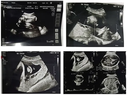
Given the confirmed vasa previa and low-lying placenta and suspicious vasa previa the patient was counseled regarding the risk of sudden fetal hemorrhage with membrane rupture or labor. She received a course of antenatal corticosteroids for fetal lung maturity between 28 and 32 weeks, as recommended in anticipated preterm delivery. She was advised to report immediately any bleeding, contractions or fluid loss and was followed with serial ultrasounds every 2–3 weeks to monitor vessel position and fetal growth. [ Because of the combination of antepartum hemorrhage, persistent placenta previa, suspected vasa previa and marginal cord insertion, the patient underwent an emergency lower‑segment cesarean section under general anesthesia. Concomitant sterilization was performed. To control intraoperative bleeding and reduce the risk of postpartum hemorrhage, bilateral uterine artery ligation and Hayman compression sutures were placed. A live male infant weighing 2.640 kg was delivered at 10:35 AM. The postoperative course was uneventful; both mother and neonate remained stable and were discharged on postoperative day five.
DISCUSSION:
This case underscores several key principles in the management of vasa previa.
CONCLUSION
Clinicians should suspect vasa previa in pregnancies complicated by placenta previa or abnormal cord insertion. Transvaginal ultrasound with color Doppler is essential for diagnosis. Early detection, appropriate antenatal steroids when indicated, vigilant monitoring and planned cesarean delivery improve perinatal outcomes.
REFERENCES

Vishnumaya A. M., Amritha Krishna, Grace N. Raju*, Shaiju S. Dharan, A Time Sensitive Obstetric Emergency- Vasa Previa: A Case Report, Int. J. Med. Pharm. Sci., 2026, 2 (9), 368-371. https://doi.org/10.5281/zenodo.22891946
 10.5281/zenodo.22891946