

We use cookies to ensure our website works properly and to personalise your experience. Cookies policy
Professor, Department of optometry, Haldia institute of management, (Unit of ICARE), Haldia, West Bengal
Background: Pterygium is a common fibrovascular growth of the conjunctiva extending onto the corneal surface. Progressive pterygium may produce ocular irritation, induce corneal astigmatism, and cause significant visual impairment when it approaches or encroaches upon the visual axis. Case Presentation: This case study describes a patient presenting with progressive pterygium associated with ocular discomfort and reduced visual acuity. A comprehensive ophthalmic examination was performed to assess the extent of the pterygium, corneal involvement, visual acuity, and associated ocular findings. Relevant environmental and occupational risk factors, including prolonged exposure to ultraviolet radiation, dust, wind, and other outdoor conditions, were also considered. Management: The patient was evaluated for appropriate treatment based on the size, progression, symptoms, and effect on visual function. Conservative measures were considered for symptomatic relief, while surgical excision with appropriate conjunctival management was considered where progressive growth and visual impairment warranted intervention. Conclusion: Progressive pterygium can adversely affect visual function and ocular comfort if left untreated. Early detection, regular ophthalmic evaluation, reduction of environmental risk factors, and timely management are important for preventing progression and minimizing visual impairment. This case highlights the importance of recognizing progressive pterygium before significant corneal involvement and visual-axis obstruction occurs.
Case Presentation
A 34-year-old male patient residing in a rural area of West Bengal presented with a progressively increasing fleshy growth over the right eye associated with ocular discomfort and gradual deterioration of vision. The patient was a farmer by occupation and reported prolonged outdoor exposure during agricultural activities. His occupational environment involved regular exposure to sunlight, dust, wind and other environmental irritants.
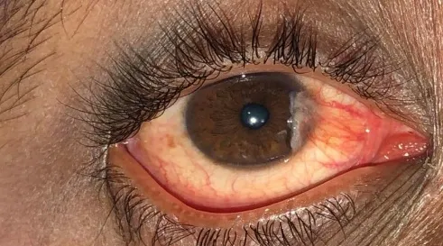
Grade I Pterygium
The patient reported that the ocular growth had gradually increased in size over time and had become increasingly noticeable. He experienced symptoms such as foreign-body sensation, irritation, watering and intermittent redness. A subjective reduction in visual clarity was also reported, particularly as the lesion progressed toward the central corneal region. On ophthalmic examination, the pterygium should be documented according to its laterality, site, morphology and extent.
Visual acuity: Right eye 6/12(p); Left eye 6/6.
Best-corrected visual acuity: Right eye 6/9(p); Left eye 6/6. Slit-lamp examination demonstrated a triangular, fibrovascular conjunctival growth extending from the nasal bulbar conjunctiva toward the cornea. The lesion was clinically assessed as progressive pterygium based on its vascularity, thickness and extension toward the cornea.
A complete ocular examination should also include intraocular pressure, corneal examination, tear-film/ocular-surface assessment and fundus examination. Refraction should be performed to determine whether induced astigmatism contributes to the patient's visual impairment. Pterygium can produce corneal distortion and irregular astigmatism, which may contribute to reduced visual acuity. The patient's history of farming and prolonged outdoor work is clinically relevant because cumulative ultraviolet exposure is an important environmental factor associated with pterygium. His rural occupation may therefore represent an important occupational exposure in the development and progression of the lesion.
REVIEW OF LITERATURE
Pterygium is a common ocular-surface disorder characterized by a triangular fibrovascular proliferation of conjunctival tissue that extends onto the cornea. It occurs more frequently in populations living in regions with substantial ultraviolet exposure and is particularly relevant to individuals who spend prolonged periods outdoors. The disease may remain relatively asymptomatic initially but can produce redness, irritation, foreign-body sensation, tearing and cosmetic concerns as it progresses. Progressive pterygium may affect vision through several mechanisms. Direct encroachment toward the visual axis can interfere with the optical pathway, while traction and changes in corneal curvature may produce astigmatism and optical distortion. Contemporary reviews have emphasized the relationship between pterygium and corneal irregularity as well as the refractive effects associated with pterygium and its removal. Environmental and occupational exposure is particularly important in rural agricultural workers. Prolonged exposure to sunlight, ultraviolet radiation, wind and dust may contribute to chronic ocular-surface irritation and the development of pterygium. Therefore, preventive measures such as ultraviolet-protective eyewear and reduction of direct environmental exposure are relevant for outdoor workers. The principal aim of treatment is to control symptoms, preserve vision and prevent or reduce recurrence. Small, asymptomatic lesions may generally be managed conservatively with ocular-surface lubrication and protection from environmental exposure. However, progressive lesions causing significant symptoms, visual impairment, induced astigmatism, restriction of ocular movement or threatening the visual axis may require surgical excision. Historically, bare-sclera excision has been associated with substantial recurrence and is no longer considered an optimal standalone approach. Evidence from systematic reviews and meta-analyses supports the use of conjunctival or limbal-conjunctival autografting to reduce recurrence compared with bare-sclera excision.
Management
The patient was counselled regarding the progressive nature of the condition and the importance of reducing exposure to environmental risk factors. Protective measures, including the regular use of UV-protective sunglasses, a wide-brimmed hat, and avoidance of direct exposure to dust and wind whenever possible, were advised. For symptomatic relief, appropriate lubricating/artificial tear preparations were recommended to reduce ocular surface dryness and irritation. The patient was advised to maintain regular ophthalmic follow-up to monitor the size of the pterygium, corneal involvement, and visual acuity. Because the pterygium was progressive and associated with visual impairment, surgical excision may be considered when there is significant encroachment onto the visual axis, visually significant induced astigmatism, persistent symptoms, restricted ocular motility, or significant cosmetic concern. If surgery is undertaken, excision with conjunctival autografting is commonly used to reduce recurrence compared with bare-sclera excision. Postoperative follow-up is important for detecting recurrence and managing ocular-surface inflammation.
DISCUSSION
This case illustrates the possible relationship between occupational outdoor exposure and progressive pterygium in a young rural agricultural worker. The patient's age and farming occupation are clinically relevant because prolonged exposure to sunlight and environmental irritants may contribute to chronic ocular-surface damage. The visual impairment in this patient should not automatically be attributed solely to the pterygium. A complete ophthalmic examination, including refraction and assessment of corneal topography or keratometry where available, is important to establish whether the reduction in vision is related to pterygium-induced astigmatism, direct involvement of the visual axis, dry-eye disease or another ocular condition. Early recognition and appropriate follow-up are particularly important in young outdoor workers. Preventive measures, including ultraviolet protection and regular eye examinations, may help reduce ocular-surface morbidity. When a pterygium becomes progressive and visually significant, appropriate surgical management can provide both functional and symptomatic benefits while minimizing the risk of recurrence.
CONCLUSION
Progressive pterygium can cause significant ocular discomfort, corneal distortion, refractive changes and visual impairment. In this 34-year-old rural male farmer, prolonged outdoor occupational exposure represents an important environmental consideration. The case highlights the importance of early detection, ultraviolet protection and regular ophthalmic assessment among individuals who work outdoors. Early detection, regular ophthalmic examination, appropriate symptomatic treatment, and preventive measures such as UV-protective eyewear and hats are important in reducing progression and protecting vision. Awareness among rural agricultural workers regarding occupational eye protection and the importance of early eye-care consultation may help reduce the burden of pterygium-related visual impairment. When a pterygium becomes progressive or produces clinically significant visual impairment, surgical excision with ocular-surface reconstruction, particularly conjunctival or limbal-conjunctival autografting, is an established management approach. Evidence indicates that graft-based techniques have lower recurrence rates than bare-sclera excision. This case emphasizes that pterygium should not be regarded merely as a cosmetic ocular condition. In occupationally exposed individuals, progressive disease may have meaningful functional consequences and may require timely ophthalmic intervention.
REFERENCES

Dr. Goutam Datta*, A Case Study of Progressive Pterygium with Visual Impairment, Int. J. Med. Pharm. Sci., 2026, 2 (9), 256-259. https://doi.org/10.5281/zenodo.22668809
 10.5281/zenodo.22668809