

We use cookies to ensure our website works properly and to personalise your experience. Cookies policy
1Pharm. D. Student, Ezhuthachan College of Pharmaceutical Sciences, Marayamuttom, Neyyatinkara, Thiruvananthapuram, Kerala, India
2Assistant Professor, Department of Pharmacy Practice, Ezhuthachan College of Pharmaceutical Sciences, Marayamuttom, Neyyatinkara, Thiruvananthapuram, Kerala, India
3Senior Consultant, Department of General Medicine, NIMS Medicity, Aralumoodu, Neyyatinkara, Thiruvananthapuram, Kerala, India
4Principal / Head of Department, Department of Pharmacy Practice, Ezhuthachan College of Pharmaceutical Sciences, Marayamuttom, Neyattinkara, Thiruvananthapuram, Kerala, India
Obesity Hypoventilation Syndrome (OHS) is an underrecognized cause of chronic hypercapnic respiratory failure and is frequently associated with obstructive sleep apnea and obstructive airway disease, particularly among elderly individuals with multiple cardiometabolic comorbidities. Delayed recognition may result in recurrent hospitalizations, worsening respiratory compromise, and increased morbidity. We report the case of an elderly female with a history of asthma, hypertension, dyslipidemia, and type 2 diabetes mellitus who presented with slurring of speech and progressive breathing difficulty. Clinical evaluation, laboratory investigations, and blood gas analysis were suggestive of chronic hypoventilation, leading to the diagnosis of obesity hypoventilation syndrome with obstructive airway disease. The patient was managed with pharmacological therapy, oxygen supplementation, bronchodilator nebulization, corticosteroids, antimicrobial therapy, and supportive care, resulting in gradual clinical improvement and successful discharge. This case highlights the importance of maintaining a high index of suspicion for OHS in obese patients presenting with unexplained hypercapnia and respiratory symptoms. Early diagnosis, comprehensive evaluation, and multidisciplinary management are essential to improve clinical outcomes and reduce disease-related complications.
Obesity hypoventilation syndrome (OHS) is a clinically important disorder characterized by obesity-associated alveolar hypoventilation resulting in awake hypercapnia, conventionally defined by an arterial partial pressure of carbon dioxide (PaCO₂) ≥45 mmHg, after exclusion of alternative causes of hypoventilation. OHS represents one of the major obesity-related respiratory disorders and is frequently associated with sleep-disordered breathing, particularly obstructive sleep apnoea (OSA). Despite its substantial clinical consequences, OHS remains frequently underrecognized, and diagnosis may be delayed until patients develop significant respiratory or cardiovascular complications [1,2]. Recent evidence further emphasizes that OHS is a heterogeneous disorder with different clinical phenotypes and disease trajectories, making early recognition and individualized assessment particularly important [3]. The pathophysiology of OHS is multifactorial and involves the interaction of excessive body mass, altered respiratory mechanics, impaired ventilatory drive and sleep-disordered breathing. Increased adipose tissue imposes a mechanical load on the respiratory system, reducing functional residual capacity and increasing the work of breathing, while abnormalities in ventilatory control may impair the compensatory response to carbon dioxide retention [1,3]. Approximately 90% of individuals with OHS have coexisting OSA, whereas a smaller proportion exhibit sleep-related hypoventilation without significant OSA [1]. The coexistence of these abnormalities may result in sustained or intermittent hypoxemia and chronic hypercapnia, contributing to progressive cardiopulmonary dysfunction [2,3]. The clinical significance of OHS extends beyond respiratory symptoms. Chronic hypoxemia and hypercapnia are associated with pulmonary vascular and cardiovascular consequences, including pulmonary hypertension and right-sided cardiac dysfunction. The relationship between OHS, OSA and pulmonary hypertension is complex, with sustained hypoxemia, recurrent nocturnal oxygen desaturation, hypercapnia and obesity acting through several interrelated mechanisms to increase pulmonary vascular stress [4]. Consequently, patients with OHS frequently present with multiple cardiometabolic comorbidities, which may complicate clinical recognition of the underlying ventilatory disorder. The diagnostic evaluation of suspected OHS requires careful assessment because hypercapnia may occur secondary to several other conditions, including chronic obstructive pulmonary disease, neuromuscular disorders, chest-wall abnormalities, medications that suppress respiratory drive and other causes of sleep-related hypoventilation [5]. In patients with coexisting obstructive airway disease, distinguishing the contribution of airway obstruction from obesity-related hypoventilation can be particularly challenging. Contemporary clinical reviews therefore emphasize systematic assessment of obesity, daytime gas exchange, sleep-disordered breathing and alternative causes of hypoventilation rather than attributing hypercapnia solely to an underlying obstructive airway disorder [5,6]. Serum bicarbonate may provide a useful initial screening marker for chronic hypoventilation because renal bicarbonate retention occurs as a physiological response to sustained carbon dioxide elevation. However, elevated bicarbonate alone is insufficient to establish OHS, and arterial blood gas assessment remains important when clinical suspicion is high [5,6]. This distinction is particularly relevant in elderly patients, in whom respiratory symptoms may be attributed to common cardiopulmonary diseases and multiple comorbidities may obscure the presence of chronic hypoventilation. OHS is increasingly recognized in older adults, although the elderly population remains comparatively less characterized than younger patients. A single-centre study of elderly individuals with OHS demonstrated a substantial burden of associated comorbidities, emphasizing the clinical complexity of OHS in this population [7]. In an elderly patient presenting with acute respiratory symptoms, therefore, OHS may coexist with chronic airway disease, hypertension, diabetes mellitus and cardiovascular abnormalities, creating diagnostic overlap and potentially delaying recognition of chronic ventilatory failure. Management of OHS is directed toward correcting hypoventilation, treating associated sleep-disordered breathing, reducing obesity-related respiratory load and controlling comorbid disease. Positive Airway Pressure (PAP) therapy is an important component of treatment, and evidence from systematic review and meta-analysis indicates that PAP can improve carbon dioxide levels, oxygenation and several objective sleep parameters in patients with OHS [8]. Current clinical practice also emphasizes weight management as a fundamental component of long-term treatment, together with appropriate selection of continuous positive airway pressure or non-invasive ventilation according to the patient's respiratory and sleep-disordered breathing phenotype [6,9]. More recent literature highlights the need for an integrated, multidisciplinary approach because OHS may follow diverse clinical trajectories and coexist with multiple metabolic and cardiopulmonary conditions [3]. The case is clinically relevant because it illustrates the diagnostic complexity of recognizing an OHS phenotype in an elderly patient with concomitant obstructive airway disease and multiple cardiometabolic comorbidities. It also highlights the importance of considering chronic hypoventilation when respiratory deterioration occurs in patients whose symptoms may initially be attributed to obstructive airway disease alone.
Case Presentation
A 78-year-old woman was admitted to the Department of General Medicine with acute onset of progressive breathlessness accompanied by slurring of speech of one day's duration. The respiratory symptoms had gradually worsened prior to admission and were associated with reduced exercise tolerance and intermittent episodes of oxygen desaturation. There was no documented history of fever, chest pain, palpitations, hemoptysis, or recent respiratory tract infection. Her past medical history was significant for bronchial asthma diagnosed 10 years previously, hypertension for approximately two decades, dyslipidemia for 20 years, and type 2 diabetes mellitus managed with oral hypoglycemic agents. She denied tobacco smoking, alcohol consumption, or exposure to biomass fuels. Family history was notable for maternal type 2 diabetes mellitus. The patient led a predominantly sedentary lifestyle and consumed a mixed diet. She had no known drug allergies. On admission, the patient was conscious, alert, and oriented to time, place, and person. Her temperature was 98.6°F, blood pressure was 120/80 mmHg, pulse rate was 81 beats/minute, respiratory rate was 14 breaths/minute, and peripheral oxygen saturation was initially 99% on pulse oximetry. Physical examination revealed bilateral normal vesicular breath sounds without focal crepitations or wheeze at rest. Cardiovascular examination demonstrated normal first and second heart sounds without audible murmurs. Abdominal examination revealed a distended abdomen without tenderness or organomegaly, while neurological examination showed no focal neurological deficit despite the initial complaint of slurred speech. During hospitalization, serial monitoring demonstrated progressive fluctuations in oxygen saturation ranging from 99% to 85%, reflecting intermittent hypoxemia requiring close respiratory monitoring. Anthropometric assessment revealed a body mass index (BMI) of 34.1 kg/m², consistent with Class I obesity according to the World Health Organization classification. The presence of obesity in an elderly patient presenting with unexplained chronic hypercapnia prompted further evaluation for obesity hypoventilation syndrome (OHS), particularly considering the coexistence of chronic obstructive airway disease and the absence of alternative neuromuscular or chest wall disorders. Initial laboratory investigations demonstrated a hemoglobin concentration of 12.9 g/dL, total leukocyte count of 9,280 cells/mm³ with neutrophilic predominance (79.1%) and relative lymphopenia (13.6%), while platelet count remained within normal limits. Glycemic assessment revealed suboptimal long-term diabetic control with an HbA1c of 7.9%, although the admission random blood glucose level was 111 mg/dL. Liver function testing showed elevated serum aminotransferases (AST 88 U/L and ALT 148 U/L) with preserved bilirubin, albumin, and alkaline phosphatase levels. Renal function was largely maintained with serum creatinine ranging between 0.70 and 0.92 mg/dL despite mildly elevated blood urea levels. Persistent mild hyponatremia (130 mmol/L) was observed during the early hospital course, whereas potassium concentrations remained within normal limits. C-reactive protein was elevated at 14.1 mg/L, suggesting an underlying inflammatory response. Cardiac evaluation demonstrated markedly elevated brain natriuretic peptide (4420 pg/mL) with negative cardiac troponin I levels, indicating volume overload without biochemical evidence of acute myocardial injury. Neuroimaging with a non-contrast computed tomography scan of the brain, performed prior to transfer from an outside hospital, demonstrated bilateral periventricular and frontoparietal deep white matter hypodensities suggestive of chronic small-vessel ischemic changes without evidence of acute intracranial pathology, thereby making a primary cerebrovascular event an unlikely explanation for the presenting symptoms. Assessment of gas exchange proved pivotal in establishing the diagnosis. Arterial blood gas analysis demonstrated PaCO₂ 56 mmHg, PaO₂ 58 mmHg, HCO₃⁻ 33.9 mmol/L, and arterial oxygen saturation of 89%, consistent with chronic compensated hypercapnic respiratory failure accompanied by moderate hypoxemia. These findings were supported by the available venous blood gas analysis, which similarly demonstrated hypercapnia (pCO₂ 56 mmHg), elevated bicarbonate (33.9 mmol/L), increased base excess (8.9 mmol/L), and normal pH (7.39), indicating chronic renal compensation for sustained carbon dioxide retention rather than an acute ventilatory disturbance. Collectively, the presence of obesity (BMI 34.1 kg/m²), daytime hypercapnia (PaCO₂ >45 mmHg), chronic bicarbonate retention, and persistent hypoxemia strongly supported the diagnosis of obesity hypoventilation syndrome after exclusion of alternative causes of alveolar hypoventilation. The patient was initially managed under the provisional diagnosis of acute exacerbation of obstructive airway disease. Empirical intravenous CEFOPERAZONE-SULBACTAM 1.5 G Twice Daily And AZITHROMYCIN 500 Mg Once Daily Followed By Oral AZITHROMYCIN Were Initiated To Address Possible Lower Respiratory Tract Infection. Intravenous METHYLPREDNISOLONE 40 Mg Every 8 Hours, Bronchodilator Nebulization With IPRATROPIUM BROMIDE 250 µg Every 6 Hours Followed By Combination IPRATROPIUM–LEVOSALBUTAMOL (500 µg/1.25 Mg) Every 6 Hours Therapy, Inhaled BUDESONIDE 0.5 Mg Every 8 Hours, And DOXOFYLLINE 200 Mg With A Half-Tablet Regimen Twice Daily Were Administered To Optimize Airway Patency And Reduce Bronchial Inflammation. PANTOPRAZOLE 40 Mg Was Prescribed For Gastrointestinal Prophylaxis, While Chronic Antihypertensive, Lipid-Lowering, Antiplatelet, And Antidiabetic Therapies Were Continued With Appropriate Dose Adjustments. Hyperglycemia Observed During Hospitalization, Likely Exacerbated By Systemic Corticosteroid Therapy, Was Managed Using Short-Acting Insulin According To Serial Capillary Blood Glucose Measurements. LOSARTAN, initially 25 mg once daily and subsequently increased to 50 mg once daily to improve blood pressure control, Diuretic therapy was changed from FUROSEMIDE 40 mg twice daily to TORSEMIDE 10 mg once daily in view of clinical evidence of fluid overload and markedly elevated BNP levels. Following comprehensive medical management, the patient's respiratory symptoms gradually improved with stabilization of oxygen saturation, reduction in dyspnea, and improvement in overall clinical status. She remained hemodynamically stable throughout the remainder of her hospital stay without progression to invasive ventilatory support. After six days of inpatient treatment, she was discharged in stable condition on oral antibiotics, tapering corticosteroids, bronchodilator therapy, antihypertensive medications, lipid-lowering therapy, diuretics, and optimized antidiabetic treatment. She was advised regular follow-up in the general medicine outpatient department with blood glucose monitoring and continued evaluation of her chronic respiratory condition. The final diagnosis at discharge was obesity hypoventilation syndrome with coexisting obstructive airway disease.
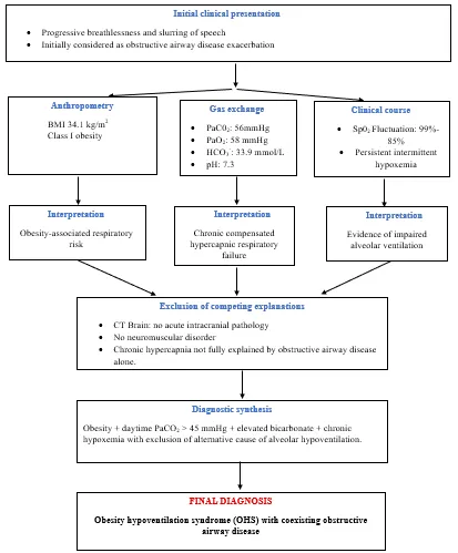
Figure 1. diagnostic reasoning pathway leading to the recognition of OHS
This presentation is clinically noteworthy because the diagnosis of OHS was established through integration of anthropometric assessment, chronic daytime hypercapnia with metabolic compensation, persistent hypoxemia, and exclusion of alternative causes of hypoventilation in an elderly woman initially suspected to have isolated obstructive airway disease, underscoring the importance of recognizing OHS as a frequently underdiagnosed contributor to chronic respiratory failure in obese patients.
DISCUSSION
Obesity hypoventilation syndrome (OHS) is a distinct clinical entity characterized by the coexistence of obesity (body mass index [BMI] ≥30 kg/m²), chronic daytime alveolar hypoventilation resulting in arterial hypercapnia (PaCO₂ ≥45 mmHg), and sleep-disordered breathing after exclusion of other causes of hypoventilation. Despite increasing recognition over the past decade, OHS remains considerably underdiagnosed because its clinical manifestations often overlap with chronic obstructive pulmonary disease (COPD), asthma, heart failure, and other causes of chronic respiratory insufficiency. Consequently, many patients are initially managed for acute exacerbations of obstructive airway disease without recognition of the underlying ventilatory disorder. This diagnostic delay contributes to repeated hospitalizations, progression of chronic respiratory failure, pulmonary hypertension, cardiovascular complications, and increased mortality [10]. The present case illustrates several important clinical aspects of OHS encountered in routine practice. Although the patient was admitted with breathlessness and slurring of speech and was initially suspected to have an acute exacerbation of obstructive airway disease, further evaluation demonstrated obesity (BMI 34.1 kg/m²), chronic daytime hypercapnia (PaCO₂ 56 mmHg), elevated serum bicarbonate (33.9 mmol/L), persistent hypoxemia (PaO₂ 58 mmHg), and oxygen desaturation during hospitalization. These findings collectively fulfilled the established diagnostic criteria for OHS after exclusion of acute neurological pathology and other causes of chronic hypoventilation. The presence of metabolic compensation, reflected by markedly elevated bicarbonate with near-normal blood pH, further indicated chronic rather than acute carbon dioxide retention, thereby supporting the diagnosis of chronic alveolar hypoventilation. Although OHS is traditionally associated with severe obesity, accumulating evidence indicates that the syndrome may also occur in patients with Class I or Class II obesity, particularly among elderly individuals with reduced respiratory reserve and coexisting cardiometabolic diseases [11]. The BMI of 34.1 kg/m² observed in the present patient therefore does not exclude OHS but instead highlights that clinicians should maintain a high index of suspicion even in moderately obese individuals presenting with unexplained hypercapnic respiratory failure. Recent studies suggest that reliance solely on BMI thresholds may contribute to delayed diagnosis because physiological impairment correlates more closely with ventilatory mechanics and chronic carbon dioxide retention than with obesity severity alone [12]. Progressive respiratory muscle loading eventually results in alveolar hypoventilation with impaired carbon dioxide elimination. In addition, many patients exhibit a diminished central ventilatory response to hypercapnia and hypoxemia, resulting in inadequate compensatory increases in minute ventilation. Leptin resistance, increasingly recognized as an important contributor to ventilatory dysregulation, further impairs respiratory drive despite elevated circulating leptin concentrations in obese individuals. Together, these abnormalities culminate in chronic hypercapnic respiratory failure characteristic of OHS [13, 14]. An important feature of this case was the coexistence of obstructive airway disease. Differentiating OHS from isolated asthma or COPD exacerbations may be clinically challenging because both conditions present with dyspnea, hypoxemia, and respiratory distress. Nevertheless, chronic daytime hypercapnia accompanied by elevated serum bicarbonate should prompt evaluation for OHS rather than attributing respiratory failure solely to airway obstruction. In the present patient, the markedly elevated bicarbonate concentration of 33.9 mmol/L suggested longstanding renal compensation for chronic respiratory acidosis, a finding unlikely to occur during an isolated acute asthma exacerbation. This biochemical profile therefore served as an important diagnostic clue supporting chronic hypoventilation [15]. Another noteworthy finding was the progressive decline in peripheral oxygen saturation from 99% to 85% during hospitalization. Although oxygen desaturation frequently accompanies obstructive airway disease, persistent daytime hypoxemia combined with chronic hypercapnia should raise suspicion for alveolar hypoventilation. The arterial blood gas findings of PaO₂ 58 mmHg and PaCO₂ 56 mmHg demonstrated impaired alveolar ventilation rather than isolated ventilation–perfusion mismatch. Furthermore, preservation of near-normal arterial pH despite substantial hypercapnia reflected chronic physiological adaptation, emphasizing that a normal pH should never exclude clinically significant respiratory failure in obese patients [16]. The markedly elevated brain natriuretic peptide (BNP 4420 pg/mL) observed in this patient may also be interpreted within the pathophysiological spectrum of OHS. Chronic nocturnal and daytime hypoxemia promotes pulmonary vasoconstriction, pulmonary vascular remodeling, and increased right ventricular afterload, eventually leading to pulmonary hypertension and right-sided cardiac dysfunction. Although echocardiographic data were unavailable in this case, the elevated BNP together with chronic hypoxemia raises the possibility of underlying cardiopulmonary strain secondary to longstanding untreated hypoventilation. Similar associations between OHS, pulmonary hypertension, right ventricular dysfunction, and increased cardiovascular morbidity have been consistently reported in contemporary literature [17]. One of the most clinically significant observations in this case is that the diagnosis was established only after careful interpretation of arterial blood gas analysis in conjunction with anthropometric assessment and biochemical evidence of chronic respiratory compensation. Current American Thoracic Society recommendations emphasize that obese patients with elevated serum bicarbonate concentrations (≥27 mmol/L) should undergo arterial blood gas analysis because bicarbonate serves as a valuable screening marker for chronic hypercapnia. In the present patient, the bicarbonate concentration of 33.9 mmol/L substantially exceeded this threshold, while PaCO₂ remained persistently above 45 mmHg, thereby strongly supporting the diagnosis of OHS [18]. Early recognition of OHS has important therapeutic implications because treatment extends beyond conventional bronchodilator therapy used for obstructive airway disease. Contemporary management focuses on correcting chronic alveolar hypoventilation through positive airway pressure therapy, optimizing comorbid respiratory disease, promoting sustained weight reduction, and preventing recurrent episodes of acute-on-chronic hypercapnic respiratory failure. Failure to recognize OHS often results in repeated hospital admissions and progressive cardiopulmonary deterioration, whereas timely diagnosis substantially improves gas exchange, quality of life, and long-term survival [19].
CONCLUSION
This case highlights the importance of considering obesity hypoventilation syndrome in elderly patients presenting with respiratory symptoms and evidence of hypercapnia, particularly when obstructive airway disease and multiple comorbidities coexist. The observed hypercapnia with elevated bicarbonate and episodes of hypoxemia supported chronic hypoventilation, although definitive confirmation of OHS would require documented obesity and arterial blood gas assessment. Early recognition, appropriate diagnostic evaluation, and individualized long-term management including positive airway pressure and weight-management strategies are essential to reduce complications and improve outcomes.
REFERENCES


Iswarya Chandran, Chintha Chandran*, E. Velayudhan, Shaiju S. Dharan, Beyond Obesity: A Case Report of Obesity Hypoventilation Syndrome Presenting as Chronic Hypercapnic Respiratory Failure, Int. J. Med. Pharm. Sci., 2026, 2 (9), 428-435. https://doi.org/10.5281/zenodo.22915927
 10.5281/zenodo.22915927