

We use cookies to ensure our website works properly and to personalise your experience. Cookies policy
1Research Scholar, QIS College of Pharmacy, Vengamukkapalem, Ongole
2Professor, QIS College of Pharmacy, Vengamukkapalem, Ongole
3Professor & Principal, QIS College of Pharmacy, Vengamukkapalem, Ongole
Objective: To develop and evaluate herbal transdermal patches containing phytopharmaceuticals with Naringenin as a natural bioenhancer to improve transdermal drug permeation and anti-inflammatory efficacy. Methods: A reservoir-type transdermal delivery system (TDS) containing 18β-glycyrrhetinic acid (GA) was developed using a 2 × 3 factorial design by optimizing penetration enhancers, formulation matrix, and rate-controlling membranes. In addition, a matrix-type transdermal patch incorporating boswellic acids was prepared by the solvent-casting technique. Naringenin was incorporated as a bioenhancer in both formulations. The prepared patches were evaluated for physicochemical properties, in vitro drug release, ex vivo skin permeation, and in vivo anti-inflammatory activity using the rat paw edema model. Results: Both reservoir and matrix-type patches exhibited satisfactory physicochemical characteristics and uniform appearance. Among the reservoir formulations, F4 containing 5% menthol, 42% ethanol, 2% Carbopol 934 gel base (50 g), and 0.5% Naringenin demonstrated the highest drug release, with 95.55% in vitro release and 91.58% ex vivo permeation within 10 hours. For the matrix-type formulations, F10, composed of 200 mg HPMC E50, 5% menthol, 30% glycerine, and 25% Naringenin, achieved 97.80% in vitro drug release and 93.20% ex vivo permeation over the same period. In vivo evaluation revealed significant anti-inflammatory activity, with F4 and F10 producing 87.36% and 89.77% inhibition of carrageenan-induced rat paw edema, respectively, after 10 hours. Conclusion: The optimized reservoir- and matrix-type herbal transdermal patches incorporating Naringenin effectively enhanced drug permeation and demonstrated promising anti-inflammatory activity. These findings support the potential of Naringenin as a natural bioenhancer and indicate that both transdermal systems are suitable candidates for further preclinical investigations and future clinical evaluation.
Transdermal drug delivery system
It generally refers to topical application of drug to healthy skin either for localized treatment of tissues underlying the skin or for systemic therapy. In this type of therapy, percutaneous absorption of drug occurs through the skin into the general circulation for systemic effects3-6.
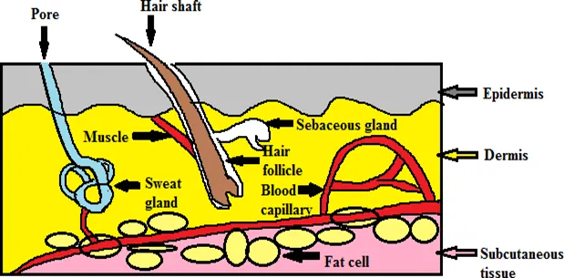
Skin Structure
Skin components and layers
The skin made up of four distinct layers of tissues.
1. Non-viable epidermis (stratum corneum)
2. Viable epidermis
3. Viable dermis
4. Subcutaneous connective tissue (hypodermis)
Pathway of Transdermal Permeation
Permeation can occur by diffusion through
1. Sebaceous and sweat glands (trans appendaged) permeation
2. Transdermal (intercellular) permeation through the stratum corneum
3. Hair follicle (trans appendaged) permeation8, 9
Basic Principle Behind TDDS
Stratum corneum is the most important layer for TDDS. If the drug is able to penetrate the stratum corneum, it can enter the blood stream. A process known as passive diffusion, which occurs too slowly for practical use, is the only means to transfer normal drugs be both water soluble and lipid soluble. Through a diffusion process, the drug directly enters in the blood stream through the skin. Since there is a high concentration on the patch and low concentration in the blood, the drug will take long time for diffusing into the blood. The ideal mixture is approximately fifty percent hydrophilic and fifty percent lipophilic. This is because lipid soluble substances readily pass through the intercellular lipid bi-layer of the cell membranes whereas water soluble drugs are able to pass limiting steps in transdermal drug delivery system. Sweat ducts and hair follicles are paths of entry of drugs, but are considered rather insignificant10.
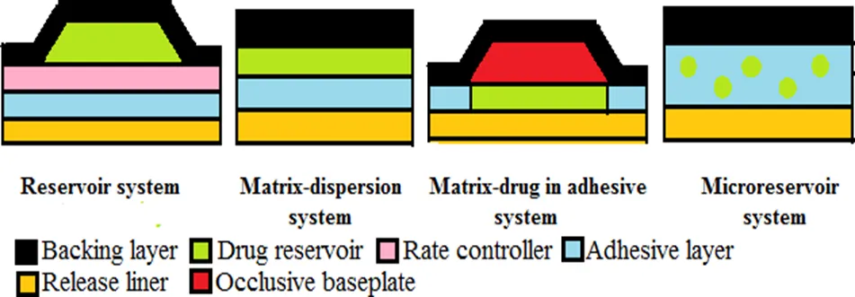
Types of Transdermal Patch
Recent Techniques For Enhancing TDDS
1. Structure-based
Micro needles: It is hybrids of the hypodermic needle (silicon needles with radius is <1μm, 150μm long and 80μm diameter) and bioadhesive patch. Due to their small sizes delivers the large molecular drug (calcein, insulin) effectively across epidermis without pain.
Macro flux®: The system incorporates a titanium micro projection array that creates superficial pathway through the skin barrier layer to allow transportation of therapeutic proteins and vaccines or ovaalbumin. It has an area of up to 8cm2 and contains as many as 300μ projection per cm2 with individual micro projection length being < 200μm. The maximal adhesive patch size is 10cm2 23,24.
MDTS (metered dose topical solution): It is made up by dissolving drug into volatile vehicle. After application over unbroken skin results into evaporation of the volatile component, leaving non-volatile drug and enhancer into the stratum corneum25.
2. Electrically based
Iontophoresis: Ionisable API permeation across the skin by appling electrical potential (0.5mA/cm2. Iontophoresis device consists of external power source, micro controller, drug compartment and electrodes26. e.g. lidocaine, ketorolac, dexamethasone, etofenamate, naproxen, vincristine, cortisone, fentanyl27, 28.
Ultrasound (sonophoresis): Skin permeation of active ingredients increase using ultrasound physical force. Here, active ingredient is mixed with gel, cream or ointment which transfers ultrasonic energy from the machine selected sites of the skin 29.
Electroporation: Application of high voltage in the form of direct current (100volts) with short durations (milliseconds) to epidermal layer of skin forms temporary pores. From these pores drug with molecular weight up to 39 kilo dalton (insulin, lidocaine, heparin and hormones) passed out30-33.
3. Velocity based
Needle-free injections:
Intraject® is prefilled injector containing nitrogen gas with blank drug capsule. It is needle free devices developed for drugs like insulin and growth hormone The patient break the tip, pull apart the safety end and full the syringe with pressurized gas. Then push the liquid formulation through a narrow orifice into the skin34, 35.
4. Others
Medicated tattoos: It is produced by Lipper–Man Ltd. It is conversion of ordinary tattoo which contains active ingredient; applied to clean, dry skin36.
Skin abrasion: The abrasion technique involves the direct removal or disruption of the upper layers of the skin to facilitate the permeation of topically applied medicaments. Some of these devices are based on techniques employed by dermatologists for superficial skin resurfacing (e.g. micro dermal abrasion) are used in the treatment of acne, scars, hyper pigmentation and other skin blemishes. Med Pharm Ltd. (Charlbury, United Kingdom) had recently developed a novel dermal abrasion device (D3S) for the delivery of difficult-to-formulate therapeutics ranging from hydrophilic low molecular weight compounds to biopharmaceuticals. With this device, in-vitro angiotensin release was increased 100-fold as compared with untreated human skin.
METHODOLOGY:
Identification of phytoconstituents
18 β-glycyrrhetinic acid
18 β-glycyrrhetinic acid was purchased from Yucca Enterprises, Mumbai-37. It was identified on the basis of physicochemical properties and HPTLC method given in the literature.
By physicochemical properties1,2
By HPTLC method Standard: 18 β-glycyrrhetinic acid
Preparation of Standard Solution: 10mg sample dissolved in 10ml methanol.
Preparation of Test Solution: 1g powder sample was refluxed for 5h with 20ml of 5M hydrochloric acid and extracted with 3x15ml chloroform. The chloroform layer was concentrated, and the residue was dissolved in 10ml chloroform.
Chromatographic condition:
Preformulation study
Investigation of physicochemical compatibility of drug and polymer-drug-excipients play a vital role with respect to release of drug from the formulation amongst others. FTIR and DSC techniques have been used here to study the physical and chemical interaction between drug and excipients. For reservoir type patch drug and polymer selected were 18 β-glycyrrhetinic acid and Carbopol 934.
Preparation of Reservoir type patch
Calculation of dose
According to the review of literature liposomal gel with 18 ß-glycyrrhetinic acid 0.9% (9mg in 1g gel base) showed a stronger anti-inflammatory activity. So dose was selected is 9mg in 1g gel base7.
Selection of batches
23 factorial design was employed to study the effect of independent variables (gel base, penetration enhancer, rate controlling membrane) on dependent variable (% drug release)8.
Formulation of reservoir type patch of 18 ß-glycyrrhetinic acid
|
Formulation of gel base |
||||||||||||
|
Sr. No. |
Ingredients |
Formulations |
||||||||||
|
F1 |
F2 |
F3 |
F4 |
F5 |
F6 |
F7 |
F8 |
F9 |
F10 |
F11 |
||
|
1 |
Carbopol 934 (%) |
4 |
4 |
4 |
4 |
4 |
4 |
4 |
4 |
4 |
4 |
4 |
|
2 |
Distilled water (ml) |
100 |
100 |
100 |
100 |
100 |
100 |
100 |
100 |
100 |
100 |
100 |
|
Formulation of medicated gel |
||||||||||||
|
1 |
Gel base (%) |
50 |
50 |
50 |
50 |
60 |
60 |
60 |
60 |
50 |
50 |
50 |
|
2 |
Benzyl alcohol (%) |
1 |
1 |
1 |
1 |
1 |
1 |
1 |
1 |
1 |
1 |
1 |
|
3 |
18 ß-glycyrrhetinic acid (%) |
0.9 |
0.9 |
0.9 |
0.9 |
0.9 |
0.9 |
0.9 |
0.9 |
0.9 |
0.9 |
0.9 |
|
4 |
Naringenin (%) |
0.5 |
0.5 |
0.5 |
0.5 |
0.5 |
0.5 |
0.5 |
0.5 |
------- |
0.25 |
1 |
|
5 |
Menthol (%) |
2 |
2 |
5 |
5 |
2 |
2 |
5 |
5 |
5 |
5 |
5 |
|
6 |
Alcohol (%) |
45 |
45 |
42 |
42 |
35 |
35 |
32 |
32 |
42.5 |
42.3 |
41.5 |
|
7 |
Triethanolamine |
q.s |
q.s |
q.s |
q.s |
q.s |
q.s |
q.s |
q.s |
q.s |
q.s |
q.s |
|
Rate-controlling membrane |
||||||||||||
|
1 |
EVA with % VA |
9 |
19 |
9 |
19 |
9 |
19 |
9 |
19 |
19 |
19 |
19 |
Here, benzyl alcohol used as preservative, triethanolamine as pH adjuster, menthol as penetration enhancer and ethyl vinyl acetate (EVA) as rate controlling membrane.
Fabrication of patch
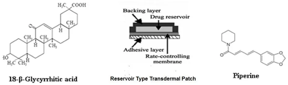
Reservoir-type transdermal patch of 18 β-glycyrrhetinic acid
Fabrications of reservoir patches were done using heat seal method.
Formulation of medicated gel base
4g polymer carbopol 934 was diffused in 100ml distilled water and set aside overnight to get a smooth gel. Preservative benzyl alcohol was incorporated into gel base. Penetrating enhancer menthol and drug were dissolved in the solvent ethanol. Drug solution poured into gel base with continuous stirring. Triethanolamine was added drop wise to the formulation for to obtained normal skin pH to 7.
Formation of reservoir patch
1g medicated gel was placed on a sheet of backing layer (pedlite polyester) covering (2x2) cm2 area. Placed rate controlling membrane over the gel and the edges of the membrane were heat sealed to obtain a leak proof device. For adhesion of the patch to the skin, a pressure sensitive adhesive, polyisobutylene was applied onto rate controlling membrane (3ml; 10%w/v in petroleum ether). Finally release liner was finally placed over the adhesive8.
Evaluation parameters of Reservoir type patch
Drug content uniformity 16, 17
The patch (2x2) cm2 was put into borosilicate glass beaker containing 100ml of phosphate buffered pH 7.4. The solvent was stirred (50rpm) with magnetic stirrer for 24 hours. The content was filtered using what man filter paper and 0.5ml filtrate was extracted with 5ml solvent chloroform. Chloroform layer was evaporated on water bath. Then residue was reconstituted in 5ml methanol and analysed for 18 ß-glycyrrhetinic acid and Naringenin at wave length maxima 250nm and 342.5nm using simultaneous UV method against the solution containing placebo patch.
In-vitro permeation study by Franz diffusion cell16, 17, 18
The formulated patch (2x2) cm2 was located on cellulose acetate membrane previously treated with 0.1N sodium hydroxide and soaked overnight in the phosphate buffer 7.4. Then put into the Franz diffusion cell such that the cell’s drug releasing surface remained towards the receptor compartment; which containing 50ml of phosphate buffer pH 7.4 at 37±0.5°. The cell was placed on a magnetic stirrer, and the solution in the receptor compartment was continuously stirred using magnetic bead at 50rpm at 37±0.5°C. 5ml solution was withdrawn at predefine time intervals and changed with same volume of phosphate buffer pH 7.4. Then the solution was extracted with 5 ml chloroform. Chloroform layer evaporated on water bath and residue was reconstituted in 5ml methanol. Finally test solutions were quantified for 18 ß-glycyrrhetinic acid and Naringenin at maximum wavelength 250nm and 343.17nm using simultaneous UV method against the standard solution containing placebo patch.
Ex-vivo permeation study by Franz diffusion cell (For F4 formulation)
Abdominal side hairs of Wister albino rat (200-210g) was removed by shaving. The rats were sacrificed, full thickness skin of the abdomen was surgically removed and adhering subcutaneous fat was cleaned using wetted cotton in isopropyl alcohol solution. Finally skin washed with distilled water and afterward with saline19. Ex-vivo permeation was performed using Franz diffusion cell which was filled with freshly prepared phosphate buffer solution of pH 7.4. Put the patch on stratum corneum side of skin in the donor part and dermis side of skin was facing towards receptor part. From the receptor part solution was withdrawn at predefine time intervals and replaced with same volume of fresh phosphate buffer solution of pH 7.4 18. Finally these test solutions were quantified for 18 ß-glycyrrhetinic acid and Naringenin with wavelength maxima at 250nm and 343.17nm using simultaneous UV method against the standard solution containing placebo patch.
Kinetic modelling of ex-vivo drug release (For F4 formulation)
Various models were tested for explaining the kinetics of drug release.
Zero order release

First order release

Higuchi model

Korsmeyer-Peppas model

Skin irritancy test (For F4 formulation)
The irritancy of formulated patches was evaluated on Wister albino rats (200-210g) according to Draize et al method20. The animals were anesthetized with thiopental sodium i.p injection (60mg/kg) then dorsal side was shaved with blade 24h before starting the experiment. The animals were separated into 3 groups, each group was containing 6 rats. Group A was control (standard), Group B was disease control which received 0.5ml of a 0.8%v/v aqueous formalin solution as a standard irritant 21 and Group C was test received F4 formulation (18 ß-glycyrrhetinic acid with Naringenin) for 3 days, every day new patch applied. After 24 and 72h; the application site of patch examined for edema and erythema. 0-4 grade was given using visual scoring method by same examiner; the final score was the mean of the 12h reading. The erythema and edema scale was 0 not any; 1 minor; 2 distinct; 3 modest and 4 harsh formulations. The primary irritancy index (PII) was calculated for each preparation according to edema and erythema scores and were classified. For non-irritant formulations PII was less than 2, for irritant formulations, PII was in between 2 to 5 and for highly irritant formulation PII was 5 to 820.
In-vivo anti-inflammatory action
Carrageenan induced rat hind paw edema animal model was used for carried out of anti inflammatory activity of the prepared formulations as per Swingle et al method22. Wistar rats were used after 2 weeks of accommodation. Wister albino rats were fasted overnight but allowed access to water ad libitum and backsides of rats shaved before the experiment. The animals were separated into 3 groups each group contained 6 rats. In disease control group, paw edema was produced by injecting 0.1ml 1%w/v of carrageenan suspension prepared in double-distilled water. The volume of injected paw was measured at 0, 1, 2, 4, 6, 8, 10, 12h using a plethysmometer. The paw swelling volume was obtained by subtracting initial volume at 0h from volume at different times. In standard group (control group), Aceclofenac patch was applied half an hour before sub plantar injection of carrageenan. In test groups 1 and 2, formulated patches were applied half an hour before subplantar injection of carrageenan. % Inhibition of edema was calculated using the following formula23, 24.
% Inhibition of edema = (1- Vt / Vc) X 100
Where, Vt = edema volume of test groups; Vc = edema volume of control
Carrageenan induced paw edema model of the reservoir patch
|
Adult albino Wistar rat total No. of animals required = 24 |
Groups |
Treatment |
|
Disease control |
Rat chow diet |
|
|
Control group |
Aceclofenac (9mg) with Naringenin (5mg) patch (2x2) cm |
|
|
Test group -1 |
Formulation F4: 18 ß-glycyrrhetinic acid (9mg) with Naringenin (5mg) patch (2x2) cm |
|
|
Test group -2 |
Formulation F4: 18 ß-glycyrrhetinic acid (9mg) patch (2x2) cm |
Stability study
Performed as per international conference of harmonization Q1A(R2) guidelines by storing the prepared patch (F4 for 18 ß-glycyrrhetinic acid and F10 for boswellic acids) at different atmospheric conditions 25oC (60±5%RH), 30oC (65±5%RH) and 40oC (75±5%RH) in stability chamber for 6 months. Then samples were taken out and examined for physical properties, drug quantity and in-vitro drug release27, 28.
RESULTS & DISCUSSION:
Identification of phytoconstituents
18 β-glycyrrhetinic acid
By Physicochemical properties White colour powder, tasteless, odourless. It is freely soluble in ethanol, chloroform. Melting point: 294oC.
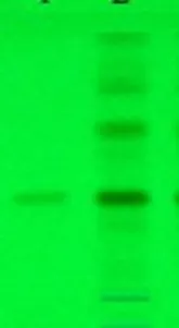
HPTLC of 18 β-glycyrrhetinic acid (Rf 0.4)
Preformulation study
18 β-glycyrrhetinic acid
By Fourier transform infrared spectroscopy (FTIR)
Infrared (IR) spectra of drug, polymer and physical mixture of drug with excipients was shown in Fig respectively. Infrared absorption spectroscopy (IR) of 18 β-glycyrrhetinic acid showed sharp band at 603, 1380, 1612, 1715 and 1760 cm-1 due to stretching vibration bands of aromatic ring, -CH3, C=C (Cyclic), C=O and –COOH respectively.
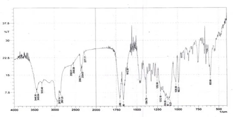
FTIR of 18 β-glycyrrhetinic acid
By Differential Scanning Calorimetry (DSC)
DSC studies were performed to test the compatibility between the drug and polymer. DSC thermograms of drug, polymer, and physical mixture (drug and excipients). API (18 β-glycyrrhetinic acid) exhibited a peak at 293.90C accordance with its melting point (292-2970C).
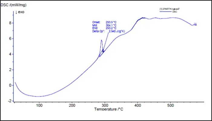
DSC of 18 β-glycyrrhetinic acid
Evaluation parameters of Reservoir type patch
Drug content uniformity
Drug content uniformity of 18 β-glycyrrhetinic acid patches
|
Formulations |
% Drug content |
|||
|
Patch 1 |
Patch 2 |
Patch 3 |
Mean± S.D. |
|
|
F1 |
99.34 |
99.32 |
99.364 |
99.34±0.015 |
|
F2 |
99.32 |
99.35 |
99.358 |
99.34±0.015 |
|
F3 |
99.35 |
99.34 |
99.31 |
99.33±0.016 |
|
F4 |
99.31 |
99.34 |
99.343 |
99.33±0.014 |
|
F5 |
99.365 |
99.34 |
99.324 |
99.34±0.015 |
|
F6 |
99.36 |
99.32 |
99.35 |
99.34±0.016 |
|
F7 |
99.31 |
99.343 |
99.34 |
99.34±0.014 |
|
F8 |
99.365 |
99.34 |
99.322 |
99.34±0.015 |
|
F9 |
99.366 |
99.34 |
99.326 |
99.34±0.015 |
|
F10 |
99.35 |
99.349 |
99.316 |
99.34±0.015 |
|
F11 |
99.303 |
99.33 |
99.35 |
99.33±0.016 |
|
Batch |
In-vitro % cumulative release of 18 β-glycyrrhetinic acid (Mean ±S.D.) |
|||||
|
0.5h |
2h |
4h |
6h |
8h |
10h |
|
|
F1 |
2.5±0.01 |
11±0.02 |
22±0.02 |
33.44±0.03 |
44±0.02 |
55.88±0.02 |
|
F2 |
3.9±0.01 |
15±0.01 |
30±0.01 |
45±0.01 |
60±0.01 |
75±0.01 |
|
F3 |
4.2±0.01 |
13.11±0.01 |
26.23±0.01 |
39.3±0.01 |
50.22±0.01 |
65.5±0.03 |
|
F4 |
5.11±0.02 |
19±0.01 |
38.22±0.01 |
57.33±0.02 |
76.44±0.01 |
95.55±0.03 |
|
F5 |
1.4±0.01 |
7.77±0.02 |
15.55±0.01 |
23.33±0.02 |
31.11±0.01 |
38.89±0.02 |
|
F6 |
3.11±0.03 |
11.67±0.03 |
23.33±0.02 |
34.78±0.01 |
46.33±0.01 |
59.11±0.01 |
|
F7 |
2.3±0.02 |
9.9±0.02 |
19.66±0.03 |
29.44±0.02 |
39.22±0.02 |
49±0.02 |
|
F8 |
4.82±0.02 |
14.11±0.01 |
28.11±0.01 |
43.33±0.02 |
56.22±0.02 |
70.3±0.02 |
|
F9 |
1.2±0.01 |
7.57±0.02 |
16.56±0.01 |
24.32±0.01 |
32.11±0.01 |
40.11±0.01 |
|
F10 |
4.7±0.02 |
14.11±0.02 |
27.92±0.01 |
42.33±0.03 |
56.22±0.02 |
70.11±0.02 |
|
F11 |
4.9±0.01 |
18.9±0.02 |
37.89±0.01 |
58.11±0.01 |
75.99±0.01 |
94.89±0.01 |
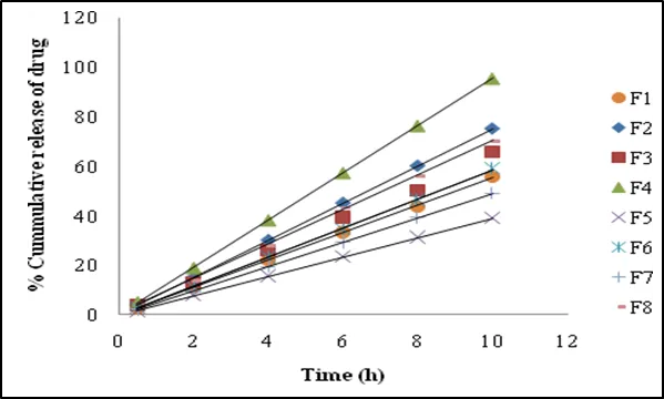
In-vitro % cumulative drug release of 18 β-glycyrrhetinic acid patch
% Cumulative drug release of 18 β-glycyrrhetinic acid patches
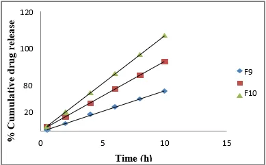
% Cumulative drug release of 18 β-glycyrrhetinic acid patch showing bioenhancer property of Naringenin
Kinetic modelling of ex-vivo drug release
Drug release from transdermal patch is controlled by chemical properties of drug and delivery form; as well as physicochemical properties of biological membrane. The release profile for F4 fitted to zero order kinetic was linear with high regression value. The rate constants were calculated from the slope of the respective plots. Data obtained were also fitted to Korsmeyer-Peppas model. The n value described release mechanism; was between 0.5 to 1 indicating the drug release to be diffusion and non-Fickian transport.
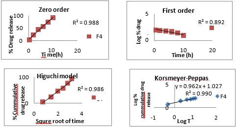
Kinetic modelling of drug release of 18 β-glycyrrhetinic acid patch
|
Formulation |
Zero order |
First order |
Higuchi |
Korsmeyer-Peppas |
|
|
R2 |
R2 |
R2 |
R2 |
n |
|
|
F4 |
0.988 |
0.892 |
0.986 |
0.990 |
0.962 |
Kinetic modelling of drug release of 18 β-glycyrrhetinic acid patch
Skin irritancy test (For F4 formulation)
Skin irritancy data of F4 formulation
|
Groups |
Erythema scale after 12h |
Mean (n=6 animals) |
|||||
|
n =1 |
n =2 |
n =3 |
n =4 |
n =5 |
n =6 |
||
|
A. Standard |
0 |
0 |
0 |
0 |
0 |
0 |
0 |
|
B. Disease control |
2 |
3 |
1 |
2 |
1 |
3 |
2 |
|
C. Formulation F4 |
0 |
0 |
0 |
0 |
0 |
0 |
0 |
|
Groups |
Edema scale after 12h |
Mean (n=6 animals) |
|||||
|
n =1 |
n =2 |
n =3 |
n =4 |
n =5 |
n =6 |
||
|
A. Standard |
0 |
0 |
0 |
0 |
0 |
0 |
0 |
|
B. Disease control |
2 |
2 |
1 |
3 |
2 |
2 |
2 |
|
C. Formulation F4 |
0 |
0 |
0 |
0 |
0 |
0 |
0 |
|
Groups |
PII |
||||||
|
A. Standard |
< 2 (Non-irritant) |
||||||
|
B. Disease control |
2 (Irritant) |
||||||
|
C. Formulation F4 |
< 2 (Non-irritant) |
||||||
In-vivo anti-inflammatory action
Carrageenan induced rat paw edema volume of standard and test groups
|
Time (h) |
Carrageenan induced rat paw edema volume (ml) |
|||
|
Disease control group (Vc) |
Standard group (Vt) |
Test group -1 (Vt) |
Test group-2 (Vt) |
|
|
0 |
0.09±0.013 |
0.09±0.013*** |
0.09±0.017*** |
0.09±0.006 |
|
1 |
0.58±0.014 |
0.31±0.011*** |
0.28±0.014*** |
0.41±0.011 |
|
2 |
1.11±0.006 |
0.51±0.028*** |
0.41±0.027*** |
0.76±0.013 |
|
4 |
1.48±0.027 |
0.41±0.016*** |
0.38±0.013*** |
0.96±0.014 |
|
6 |
1.38±0.015 |
0.28±0.006*** |
0.26±0.017*** |
0.9±0.11 |
|
8 |
1.28±0.036 |
0.24±0.013*** |
0.17±0.011*** |
0.81±0.013 |
|
10 |
0.88±0.009 |
0.13±0.014*** |
0.11±0.006*** |
0.55±0.013 |
|
12 |
0.87±0.027 |
0.13±0.014*** |
0.11±0.009*** |
0.55±0.011 |
All values were analysed using one-way ANOVA followed by Dunnett’s multiple comparison test, expressed as mean ± SEM (n = 6), ***p < 0.05. All the groups compared with control.
Anti-inflammatory effect of reservoir patches
|
Time (h) |
% Inhibition of edema |
||
|
Standard group |
Test group-1 |
Test group-2 |
|
|
0 |
0 |
0 |
0 |
|
1 |
46.55 |
51.72 |
29.31 |
|
2 |
54.05 |
63.06 |
31.53 |
|
4 |
72.30 |
74.32 |
35.14 |
|
6 |
79.71 |
81.16 |
34.78 |
|
8 |
81.25 |
86.72 |
36.72 |
|
10 |
85.23 |
87.5 |
37.50 |
|
12 |
85.06 |
87.36 |
36.78 |
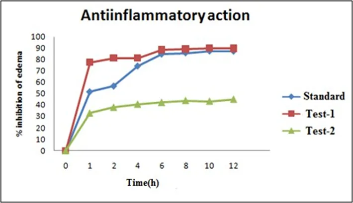
Anti-inflammatory effect of reservoir patches
|
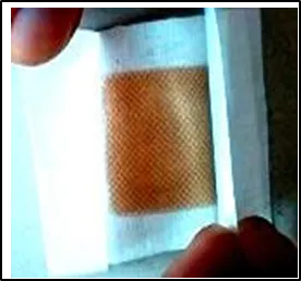
Indication: Back and Joint pain, Inflammation Directions for use: Apply to clean, dry skin Patch size: 20mm X 20mm Storage condition: Store in a cool and dry place |
Reservoir-type patch
Stability study
Stability data of the F4 patch of 18 ß-glycyrrhetinic acid
|
Parameter |
F4 patch of 18 ß-glycyrrhetinic acid |
|||||
|
25oC (60±5%RH) |
30oC (65±5%RH) |
40oC (75±5%RH) |
||||
|
% drug content |
99.31±0.015 |
99.22±0.015 |
99.33±0.015 |
|||
|
In-vitro drug release (Mean±S.D.) |
||||||
|
Temp |
Time (h) |
|||||
|
0.5 |
2 |
4 |
6 |
8 |
10 |
|
|
25oC (60±5%RH) |
5.2±0.02 |
18.9±0.03 |
38.20±0.05 |
57.14±0.04 |
76.64±0.05 |
95.48±0.03 |
|
30oC (65±5%RH) |
5.1±0.04 |
19.1±0.06 |
38.19±0.02 |
57.30±0.04 |
76.44±0.04 |
95.50±0.03 |
|
40oC (75±5%RH) |
5.3±0.05 |
19.2±0.05 |
38.22±0.06 |
57.34±0.04 |
76.45±0.02 |
95.55±0.02 |
DISCUSSION
The present study demonstrated the successful development of a reservoir-type herbal transdermal patch (F4) containing 18-β-glycyrrhetinic acid with 0.5% naringenin as a natural bioenhancer for sustained anti-inflammatory therapy. FTIR and DSC studies confirmed the compatibility of the drug with the selected formulation components, indicating the absence of any significant drug–excipient interactions. The optimized F4 formulation exhibited uniform drug content, acceptable physicochemical properties, and good stability, suggesting reliable formulation performance. The combination of 5% menthol and 0.5% naringenin significantly enhanced the percutaneous permeation of 18-β-glycyrrhetinic acid, achieving 95.55% in-vitro drug release, compared with 40.11% in the formulation without naringenin, demonstrating the remarkable bioenhancing effect of naringenin. This enhancement may be attributed to the synergistic action of menthol in disrupting the lipid structure of the stratum corneum and naringenin in improving skin permeability while reducing drug metabolism during permeation. Ex-vivo permeation studies using rat skin further confirmed the superior drug transport of the optimized formulation, and the release kinetics followed zero-order release with non-Fickian diffusion, indicating sustained and controlled drug delivery. Skin irritation studies showed that the F4 patch was non-irritant and safe for topical application. Furthermore, the optimized formulation produced significant anti-inflammatory activity in the carrageenan-induced rat paw edema model, providing prolonged suppression of inflammation compared with conventional therapy. The enhanced therapeutic efficacy observed with the naringenin-containing formulation may also be associated with the intrinsic antioxidant and anti-inflammatory properties of naringenin, which act synergistically with 18-β-glycyrrhetinic acid. Overall, the findings indicate that the optimized naringenin-bioenhanced reservoir transdermal patch (F4) is a promising strategy for improving the percutaneous delivery and sustained anti-inflammatory efficacy of herbal drugs, offering a potential alternative to conventional oral therapy for the long-term management of inflammatory disorders.
CONCLUSION
The present study successfully developed an optimized reservoir-type herbal transdermal patch (F4) containing 18β-glycyrrhetinic acid, which exhibited excellent physicochemical properties, uniform drug content, sustained drug release, effective skin permeation, and good dermal safety. The incorporation of 0.5% naringenin as a bioenhancer significantly improved percutaneous drug delivery and contributed to enhanced anti-inflammatory efficacy. Overall, the developed formulation demonstrates promising potential as a safe and effective transdermal delivery system for the sustained management of inflammatory disorders. Further pharmacokinetic and clinical studies are warranted to confirm its therapeutic applicability.
REFERENCES

M. G. Pujitha, K. Ashok*, M. Kishore Babu, Formulation and Evaluation of a Naringenin-Bio Enhanced Herbal Transdermal Patch for Improved Percutaneous Delivery and Sustained Anti-Inflammatory Therapy, Int. J. Med. Pharm. Sci., 2026, 2 (8), 148-162. https://doi.org/10.5281/zenodo.21792751
 10.5281/zenodo.21792751