

We use cookies to ensure our website works properly and to personalise your experience. Cookies policy
Dattakala College of Pharmacy, Daund-Pune
Transdermal drug delivery systems (TDDS) have emerged as an effective alternative to conventional oral and parenteral dosage forms by delivering therapeutic agents through the skin into the systemic circulation. This route offers several advantages, including avoidance of first-pass hepatic metabolism, improved patient compliance, sustained drug release, reduced dosing frequency, and minimization of gastrointestinal side effects. The present review provides a comprehensive overview of transdermal patches, covering the structure and physiology of the skin, mechanisms of drug permeation, essential components, formulation strategies, different types of transdermal patches, methods of preparation, and evaluation parameters. The review also discusses the factors influencing transdermal drug absorption, along with the advantages, limitations, and therapeutic applications of TDDS. Furthermore, recent advances in transdermal drug delivery, including microneedle-based systems, iontophoresis, sonophoresis, electroporation, nanocarrier-based formulations, and smart transdermal patches, are highlighted for their potential to enhance drug permeation and therapeutic efficacy. Future perspectives emphasize the integration of advanced biomaterials and wearable technologies for personalized drug delivery. Overall, transdermal drug delivery systems represent a rapidly evolving field with significant potential to improve clinical outcomes and expand the range of drugs suitable for non-invasive administration.
Drug delivery systems have undergone significant development over the past few decades with the aim of improving therapeutic efficacy and patient compliance. Among the various novel drug delivery approaches, the transdermal drug delivery system (TDDS) has gained considerable attention because it delivers drugs through the skin directly into the systemic circulation. This route provides an effective alternative to conventional oral and injectable dosage forms for suitable drug candidates. The oral route remains the most commonly used method of drug administration due to its convenience and patient acceptance. However, it has several limitations, including first-pass metabolism, degradation of drugs in the gastrointestinal tract, variable absorption, and the need for frequent dosing. These factors may reduce the bioavailability of many drugs and affect therapeutic outcomes. Transdermal drug delivery helps overcome many of these limitations by providing controlled and sustained drug release while avoiding the gastrointestinal tract and hepatic first-pass metabolism. A transdermal patch is a medicated adhesive dosage form that is applied to intact skin to deliver a predetermined amount of drug over a prolonged period. The drug diffuses across the skin layers and reaches the systemic circulation, maintaining relatively constant plasma drug concentrations. The success of transdermal therapy depends on factors such as the physicochemical properties of the drug, the condition of the skin, and the design of the patch, including the use of suitable polymers, adhesives, backing membranes, and permeation enhancers. Recent advances, including microneedles, iontophoresis, sonophoresis, and nanotechnology-based delivery systems, have further expanded the scope of transdermal drug delivery and improved its clinical applications. The present review discusses the basic principles of transdermal drug delivery systems, their components, types, methods of preparation, evaluation parameters, advantages, limitations, recent developments, and future prospects. It aims to provide a concise and updated overview of TDDS and its growing importance in modern pharmaceutical research.
1. Polymer Matrix
The polymer matrix forms the main body of the transdermal patch and holds the drug uniformly. It controls the rate of drug release and provides mechanical strength and flexibility to the patch. Commonly used polymers include hydroxypropyl methylcellulose (HPMC), ethyl cellulose, polyvinyl alcohol (PVA), and Eudragit.
2. Permeation Enhancers
Permeation enhancers are substances that temporarily increase the permeability of the skin, allowing the drug to pass more easily through the stratum corneum. They improve drug absorption without causing permanent damage to the skin. Examples include ethanol, dimethyl sulfoxide (DMSO), oleic acid, and propylene glycol.
3. Adhesive Layer
The adhesive layer secures the patch firmly to the skin throughout the application period. In some formulations, it also serves as a drug-containing layer. Pressure-sensitive adhesives such as acrylic, silicone, and polyisobutylene adhesives are commonly used.
4. Backing Laminate
The backing laminate is the outermost layer of the patch. It protects the formulation from moisture, oxygen, and environmental contaminants while providing support and flexibility. It should be impermeable, non-irritating, and compatible with the other components.
5. Release Liner (Release Laminate)
The release liner is a protective layer that covers the adhesive surface before application. It is removed immediately before the patch is applied to the skin. This layer prevents contamination and preserves the adhesive properties of the patch during storage.
6. Drug
The drug is the active pharmaceutical ingredient incorporated into the patch. It should possess suitable physicochemical properties, such as low molecular weight, adequate lipid and water solubility, and sufficient potency, to enable effective absorption through the skin and achieve the desired therapeutic effect.
Drugs incorporated into transdermal patches penetrate the skin through three main pathways:
The drug diffuses through the lipid matrix present between the cells of the stratum corneum. This is the most common route for the permeation of lipophilic drugs.
In this pathway, drug molecules pass directly through the corneocytes and the surrounding lipid layers. This route is generally preferred by small molecules possessing both hydrophilic and lipophilic properties.
The drug enters the skin through its appendages, including:
Hair follicles: Act as reservoirs for drug absorption and provide an alternative pathway through the skin.
Sweat glands: Provide small channels that allow limited drug permeation.
Sebaceous glands: Contribute minimally to drug transport but may facilitate the penetration of lipophilic drugs
1. Matrix type.
In the matrix-type patch, the drug is uniformly dispersed within a polymer matrix that controls the rate of drug release. The adhesive layer keeps the patch attached to the skin, while the drug diffuses gradually from the polymer into the skin. This type is simple to manufacture, flexible, and widely used in commercial products.

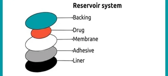
2. Reservoir type.
The reservoir-type patch contains the drug in a separate liquid or gel reservoir enclosed between a backing layer and a rate-controlling membrane. The membrane regulates the release of the drug, providing a nearly constant release rate over an extended period. However, damage to the membrane may result in dose dumping.
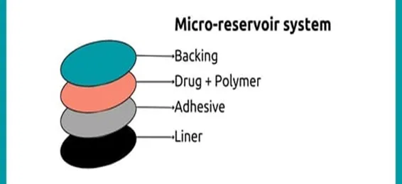
3. Micro reservoir type
The microreservoir-type patch combines the features of both matrix and reservoir systems. In this design, microscopic drug reservoirs are uniformly dispersed within a polymer matrix. This system offers better control over drug release while maintaining the stability of the formulation.
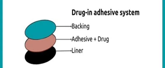
4. Drug in adhesive
In the drug-in-adhesive system, the drug is incorporated directly into the pressure-sensitive adhesive layer, which also attaches the patch to the skin. This design is thin, flexible, easy to manufacture, and is one of the most commonly used transdermal patch systems. It is available as single-layer and multi-layer drug-in-adhesive patches.
There are two types of drug in adhesive
Single-layer drug-in-adhesive
The drug-filled sticky layer. In this kind of patch, the adhesive layer is in charge of the medication release from the patch in addition to keeping the different layers of patch attached to one another and the skin.
Multilayer drug-in-adhesive
It resembles single-layer drug-in-adhesive technology. In this case, the medication is released by both sticky layers. The multilayer patch, on the other hand, differs slightly in that it incorporates an additional layer of drug-in-adhesive and, in certain situations, is divided by a membrane. This patch has both a permanent backing and a transient liner layer.
5. Miscellaneous
This category includes advanced transdermal systems such as vapour patches, hybrid matrix patches, and other modified designs developed for specific therapeutic applications. These systems are intended to improve drug delivery, enhance patient comfort, and increase therapeutic effectiveness.
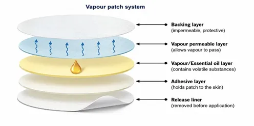
i) Vapour patch
In addition to holding the different surfaces together, the adhesive layer-containing patch also lets go of the vapour. Recently introduced to the market, vapor patches are frequently utilized to release essential oils for decongestion. There are numerous other kinds of vapor patches on the market that are intended to lessen the effects of cigarette smoking and enhance sleep quality.
1. Assymetric TPX membrane method.
2. Circular Teflon mould method.
3. By using “IPM Membrane” method.
4. Mercury substrate method.
5. Preparation of TDDS by using proliposomes.
6. By using “EVAC Membrane” method.
7. By using free film method.
There are a variety of factors that are affecting on the Action Of Transdermal patches
A. Biological Factors: -
B. Formulation Factors: -
C. Physicochemical Factors: -
A. Physicochemical evaluation
B. In vitro evaluation
C. In vivo evaluation
A. Physicochemical evaluation: -
Physicochemical evaluation is done by considering the following parameters -
1. Thickness
At various locations along the film, the screw gauge, dial gauge, and microscope were used to measure the thickness of the transdermal patches.
2. Uniformity of weight
Another name for it is weight fluctuation. Ten randomly chosen patches can be weighed in order to study it separately. Next, the patches’ average weight was determined. The weight of each individual should not differ from the weight of the average.
3. Drug content determination
Weigh the film precisely, then dissolve it in 100 mL of a suitable solvent that the medication will dissolve in. After that, the shaker incubator is used to continually shake this solution for a full day. This solution can then be requested. Following mixing and filtering, the drug’s concentration in the solution is calculated using spectroscopy and the proper dilutions.
4. Drug content uniformity test
Ten patches were chosen at random, and each patch’s content is different. The results should look like this: of these 10 patches, 9 should fall between 85% and 115% of the given value, and the remaining patch should fall between 75 and 125%. At that point, the patches are deemed to have passed the test. To pass the test, further 20 patches must be taken with content in the range of 85–125% if the first three have content in the 75–125% range.
5. Moisture content
The prepared films were removed, weighed separately, and then stored in a desiccator that holds calcium chloride. Following a 24-hour period at room temperature, each film needs to be weighed once again. The moisture content percentage is calculated using the formula provided.
6. Flatness
The transdermal patch should be smooth and should not be constricted with the time. Hence, this study was performed. For the determination of the flatness, one patch is cut down from the center and the two from each side of the patch.
7. Folding endurance
The process of evaluating folding endurance for films is figuring out how well they fold after putting them under rigorous folding circumstances. Folding endurance can be assessed by folding the film repeatedly without breaking at the same spot. That is the patch’s folding endurance value.
B. In vitro evaluation
The drug’s release from the polymeric transdermal film determines how much of it is available for blood absorption. The medication that reaches the skin’s surface is next subjected to standard penetration tests, which were carried out by affixing the transdermal patch to the skin of rats or to the artificial membrane found in vertical diffusion cells between the donor and receptor. The hydrophilic side of the membrane receives application from the transdermal system, while the lipophilic side comes into touch with the receptor fluid. The receiver compartment is constantly stirred and kept at a set temperature, often 32°C. At regular intervals, the samples were removed, and each time, the same volume of buffer was added. A UV spectrophotometer is used to measure the absorbance after the samples were diluted. Calculations are made to determine how much medication has permeated each square centimeter at each interval. The system’s architecture, the patch’s size, the skin’s surface area, thickness, temperature, and other factors all affect how much drug is released.
C. In Vivo evaluation
The medication performance can be accurately represented in these study evaluations. The TDDS in this study can be completed using –
• Animal models.
• Human volunteers or human models.
In the future, drug delivery systems may incorporate microemulsion, niosomes, and liposomes. The purpose of this discovery is to enhance medication delivery, as the majority of classical formulation excipients have limited intrinsic solubility. Numerous medications, including steroids, methotrexate, interferon, antifungals, and antibiotics, are being developed for possible administration. Transdermal patch sales are expected to rise in the future and have grown at a rate of 25% annually in previous years. As new devices are developed and the number of transdermal drugs that are marketed rises, this number will rise in the future. As long as design advancements are made, transdermal analgesic administration is expected to gain more and more traction. Studies are being conducted to improve efficacy and safety. To enhance practical aspects such as the patch wearer’s experience and to offer more accurate medication delivery linked to longer duration of action. Other possible advancements include enhanced transdermal technology, which raises the energy of the drug molecules or modifies the skin barrier to increase drug flux through the skin by using mechanical energy. Many “active” transdermal technologies are being researched for a variety of medications following the successful construction of patches utilizing iontophoresis. These include sonophoresis, which uses low frequency ultrasonic energy to disrupt the stratum corneum, thermal energy, which uses heat to increase the energy of drug molecules and make the skin more permeable, and electroporation, which uses short electrical pulses of high voltage to create transient aqueous pores in the skin. Drug flux over the skin has been studied in relation to magnetic energy, or magnetophoresis. It’s possible that the transdermal patch is a neglected method for treating both acute and chronic pain.
Several transdermal patches have been successfully commercialized for the treatment of various acute and chronic conditions. These products provide controlled drug release, improve patient compliance, and reduce dosing frequency. Some commonly marketed transdermal patches are listed in Table 1.
Table 1. Commonly Marketed Transdermal Patches
|
Drug |
Brand Name |
Therapeutic use |
Duration |
|
Nicotine |
Nicoderm CQ |
Smoking cessation |
24 hours |
|
Nitroglycerin |
Nitro-Dur |
Angina pectoris |
12-24hours |
|
Rivastigmine |
Exelon Patch |
Alzheimer’s Disease |
24 hours |
|
Fentanyl |
Duragesic |
Chronic pain |
72 hours |
|
Clonidine |
Catapres-TTS |
Hypertension |
7 days |
CONCLUSION
Transdermal drug delivery systems have emerged as an effective alternative to conventional drug administration by providing controlled drug release, improved bioavailability, and enhanced patient compliance. Continuous advances in polymer science, nanotechnology, microneedles, and wearable drug delivery devices have significantly expanded the therapeutic potential of transdermal systems. Although challenges such as limited skin permeability and drug selection remain, ongoing research is expected to overcome these limitations. Consequently, transdermal drug delivery systems are likely to play an increasingly important role in modern pharmaceutical therapy and personalized medicine.
REFERENCES

Ajitkumar Tidake*, Rohan Honmane, Transdermal Drug Delivery Systems: An Updated Review on Design, Evaluation, and Recent Advances, Int. J. Med. Pharm. Sci., 2026, 2 (7), 1054-1062. https://doi.org/10.5281/zenodo.21637783
Severity: 8192
Message: substr(): Passing null to parameter #1 ($string) of type string is deprecated
Filename: frontend/article.php
Line Number: 705
Severity: 8192
Message: substr(): Passing null to parameter #1 ($string) of type string is deprecated
Filename: frontend/article.php
Line Number: 740
Severity: 8192
Message: substr(): Passing null to parameter #1 ($string) of type string is deprecated
Filename: frontend/article.php
Line Number: 740
Severity: 8192
Message: substr(): Passing null to parameter #1 ($string) of type string is deprecated
Filename: frontend/article.php
Line Number: 740
 10.5281/zenodo.21637783
Severity: 8192
Message: substr(): Passing null to parameter #1 ($string) of type string is deprecated
Filename: frontend/article.php
Line Number: 924
Severity: 8192
Message: substr(): Passing null to parameter #1 ($string) of type string is deprecated
Filename: frontend/article.php
Line Number: 956