

We use cookies to ensure our website works properly and to personalise your experience. Cookies policy
1Department of Pharmacy Practice, Karpagam College of Pharmacy, Coimbatore -641 032, Tamil Nadu, India.
2Professor, Department of Pharmacy Practice, Karpagam College of Pharmacy, Coimbatore -641 032, Tamil Nadu, India.
3The Tamil Nadu Dr. MGR Medical University, Chennai- 600032, Tamil Nadu, India
Hutchinson–Gilford Progeria Syndrome (HGPS) is a rare, progressive genetic disorder characterized by accelerated aging in children due to mutations in the LMNA gene, resulting in the accumulation of the abnormal protein progerin. Although affected children appear healthy at birth, clinical manifestations such as growth retardation, alopecia, lipodystrophy, skeletal abnormalities, and premature cardiovascular disease typically develop within the first two years of life. Cardiovascular complications remain the leading cause of mortality, with an average life expectancy of approximately 14–15 years. This literature review summarizes the epidemiology, genetic basis, molecular pathophysiology, clinical manifestations, diagnostic approaches, and current management strategies for HGPS. It also highlights recent therapeutic advances, including lonafarnib, mTOR inhibitors, antisense oligonucleotide therapy, and gene-editing technologies such as CRISPR-Cas9 protein, which offer promising future treatment options. Early diagnosis through molecular genetic testing and multidisciplinary management are essential for improving survival and quality of life. Continued research into disease mechanisms and targeted therapies is crucial for developing effective long-term treatments for this devastating disorder.
Hutchinson–Gilford progeria syndrome is an ' fatal disease that affects kids.It causes aging. The first description of patients with this syndrome was in 1886 by Jonathan Hutchinson. Later his colleague Hastings Gilford described it again. They named the condition progeria, which means prematurely aged in 1904. The word progeria comes from words that mean "before" and "old age”. People with Hutchinson–Gilford progeria syndrome have problems that resemble aging. These problems start in the year, after birth. They include failure to grow, loss of fat and hair skin changes, bone and joint disease and atherosclerosis. Hutchinson–Gilford progeria syndrome is also called Hutchinson–Gilford syndrome, progeria or progeria of childhood. It is a condition that affects kids and causes them to age prematurely. Progeria is a disease that needs to be understood and studied.The Hutchinson–Gilford progeria syndrome is an area of research. It affects one in twenty million newborns. Children with this disease usually live for fourteen and a half years. When they are born these children look like any child. As they grow they start to show symptoms of the disease. These symptoms get worse over time. The main symptoms of Hutchinson-Gilford progeria syndrome are hair loss, skull and face problems short height, growth problems, bone defects, metabolic disorders, hormone problems and heart disease. Heart disease is the cause of death in these children. Hutchinson-Gilford progeria syndrome is not passed down from parents to children. It is caused by a mutation in the LMNA gene. This mutation happens when a child is growing inside the womb. The LMNA gene helps make two proteins called lamin A and lamin C. Lamin A is first made as a precursor called prelamin A. Prelamin A goes through many changes before it becomes A [1,10,13]. First prelamin A is changed by an enzyme called farnesyltransferase. Then the last three amino acids are cut off by another enzyme. After that the new end of prelamin A is methylated by an enzyme. This process is important for prelamin A to become lamin A. Lamin A becomes part of the nuclear lamina, which is like a mesh that supports the nucleus of the cell. Lamin A interacts with other proteins that are important for the cell [2,8]. The problem in Hutchinson-Gilford progeria syndrome is that the mutation in the LMNA gene causes prelamin A to not be changed properly. As a result prelamin A becomes a protein called progerin. Progerin builds up in the cell. Causes many problems. It makes the nucleus of the cell look abnormal and disrupts the interactions, between proteins. This ultimately leads to the symptoms of Hutchinson-Gilford progeria syndrome [2,8]. Researchers have been trying to find ways to treat Hutchinson-Gilford progeria syndrome. They are trying to stop the production of progerin or reduce its levels in the cell. They are also trying to block the enzymes that change prelamin A into progerin. One of these enzymes is farnesyltransferase. Another one is isoprenylcysteine carboxyl methyltransferase. By blocking these enzymes researchers hope to reduce the levels of progerin and alleviate the symptoms of the disease. There are ways to approach this problem. Some researchers are trying to fix the mistake in the LMNA gene using gene editing techniques. Others are trying to reduce the production of progerin by targeting the splicing of the LMNA gene. Some are trying to stimulate the degradation of progerin while others are trying to block the enzymes that change prelamin A into progerin. All these approaches are aimed at reducing the levels of progerin and treating Hutchinson-Gilford progeria syndrome. others Names for This Condition [1,5]
What are the Different Types of Progeria Syndrome
Progeria or what people call Benjamin button sickness is a group of genetic disorders that make kids age really fast. The well-known type is HGPS or Hutchinson-Gilford Progeria Syndrome. There are several other types of progeria syndrome. Each one has its genetic causes and symptoms [3,11]. The main types of progeria syndrome include:
* HGPS Disease: This is the common type of progeria syndrome. It happens when there is a problem with the LMNA gene. This gene problem leads to the production of a protein called progerin. This bad protein makes the cell nucleus weak. As a result kids with HGPS disease start showing signs of aging when they're really young. They may look normal when they are born. Within the first two years of life they start to show signs of aging. These signs include not growing losing hair having skin that looks old and having stiff joints. Kids with HGPS disease often have bad heart problems. They usually do not live very long only about 13 to 15 years [3,10].
* Werner Syndrome: This is also known as adult progeria syndrome. It usually starts when people are teenagers or young adults. It happens when there is a problem with the WRN gene. This gene helps fix damaged DNA. People with Werner Syndrome start to show signs of aging early. These signs include hair, cloudy eyes, thin skin, diabetes and weak bones. They are also more likely to get cancer and have heart problems. They usually do not live very long only into their 40s or 50s [3].
* Cockayne Syndrome: This is not always considered a type of progeria syndrome. It has some similar features. These features include not growing aging too fast and having problems with brain development. It happens when there is a problem with genes that help fix damaged DNA. People with Cockayne Syndrome may have problems with their skin, hearing, vision and brain. They are not as likely to have heart problems like people with HGPS disease. They still do not live very long usually only into childhood or early adulthood [3].
* Rothmund-Thomson Syndrome: This is a rare type of progeria syndrome. It happens when there is a problem with the RECQL4 gene. Kids with this syndrome start to show signs of aging when they're babies. These signs include skin problems, not having hair and having problems with their bones. They are also more likely to get cancer [3].
* Mandibuloacral Dysplasia: This is a type of progeria syndrome. It happens when there is a problem with the LMNA or ZMPSTE24 genes. People with this syndrome have problems with their bones, skin and body. They may live longer than people, with HGPS disease. They still show signs of aging really early [3]. In summary there are types of progeria syndrome. Each one is different. Has its own causes and symptoms. They all help us learn more about how we age and how our genes work.
Epidemiology
HGPS is a rare genetic disorder. The number of people born with HGPS is very low it is one in four to eight million newborns. HGPS affects boys and girls equally. It affects people of all races. The number of people who get HGPS is the same over the world. It does not matter if you are a boy or a girl or where you live you have the chance of getting HGPS [4,9]. There are 200 to 250 children living with HGPS in the world at any given time. HGPS is found in countries. Now there are about 114 children with HGPS who have been diagnosed in 39 countries. In the United States HGPS is very rare it is one in eight million births. This is based on the number of cases that have been reported. HGPS is the thing, as progeria and it is very rare everywhere. One in four to eight million babies are born with Hutchinson Gilford Progeria Syndrome, a genetic disorder. Every ethnicity and gender are equally affected by Hutchinson Gilford Progeria Syndrome. People think that Progeria is just as common everywhere no matter what gender, where you live or what your ethnicity is. Now there are 300 to 350 children with Progeria all over the world. Far 39 countries and about 114 children have been diagnosed with Hutchinson Gilford Progeria Syndrome. Based on the number of cases people think that Progeria affects one in eight million babies in the United States [9,11].
Sex: Hutchinson Gilford Progeria Syndrome affects boys than girls with a ratio of 1.5 boys to one girl, which means it affects men a little more often [10,11].
Age: Some children with Hutchinson Gilford Progeria Syndrome have skin problems. You may not notice anything is wrong with them when they are born. It usually takes six to twelve months. Even longer to see the signs of Hutchinson Gilford Progeria Syndrome like the way their face and skin look and problems with their muscles and bones. At that point the child is not developing like they should. The doctor needs to do a more complete evaluation of the child, with Hutchinson Gilford Progeria Syndrome [10].
Causes
A problem with one gene causes progeria. This gene is called lamin A. It helps make a protein that keeps the center of a cell together. When this gene has a problem it makes a protein called progerin. Progerin makes cells weak. It seems to make people with progeria age very fast. The problem with the gene that causes progeria does not usually run in families. Most of the time the problem with the gene just happens by chance [8,13]. Mutations in the LMNA gene cause Hutchinson-Gilford progeria syndrome. The LMNA gene tells our bodies how to make a protein called lamin A. Lamin A helps decide the shape of the nucleus in our cells. This protein is a part of the nuclear envelope, which is like a protective layer, around the nucleus. When LMNA gene mutations happen they make a version of lamin A. This weird lamin A makes the nuclear envelope unstable. Slowly hurts the nucleus. As a result cells are more likely to die early [7,8]. Researchers are trying to figure out how these changes cause the symptoms of Hutchinson-Gilford progeria syndrome and LMNA gene mutations. They want to know how gene and lamin A problems lead to this condition. The LMNA gene tells our body how to make proteins called lamins. It makes two proteins lamin A and lamin C in almost every cell of our body. Lamin A and lamin C are made from the building blocks but lamin A is a little bit longer than lamin C. Lamins A and C are like the framework of our cells. They help keep our cells strong and stable. These proteins are part of the envelope, which is like a cover around the nucleus in our cells. The nuclear envelope is made up of a mesh- layer with lamins A and C and it controls what goes in and out of the nucleus. We can also find lamins A and C inside the nucleus. Scientists think they might help control which genes are turned on or off. Before lamin A can do its job it has to be changed inside the cell. It starts as prelamin A. Goes through a lot of steps before it becomes part of the lamina. Lamin C does not need to go through all those steps to become part of the lamina. The LMNA gene and the proteins it makes, like lamin A and lamin C are very important for our cells to work properly [8].
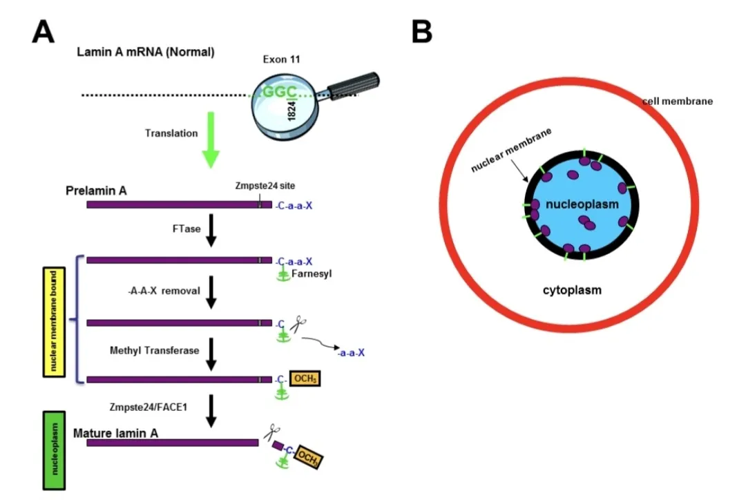
Figure 1: Formation of lamin A in normal.
First the mRNA for lamin A is translated into A. Then prelamin A gets a special tag called a farnesyl group. This tag helps prelamin A stick to the nuclear membrane. The enzyme Zmpste24 or FACE1. Cuts off some extra pieces from the end of prelamin A. This cutting lets prelamin A go, from the membrane and move into the nucleoplasm. In the process prelamin A stays stuck to the nuclear membrane for a while. Then Zmpste24 or FACE1 cuts it. It becomes mature lamin A. This mature lamin A mostly stays in the nucleoplasm [8].
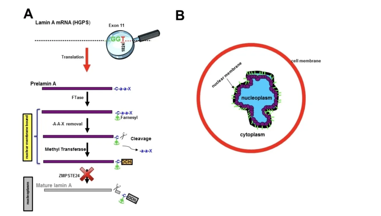
Figure 2: Formation of lamin A in abnormal.
shows what happens in a patient with progeria. The problem is with the processing of lamin A in the cells of a patient with progeria. The lamin A is mutant. This means that the mutated lamin A mRNA is translated into A. This prelamin A does not have the site where Zmpste24 or FACE1 can cut it. So prelamin A gets farnesylated. This causes it to stick to the nuclear membrane. The reason is that Zmpste24 cannot cut the end amino acids. As a result the cell cannot make lamin A. In patients, with HGPS prelamin A stays farnesylated in the cell. It builds up at the nuclear membrane. [8].
Other similar syndromes
There are health problems that are like progeria. These problems are called syndromes. They are caused by problems with genes that are passed down from parents. These syndromes make people age fast and do not live as long as they should: [3]
• Wiedemann-Rautenstrauch syndrome is also known as neonatal progeroid syndrome. It starts when a baby is still, in the womb. The baby is born with signs of aging [3].
• Werner syndrome is also known as adult progeria. It starts when people are teenagers or young adults. It makes them age fast and they get health problems that usually happen when people are old like cloudy eyes and diabetes [3].
Pathophysiological mechanism underlying the development of HGPS
HGPS is a disorder that affects the bodys cells. It is caused by a change in the LMNA gene, which is located on chromosome 1. This change happens entirely by a new point mutation in codon 608 of exon 11. The LMNA gene provides instructions for making three proteins called lamin A lamin C. Lamin 10. These proteins are important for the structure and function of the cells nucleus. The nucleus is like the control center of the cell [8,13]. The lamin A, lamin C and lamin 10 proteins help keep the nucleus in its shape. The LMNA mutation in HGPS is a change in the DNA sequence. This change creates a splice site, which leads to a missing piece of RNA. The resulting protein, called progerin is abnormal. Has a deletion of 50 amino acids. Progerin is thought to interfere with the function of the nucleus. It may also disrupt important processes such as cell division, DNA replication and gene transcription. Progerin has a fatty tail that helps it attach to the nucleus. This attachment can cause the nucleus to change shape and function. The LMNA gene has a sequence called CAAX, which is important for the processing of the lamin A protein. The CAAX sequence helps add a fatty group to the protein, which's necessary for its function. The fatty group helps the protein attach to the nucleus. The attachment is important for the proteins role in maintaining the nucleuss shape and function. In HGPS the progerin protein cannot be properly processed. It keeps its fatty tail, which causes it to accumulate in the nucleus and disrupt its function. The accumulation of progerin causes the nucleus to change shape. This change can lead to problems with cell division, DNA replication and gene transcription. The ZMPSTE24 enzyme is important for the processing of the lamin A protein. It helps remove the fatty tail from the protein, which's necessary for its function. The ZMPSTE24 enzyme is like a scissors that helps cut the protein. The cut is necessary for the protein to function properly. In HGPS the progerin protein cannot be properly processed by the ZMPSTE24 enzyme. This leads to its accumulation in the nucleus and disruption of its function. The accumulation of progerin causes problems, with the nucleuss shape and function. This can lead to the symptoms of HGPS [2,8].
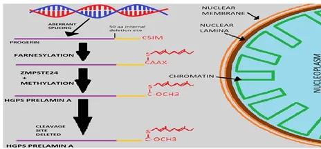
Figure 3: Pathophysiology of Hutchinson Gilford Syndrome.
Symptoms
within the first year of your child’s life you will notice that your child’s growth has slowed down.. Your child’s motor development and intelligence are not affected by this [10].
The symptoms of this disorder cause a very distinctive appearance in your child. These symptoms include: [10]
The symptoms of this disorder also include some health issues:
Clinical manifestations caused by HGPS
Typically, the ability of cells to divide in an aging syndrome is reduced. A study wanted to know if telomeres are shortened in diseases. To test this they measured the length of telomeres in cells from patients with HGPS. They compared it with individuals of the same age. They found that all five cell samples from HGPS patients had an ability to divide compared to five young healthy donors. Telomere length and ability to divide were both reduced in cells from HGPS patients. This shows that telomere length is a sign of aging [12]. In cells from HGPS patients the patients parents showed no signs of aging. The telomere length was comparable to that of age-matched donors. This supports the idea that Hutchinson-Gilford progeria is caused by an autosomal dominant mutation [12]. The irregular telomere length regulation, a high cell turnover rate or a high rate of telomere deficit could all contribute to short telomeres in HGPS patients. A study compared telomere deficit during cell aging from HGPS patients to that of donors of the same age. In HGPS patients cells there was a rise in the incidence of telomere deficit. Patients with HGPS appear normal at birth. By the age of one year they develop signs and symptoms. These include hair loss, severe loss of fat, lack of weight gain and skeletal manifestations [12]. Rigidity is a property that determines a bones ability to withstand stress. It was found to be significantly irregular in HGPS patients compared to controls. A study used techniques to assess bone-building strength. It found that HGPS tends to influence the structural geometry. This is suggestive of dysplasia. Further evidence includes the arrangement of decreased mineralization of the long bones [12]. In HGPS patients the bone formation was in the range. The bone resorption markers were also in the range. Traditional biomarkers for osteoporosis do not apply in HGPS. A study found that HGPS patients have a spectrum of to-late stage plaques similar to geriatric cardiovascular disease. Inflammation, calcification and plaque erosion were exhibited on the lesions of both typical atherosclerosis and HGPS patients [12]. Progerin accumulation can contribute to the development of thickening of the adventitia in arteries. This ultimately results in decreased plaque formation potential decreased vascular compliance and increased vessel stiffness. As adventitial fibrosis keeps proceeding the stiffness of the aorta will cause an increase in the afterload of the muscles leading to left ventricular hypertrophy [12]. Progerin accumulation within the vasculature implies an indirect influence on progressive cardiovascular disease. There was an accumulation of progerin identified in the vascular smooth muscle cells and adventitia. By illustrating progerin exists in the arteries of healthy aging individuals and rises with age we also recognize a new factor in the traditional aging process. In fibroblast lines Progerin-positive cells manifest limitations in the mitosis that escalate with passage number. This finding supports the theory that progerin-induced mitotic defects are linked to aging. The adventitia had the number of progerin-positive cells in non-HGPS arteries. This suggests that some vessel abuse can begin in this layer of the vessel and then cause distress, to the intima indicating plaque formation [12]. The average life span is 14.5 years. With treatment it can be extended to, around 19 years [1,6].
Diagnosis
Diagnosis/testing. To diagnose HGPS we look for physical signs and a specific genetic change in the LMNA gene [10].
The doctor can tell if someone has Hutchinson-Gilford progeria syndrome when they see signs. This usually happens when the person is very young. The doctor will do some tests to confirm the diagnosis [10].
Suggestive Findings
If a person has these signs the doctor might think they have Hutchinson-Gilford progeria syndrome. They have intelligence. They have signs that can be seen on x-rays [10]
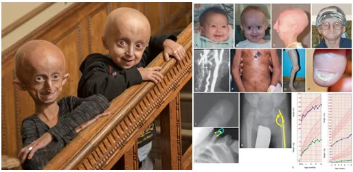
Figure 4: A patient with Hutchinson-Gilford progeria syndrome
This is a picture of a girl who's 11 years old and a boy who is 6 years old. They both have Hutchinson-Gilford progeria syndrome. You can see that they have [14,19]
Growth deficiency
Kids with Hutchinson-Gilford progeria syndrome usually start to show signs of the disease when they're very young [10].
Facial features that develop in childhood (See Figure 4) [10]
Ectodermal findings
Their teeth, skin and hair are all affected [10].
Musculoskeletal clinical and radiographic findings
Their bones and muscles are affected too [10].
Other findings
They have some other signs too [10].
Establishing the Diagnosis
Clinical Diagnosis
The diagnosis of Hutchinson-Gilford progeria syndrome or HGPS can be made when a person, known as a proband has characteristic features. These features include skin problems, severe growth failure, hair loss and a lack of fat in the body well as small collarbones. All of these features must be present in a person with intellectual development and within the first two years of life. If a person has these features it can help doctors make a diagnosis of HGPS. However if a person does not have all of these features it does not mean they do not have HGPS because some people with HGPS may not have all of these features away. To confirm the diagnosis doctors should do testing [10].
Molecular Diagnosis
There are four categories that help doctors understand disorders related to the LMNA gene. Categories 1 and 2 are used to diagnose HGPS while categories 3 and 4 are not considered HGPS: HGPS with a genotype that produces progerin. HGPS with a genotype that produces progerin. Disorders that are similar to HGPS but do not produce progerin, which can be caused by: Changes in the LMNA gene that do not produce progerin but still cause symptoms to HGPS. Changes in the ZMPSTE24 gene that cause problems with the production of lamin A protein and result in symptoms to HGPS. Disorders that are not similar to HGPS. HGPS is caused by the production of a protein called progerin. Progerin is made when there is a problem with the way the LMNA gene is read. This problem causes a piece of the gene to be missing which results in a protein that's not normal. The diagnosis of genotype HGPS can be made when a person has symptoms of HGPS and a specific change in the LMNA gene is found through molecular genetic testing. The diagnosis of genotype HGPS can be made when a person has symptoms similar to classic genotype HGPS and a change in the LMNA gene is found that produces progerin. To make a diagnosis doctors can use types of molecular genetic testing including: Single-gene testing, which involves looking at a specific gene to see if there are any changes Multigene panel testing, which involves looking at many genes at the same time to see if there are any changes. Comprehensive genomic testing, which involves looking at all of a persons genes to see if there are any changes [10]
Option 1: Single-gene testing
Doctors can start by looking at the exon 11 of the LMNA gene, which is the part of the gene that is most likely to have changes that cause HGPS. If no changes are found doctors can then look at the LMNA gene. If still no changes are found doctors can look at the ZMPSTE24 gene. A multigene panel can also be used to look at genes at the same time. This can be helpful because it can identify changes, in genes that may not have been thought to be related to HGPS. However it is important to note that the genes included in the panel and the tests used can vary depending on the laboratory [10].
Option 2
When the phenotype looks the same as other inherited disorders that have a progeroid phenotype the doctor does not need to figure out which gene is probably the cause. Comprehensive genomic testing is used to find the answer. Exome sequencing is used most of the time. Genome sequencing can also be done [10].
Table 1: Molecular Genetic Testing Used in Hutchinson-Gilford Progeria Syndrome
|
Gene |
Method |
Propotion of pathogenic variants identified by method |
|
LMNA |
Targeted sequence analysis (incl exon 11 & at least 1stbases of intron 11) |
100% |
|
LMNA |
Sequence analysis |
~ 100% |
|
LMNA |
Gene-targeted deletion/ duplication analysis |
None identified |
For information on gene variants see Molecular Genetics. 90 Percent of people with typical signs of HGPS have a specific gene change. This change is called a c.1824C>T pathogenic variant. The other 10 percent have gene changes. These include a c.1821G>A or c.1822G>A pathogenic variant. Some people have a progerin-producing variant in exon 11 or intron 11. When testing the gene sequence we look at intron 11. This test finds gene variants that're harmless, probably harmless uncertain probably harmful or harmful. The test can find types of variants. These include missense, nonsense and splice site variants. It can also find deletions or insertions within the gene. Usually it does not detect deletions or duplications of the gene. There was a case of a child with two progerin-producing variants in different cells. This is called mosaicism. It was found using a sequencing test [10]. A special test called gene-targeted deletion/duplication analysis can find deletions or duplications, within the gene. This test uses techniques. These include PCR, long-range PCR, multiplex ligation-dependent probe amplification (MLPA) and a special microarray. The microarray is designed to detect deletions or duplications of exons [10].
The urinary hyaluronic test
When doctors test the urine of patients with HGPS they find levels of hyaluronic acid. This is not a sure way to diagnose the disease. The reason for this is not fully understood [10]. When the urinary hyaluronic test is done on a patient with HGPS the results show that the level of acid in the urine is high. At the time the level of certain enzymes that protect against damage in the blood is low. The level of some fatty acids is also low. When the level of these enzymes is low it can cause the body to age faster. This is because the body has harmful substances in the blood. The urinary hyaluronic test used to be done on patients with HGPS but now it is not considered reliable and is not recommended to diagnose the disease [10]. Nowadays a genetic test can detect a gene called lamin A. This test can be done using a blood sample or a skin sample from the patient. This gives a diagnosis. On the hand if a child shows signs of HGPS this genetic test can be done to confirm the diagnosis and start treatment early [10].
Prenatal testing
To diagnose HGPS before birth doctors can test the DNA of the fetus. They can get this DNA from the fluid around the fetus usually between 15 and 18 weeks of pregnancy or from a sample of the placenta between 10 and 12 weeks. Before doing this test doctors need to identify the family members who have the disease. If the disease-causing gene has been found in a family member then other family members can have a test before getting pregnant [10].
Other tests
To check for heart disease and heart failure doctors use tests, like ECG and echocardiography. These tests can help detect problems with the heart. The urinary hyaluronic test is not used for this purpose. Instead doctors use these tests to check the heart [6,12].
Management
Current Treatment Approaches For HGPS
HGPS is a genetic disorder. Researchers have been working on finding treatments [1,5]. Following the discovery of LMNA mutations in 2003 treatment strategies for HGPS have evolved over phases: [5]
Early stage management
2003-2010: Researchers focused on using existing drugs to target progerin production and accumulation. They used farnesyltransferase inhibitors (FTIs) which were initially developed for cancer treatment to inhibit progerin farnesylation [5].
Morden therapeutic managment
2010-2020: The approach shifted to combination therapies that not targeted progerin modification but also enhanced its clearance. For example combining FTIs with statins and amino bisphosphonates aimed to prevent prenylation pathways [5].
2020-Present: Recent advancements have focused on gene-based therapies, including CRISPR/Cas9-mediated correction of the LMNA mutation. These approaches aim to address the underlying genetic defect responsible for HGPS [5]. Several treatment approaches have been explored:
1. FTIs:
FTIs were originally developed as anticancer agents. They work by binding to the farnesyltransferase CAAX motif on progerin preventing its farnesylation and subsequent accumulation. Lonafarnib (Zokinvy) is the FDA-approved drug for HGPS and progeroid laminopathy. A phase 2 clinical trial showed that Lonafarnib extended survival by 2.5 years over an 11-year follow-up period [5].
Statin and Bisphosphonate: These treatments inhibit progerin prenylation by reducing production of proteins required for farnesylation. However clinical trials showed no superiority compared to lonafarnib monotherapy [5].
ICMT inhibitor: ICMT inhibitors target the carboxymethylation step, a -translational modification of progerin. In studies have shown promising results but further studies are needed to validate its safety and efficacy [5].
MTOR. Other drugs:
mTOR inhibitors enhance autophagy-mediated progerin clearance reducing nuclear abnormalities and senescence [5,15]. Everolimus, rapamycin and temsirolimus have been investigated for progeria [15].
1. Antisense oligonucleotide (ASO) therapy:
ASO therapy corrects the LMNA splicing error. Reduces progerin production at the mRNA level [5]. Human trials are forthcoming.
Gene editing (CRISPR and base editing) : CRISPR/Cas9 and adenine base editing correct the LMNA mutation at the DNA level. Offer a potential one-time cure [5]. However, it is still experimental in humans.
Therapies for toxic effects of progerin:
Several treatments target nuclear abnormalities, inflammation and DNA damage [5]. Combination therapies with lonafarnib have been explored:
Zoledronate+ Pravastatin (ZOPRA)+Lonafarnib:
A clinical trial showed that this combination therapy did not demonstrate efficacy compared to monotherapy [5].
Everolimus+ Lonafarnib:
A combination clinical trial is ongoing [5].
Progerinin+ Lonafarnib:
A phase 2 clinical trial of combination therapy is recruiting now. multidisciplinary clinical approach is central to improving the quality of life for patients, with HGPS. Regular monitoring and supportive strategies can help maintain function [5].
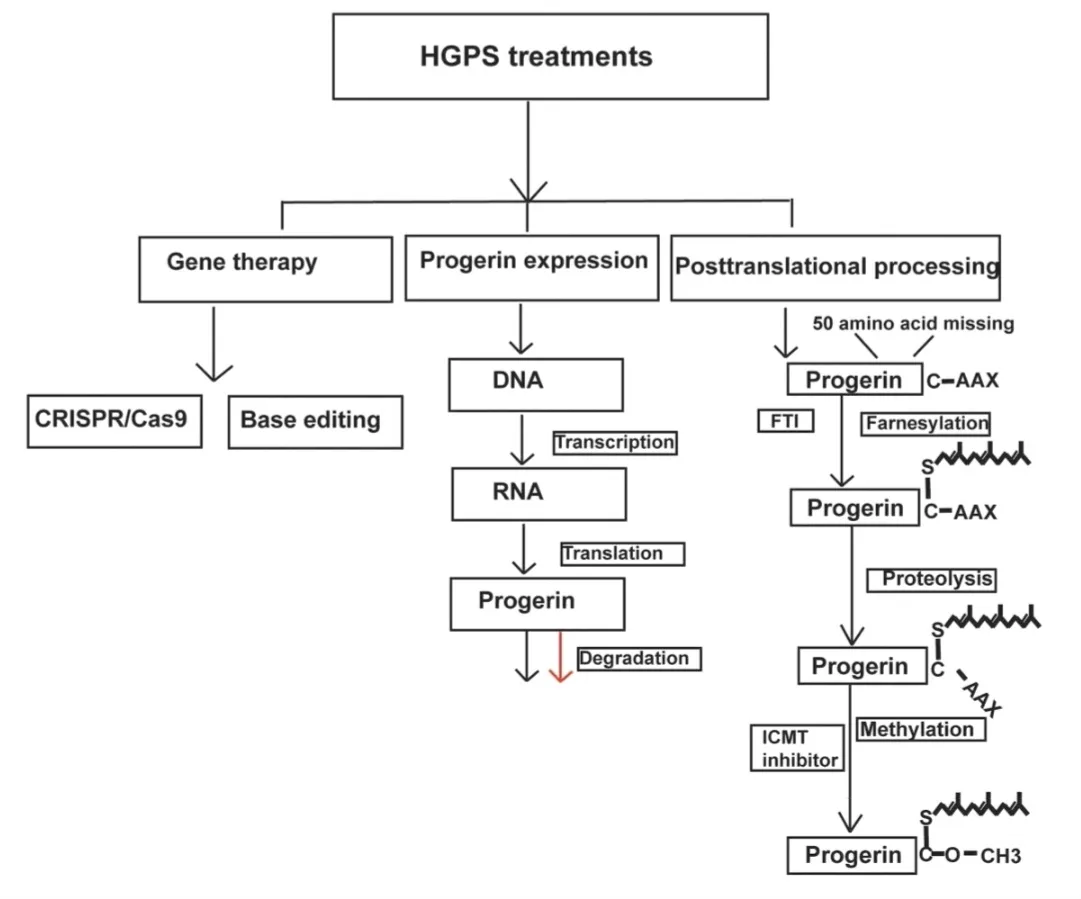
Figure 5: Overview of potential Hutchinson-Gilford progeria syndrome (HGPS) treatment strategies
Preoperative management
Atherosclerotic disease and heart failure are very common in kids with HGPS. So full ASA monitoring is a must during anesthesia. This includes using a 5-lead ECG to monitor the heart during surgery. In some cases invasive monitoring might be needed. However, it's not standard for all patients. It's also important to prevent blood volume poor blood flow and low body temperature. Some kids with HGPS have problems with blood vessels in the brain and aneurysms. These need to be considered when giving anesthesia. Because kids with HGPS often have hair loss and less body fat we need to take steps to keep them warm before anesthesia. Blood sugar levels should be controlled before during and after surgery. We also need to be careful when moving and positioning the patient to avoid injuries. If a patient has triglycerides we should be careful when using propofol for long surgeries. This is because it can increase levels and cause problems like pancreatitis. In short we need to be aware of the health changes and problems that come with aging in patients with HGPS [6].
Postoperative management
Managing pain after surgery should involve a combination of treatments. These should be tailored to the patients age and health status. Regional anesthesia can also be used. Monitoring after surgery should depend on how invasive the procedure was and how long it took [6].
Locoregional anesthesia
far there have been no reported cases of locoregional anesthesia in patients, with HGPS while they are awake. However, because of the risks of anesthesia using locoregional anesthesia techniques with mild sedation might be a good alternative. When using anesthetics, the dose should be based on the patients weight [6].
CONCLUSION
Hutchinson–Gilford Progeria Syndrome is an exceptionally rare but devastating genetic disorder that provides valuable insights into the biological mechanisms of human aging. Mutations in the LMNA gene result in the accumulation of progerin, leading to progressive cellular dysfunction and multisystem involvement, particularly affecting the cardiovascular and musculoskeletal systems. Early clinical recognition supported by molecular genetic testing is essential for timely diagnosis and management. Although current treatment options, especially lonafarnib, have improved survival and delayed disease progression, HGPS remains incurable. Recent advances in molecular medicine, including antisense Ongoing research, multidisciplinary clinical care, and international collaboration are crucial to enhancing patient outcomes and ultimately achieving effective long-term therapies for this rare disorder. [1,5,10].
REFERENCES





O. Rubina Sajjath*, P. Neelesh, M. Lakshmanan, M. Poovarasan, R. Sivasakthi, Hutchinson Gilford Syndrome - Literature Review, Int. J. Med. Pharm. Sci., 2026, 2 (8), 180-192. https://doi.org/10.5281/zenodo.21793653
 10.5281/zenodo.21793653