

We use cookies to ensure our website works properly and to personalise your experience. Cookies policy
Parul University, Vadodara
One of the most dangerous side effects of diabetes is diabetic foot ulcers, which can result in amputation or even death and have a protracted course that significantly increases patients' discomfort and financial burden. Diabetic foot ulcers are characterized by excessive blood sugar, and treating the condition is extremely difficult due to inadequate blood flow, oxidative stress problem, and high-risk bacterial infection. The treatment of diabetic foot ulcers with multisystem lesions has showed promise because to developments in hydrogel dressings. This study offers fresh viewpoints for evaluating the nursing care needs and related clinical applications while thoroughly reviewing the pathophysiology of diabetic foot ulcers and developments in hydrogel dressings for their treatment.
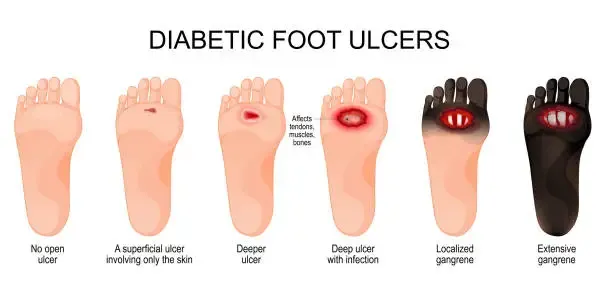
One prevalent chronic illness is diabetes mellitus. One of the most serious side effects of diabetes is diabetic foot ulcers (DFUs). Foot and lower leg ulcers are primarily caused by distal circulation disorders, poorly managed diabetes, or neurological disability. A literature review, which has certain flaws and is susceptible to subjective influences, often summarizes the present state and research hotspots in a field of study. In the meantime, academics and healthcare professionals must deal with the ongoing introduction and appearance of new ideas and technology. Therefore, in order to assess the hotspots and present stage of DFU research, a thorough analysis of DFUs is essential.[1] One of the most serious and debilitating consequences of diabetes is diabetic foot, which is characterized as an ulcerated foot in a diabetic patient that is linked to peripheral vascular disease of the lower limb and/or neuropathy. Diabetic foot ulcers are more common in older patients and affect 4–10% of the diabetic population. Diabetic foot ulcers are a serious health issue that requires a comprehensive approach to treatment. The purpose of this review is to give an overview of the current approaches to managing diabetic foot ulcers, from prevention to treatment choices. Primary care physicians, nurses, podiatrists, diabetologists, vascular surgeons, and all other healthcare professionals involved in the prevention or treatment of diabetes may find it helpful, according to the authors.[2] In this regard, wound dressing design has gradually drawn interest as a possible path toward the development of DFU treatments that are clinically successful. The ideal dressing for treating chronic wounds, like DFUs, should meet a number of crucial criteria, including providing a moist environment that promotes tissue regeneration, preventing bacterial invasion into the wound, exhibiting sufficient porosity for gas exchange, encouraging cell migration, proliferation, and neovascularization, and being sterile, biocompatible, and simple to replace (or biodegrade). In this sense, hydrogels—polymeric three-dimensional (3D) materials—have been demonstrated to be promising for the successful creation of DFU dressings because they can offer a moist environment that encourages scar formation, cell proliferation, and tissue regeneration.[3]
Pathophysiology of DFUs.
Diabetic neuropathy (DNP) and peripheral artery disease (PAD) are two well-known long-term consequences of diabetes. PAD and DNP are the primary pathophysiological conditions that predispose to DFUs, along with immunological dysfunction. DFUs are linked to PAD, DNP, and the length of DM. Eighty percent of individuals with DFUs have DNP, which promotes the development of ulcers by reducing pain and pressure perception. Additionally, DNP encourages the development of anatomic abnormalities such Charcot foot, hammertoes, and large plantar metatarsal heads. Following the diagnosis of type 2 diabetes and the fifth year of type 1 diabetes, patients with diabetes should have their DNP levels evaluated on a regular basis. It is important to inquire about burning, numbness, and pain. PAD is present in about half of DFU patients and is strongly linked to a higher incidence of adverse limb outcomes. It is important to evaluate vascular symptoms such as claudication, leg tiredness, and diminished effort capability. To determine perfusion, all peripheral pulses should be palpated together with an evaluation of the temperature and look of the extremities. The ankle-brachial index (ABI) test should be performed on patients as part of the evaluation.[4]
Foot Infection.
An ulcer is prone to infection after it has developed. Due to weakened immune systems, poor circulation, and other conditions, people with diabetes are more susceptible to infections. Furthermore, DFU creates a warm, humid atmosphere that is perfect for the growth of bacteria. Infection can worsen the healing process and raise the possibility of osteomyelitis, a bone infection that may necessitate amputation.[5]
Advantageous Hydrogel Properties for Diabetic Foot Ulcers Treatment.
New dressings meet the strict requirements of adequate wound debridement, appropriate systemic antibiotic therapy, and routine dressing changes and wound inspection, even though other types of dressings, such as hydro-colloids, foams, and alginates, are now available for DFU treatment. Because of their special qualities that promote wound healing, hydrogels are extremely promising materials for the treatment of DFU. Hydrogels' mode of action for treating DFU incorporates a number of variables. First of all, hydrogels' high water content aids in preserving a moist wound environment, which is necessary for the best possible wound healing. In turn, the hydrogel's water content makes it easier for the surrounding tissues and the wound bed to exchange nutrients and oxygen, producing an environment that is essential for cellular metabolism, tissue repair, and the ensuing healing process.[5] DWIs are a common and dangerous consequence for diabetic people. One of the main causes of DWIs is chronic hyperglycemia, which weakens the immune system and makes it more difficult for the body to identify and get rid of bacteria and other harmful microbes, raising the risk of infection. Increased vulnerability to infection is a consequence of diabetes-induced microcirculatory abnormalities and vasculopathy. The integrity of the skin is compromised by wound lesions like dry, cracked skin, which create opportunities for bacterial invasion.[6]
Prevention of foot ulceration.
Prevention is the primary treatment for lower limb problems caused by diabetes. Recent data shows a decrease in hospital admissions and avoidable amputations, supporting the use of podiatry services in the community to support diabetic foot care. The risk of amputation and hospitalization was estimated by Gibson et al. (2014) using multivariate Cox proportional hazard models for patients who had podiatry therapy in the year before developing a diabetic foot ulcer. The study showed that podiatrist treatment for diabetic foot was linked to a lower risk of lower extremity amputation and a lower likelihood of hospital admissions.[7]
Risk Factors for Developing DFU.
Peripheral vascular disease and diabetic neuropathy are the important risk factors associated with the develop- ment of DFU (Sinwar). In addition to diabetic neuropathy and peripheral vascular disease, foot defor- mities and prior history DFU are risk factors associated with the development ofDFU (Monteiro-Soares, Boyko, Ribeiro, Ribeiro, & Dinis-Ribeiro). Furthermore, oxidative stress and inflammation have an important role in the pathogenesis of DFU.[8]
Management of Diabetic Foot ulcers.
Debridement of the lesion, treatment of any infection, revascularization treatments when necessary, and off-loading of the ulcer are the gold standard for treating diabetic foot ulcers. Other techniques, like negative-pressure wound therapy (NPWT), the use of sophisticated wound care products, and hyperbaric oxygen therapy, have also been proposed as useful supplemental therapies. The effectiveness and cost-efficiency of these supplemental therapy approaches, however, have not yet been sufficiently demonstrated by data.[2]
Hydrogel Wound Dressings for DFU Treatment.
Hydrogels' exceptional capacity to retain oxygen, absorb wound exudate, and preserve the moist environment that supports regular physiological activities in the wound bed gives them a unique potential for the development of successful DFU dressings. Additionally, hydrogels have demonstrated encouraging outcomes as antibacterial substrates that prevent tissue death and speed up normal wound healing. Additionally, their porous nature encourages sufficient gas exchange, enabling the wound to breathe during the healing process.
Role of GFS in the healing of Diabetic Wounds.
GFs play a significant part in poor wound healing, particularly in diabetic wounds. Numerous processes are impacted by them, including angiogenesis, fibroblast chemotaxis, endothelial cell stimulation, proliferation and migration of various cell types, and alterations in inflammatory cells. GFs such as VEGF, PDGF, epidermal growth factor (EGF), fibroblast growth factor (FGF), transforming growth factor β (TGF-β), and hepatocyte growth factor (HGF) are primarily responsible for accelerating and promoting wound healing through their physiological actions.[9]
Functional hydrogels in Diabetic wound healing management.
In addition to being biocompatible and nontoxic, the perfect wound dressing material should be able to absorb exudate, retain moisture well, have sufficient mechanical and physical strength, and encourage cell adhesion, proliferation, and growth.18. As previously stated, the FDA has already approved a number of functional dermal matrixes. Nevertheless, these commercialized goods are costly and, in certain cases, nonresponsive, and they lack all the qualities of the perfect wound healing substance.[10]
Wound Repair and Regeneration.
Hemostasis, inflammation, proliferation, and remodeling are the four overlapping and ongoing stages of the wound repair process. Numerous cells, as well as a variety of bioactive substances, cellular compounds, and cytokines produced to promote wound healing and tissue regeneration, tightly mediate the complex process. The hemostasis stage, which is a brief period maintained by blood vessel contraction, platelet aggregation, and blood clot formation, can halt bleeding and minimize hemorrhage when tissue injury occurs. Additionally, the inflammation stage that follows tissue damage is critical in determining whether the wound will heal quickly or develop into a chronic wound.[11]
Current Evidence for prevention.
The principal preventative measures for DFUs were assessed in a number of randomized clinical trials (RCTs), however none of these studies were of good quality. Given the large number of patients and lengthy follow-up period needed, conducting RCT to identify the major preventive interventions and assess their effectiveness is a significant difficulty. However, because to the high recurrence rate, RCTs on the prevention of ulcer recurrence are technically simpler. There is strong evidence that plantar foot ulcers can be prevented from recurring or getting worse by using therapeutic shoes with the right pressure distribution.[4]
CONCLUSION
The pathophysiology of chronic wounds, multisystem lesions of diabetic foot ulcers, and current therapies have all been addressed in this article. In light of the various pathogenic variables, this study presents the corresponding therapy requirements from the perspectives of biology, physics, and chemistry. Additionally, the benefits, long-term safety, and difficulties of hydrogel dressings for their clinical applications were assessed, demonstrating the effectiveness, promise, and advancement of hydrogel dressings in the treatment of diabetic foot ulcers. Overall, the results of this work offer a theoretical foundation for the continued development of improved and more sophisticated hydrogels as well as insights into hydrogel-based therapy techniques for diabetic foot ulcers.
REFERENCES


Chandrabhan Patil*, Rajesh Dodiya, Hydrogel Dressings: An Innovative Approach to Diabetic Foot Ulcer Management, Int. J. Med. Pharm. Sci., 2026, 2 (8), 242-245. https://doi.org/10.5281/zenodo.21807648
 10.5281/zenodo.21807648