
View Article

Abstract

Diabetic wounds represent a major clinical problem because of impaired healing processes and very high risk of infection. The normal healing process impaired due to neuropathy and vascular insufficiency. Poor wound healing lead to conversion of early wound to chronic non healing wound. Diabetic foot ulcer is a very common wound in diabetic patients. Non healing diabetic wound leads to amputation of limbs. Traditional wound dressings are simple protective dressings thus failed to provide optimal environment for healing. Recent advances in nanotechnology offered development of smart nanofiber-based wound dressings with multifunctional bioactivity such as mimicking ECM, controlled drug delivery, antimicrobial activity, stimuli responsive drug delivery and smart bio sensing delivery. This review paper concludes the recent progress in smart nanofiber wound dressings for diabetic wound focuses on fabrication techniques, functionalization, and clinical potential. The future directions and Emerging opportunities to enhance diabetic wound management through smart nanofiber systems are also discussed.

Keywords

Electro spinning, Diabetic foot ulcer, Stimuli responsive Nanofiber, Smart Nanofiber

Introduction

× Popup Image

Diabetes is a complex metabolic disorder that impacting more than 340 million people globally and about 20% of them developing diabetic wounds. Diabetes is without doubt one of the major public health concern of the 21st century. There are various studies that have estimated the impact of diabetes, and it seems like numbers are increasing with an unprecedented rate. [1]

The most common wounds in diabetic patients are leg or foot ulceration. Due to hyperglycaemia normal wound healing process is affected. Most often, it is triggered by high blood sugar level, chronic inflammation, macro-and microcirculatory dysfunction, hypoxia, neuropathy, and impaired neuropeptide signalling.

In this review, the recent developments of smart nanofiber-based wound dressings for diabetic wound management are mainly discussed with special attention to diabetic foot ulcers. It describes the pathophysiological characteristics of diabetic wounds that guide the design of dressings, such as chronic inflammation, sepsis susceptibility, oxidative stress, hindered angiogenesis, abnormal protease enzyme activity, and impaired moisture and pH conditions. [2]

The review firstly describes nanofiber preparation approaches and the characteristics of natural, synthetic, and semi synthetic polymeric materials utilized in wound-dressing applications. Then, it discusses smart nanofiber systems such as stimuli-responsive mechanisms, biosensing functions, and controlled delivery of therapeutic agent. Special preference is given to nanofibers loaded with antimicrobial agents, anti-inflammatory agents, antioxidants, growth factors, peptides, stem-cell-derived products, and other bioactive molecules to regulate the diabetic wound microenvironment. [3]

2. PATHOPHYSIOLOGY OF DIABETIC WOUNDS

Peripheral neuropathy, vascular insufficiency, infections, chronic inflammation are the most common factors which inhibiting wound healing in diabetic patients. Nerve damage decreases the pain perception resulting in early wound converted into chronic wound. Due to vascular dysfunction, tissues receive less oxygen supply that impacts fibroblast activity and angiogenesis. Decrease in fibroblast activity resulting in less collagen production. Hyperglycaemia inhibits tissue regeneration and further causes cellular dysfunction, oxidative stress and ECM degradation. [4]

Prolonged inflammation with macrophage dysfunction, weak immune system increases susceptibility to infection and prolonged wound healing. Thus we need ideal dressing systems for a wound which maintain moisture levels, absorb exude, allow perspiration and protect microbial entry. Modern dressings are designed to deliver therapeutic substances (e.g. antimicrobials and antioxidants) to the site of healing. Although nanofiber-based dressings offer many structural and functional advantages, such as drug release under the stimulus, they need to be thoroughly validated in clinical trials before being used in practice. [5]

3. NANOFIBER TECHNOLOGY IN WOUND DRESSINGS

Nanofibers are biomaterial based fibrous structure having diameter of nanometres range. The structure and properties of nanofiber resembles with skin extra cellular matrix, thus provides a 3D scaffold to enhance cell attachment, migration, and proliferation during repair of skin. Nanofibers can also designed as protective barriers via multifunctional bioactivity systems for wound exude management, antimicrobial activity, controlled drug release, and stimulus-responsive therapy. [6]

3.1 Overview of Nanofiber Fabrication Methods

There are several methods for preparing nanofiber like electrospinning, template based synthesis, self-assembly, phase separation etc. Electrospinning is the most commonly used nanotechnology for fabricating nanofibrous scaffold for wound dressings because it has several advantages over conventional dressing like simple, versatile and compatible with natural and synthetic polymers.

In traditional electrospinning, a high DC voltage is applied to a polymer solution or, generating a charged jet that elongates towards collector and solidifies in the form of fibers on a collector (figure 1, 2). The fiber diameter, alignment, porosity and morphology can be controlled by the variation of polymer concentration, solvent system, applied voltage, flow rate, collection distance and environmental conditions. [7]

Table 1: Effect of Process Variables on Fiber Formation. [8]

Process Parameter Effect on fiber formation
Capillary to collector distance Too short of too long distance leads to formation of beads inside scaffold.
Flow rate of polymer solution inside syringe Optimum flow rate is necessary for perfect nanofiber diameter.
DC voltage Increase in voltage forms larger or smaller diameter fibers.
Conductivity of polymer solution Smaller diameter with an increase in conductivity with subtly beads.
M.W of polymer Increase in molecular weight leads larger fiber diameter and fewer beads.
Viscosity of polymer Increase in concentration leads larger fiber diameter and fewer beads.
Temperature Increase in temperature results in smaller diameter.

The dressing function can be improved by several electrospinning variations. Like Coaxial electrospinning generates core-shell fibers, which protect sensitive therapeutic agents and allow sustained release. Simultaneous electrospinning produces combined fibers able to deliver combination drugs. Needleless, multi-jet and centrifugal spinning methods have been developed to increase fiber production rates. Control of fiber uniformity and material properties remains important for clinical translation.
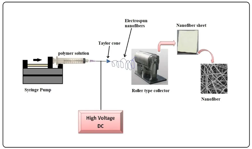
figure

Figure 1: Schematic Presentation of Nanofiber Preparation.
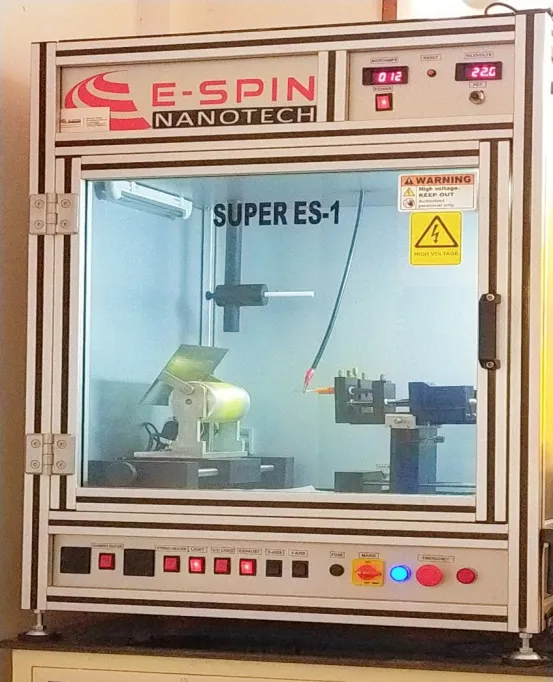
figure

Figure 2: Preparation of Nanofiber by Electrospinning Technique.

3.2 Material Types Used for Nanofiber Dressings [9, 10]

Table 2: Role of Natural Polymer in Wound Healing.

Natural Polymer Characterstics Role in wound healing
Collagen Supports cell adhesion, extracellular matrix-like interactions Promotes tissue regeneration and cell growth
Gelatin Supports cell adhesion, extracellular matrix-like interactions Enhances cell attachment and matrix mimicry
Chitosan Inherent antimicrobial and hemostatic properties Prevents infection and controls bleeding
Silk Fibroin Biocompatible, biodegradable Mimics native tissue components
Alginate Biocompatible, biodegradable Provides moist environment and supports healing
Cellulose Biocompatible, biodegradable Structural support and moisture retention
Keratin Biocompatible, biodegradable Supports cell adhesion and tissue regeneration

Limitations of natural polymer: Limited mechanical strength, variable batch properties, reduced stability in moist environments.

Synthetic polymers overcomes the limitations of the natural polymers like inhaced mechanical strength, degradation pattern, reproducibility, stability, dressing integrity, modified release behaviour etc. Examples: PCL (polycaprolactone), PLA (polylactic acid), PLGA (polylactic-co-glycolic acid), PVA (polyvinyl alcohol), PVP (polyvinylpyrrolidone), and polyethylene oxide etc.

Hence natural and synthetic polymers are blended to produce hybrid nanofibers. Resulting in improved tensile strength, good spinnability and degradation control. For more multifunctional treatment of wound these nanofibers loaded with nanoparticles, antimicrobial, antioxidant, astringents etc. [10]

3.3 Properties of Nanofibers Beneficial for Wound Healing [11]

Table 3: Beneficial Properties of Naofiber for Wound Healing

Property of nanofiber Key benefits for diabetic wound healing
High porous structure Supports gas exchange, enhance moisture-vapour transmission, absorbs wound exudes, and maintains optimum moisture level.
Interconnected pores Allow nutrient diffusion and cellular infiltration, promoting tissue regeneration
High surface area Enables loading and localized delivery of therapeutic agents (antimicrobials, antioxidants, anti-inflammatory agents, growth factors), reducing systemic administration
Similarity with ECM Provides contact guidance for cell adhesion, migration, proliferation, granulation tissue formation, and re-epithelialization
Flexible design Allows incorporation within fiber matrix, adsorption on surfaces, or encapsulation in core–shell structures for sustained release
Biocompatible and biodegradable Ensures compatibility with tissue and controlled degradation, important for safety and effectiveness

4. SMART NANOFIBER-BASED WOUND DRESSINGS

Smart wound dressings are modern/advanced dressings made to recognize the changes in the wound microenvironment and give respond accordingly. Smart nanofiber based dressings can sense wound conditions and/or release therapeutic agent in a controlled manner, in comparison to traditional dressings, which mainly offer physical protection and exude absorption only.

Nanofiber-based wound dressings are well suited for incorporating therapeutic or sensing agent due to several properties like high surface area to volume ratio, flexible structure, biocompatibility and high porosity. [12]

4.1. Main Characteristics

  • Physical protection of wound and maintain optimal moisture content inside wound.
  • Smart nanofiber dressings are able to detect changes such as pH, temperature, enzymes, and moisture in wound.
  • Provide release of drug in controlled manner when necessary.
  • It may contain some sensing molecules for monitoring and healing of wound.
  • Due to a larger surface area and porous structure nanofibers are suitable for loading drugs.

4.2. Stimuli-Responsive Nanofibers

  • pH-responsive: Microbial infection leads to change in pH of wound thus these nanofibers can detect change in wound pH and release therapeutic agents, e.g. antimicrobials, in response to abnormal pH conditions.
  • Temperature-responsive: High temperatures indicate to infection or inflammation, these nanofibers react to change in temperature of the wound area.
  • Enzyme-responsive: When some particular enzyme levels rise within wound area, these nanofibers degrade or change structurally, allowing for the release of drugs.
  • Moisture-responsive: Increase in moisture content inside wound lead to swelling of polymeric nanofiber more resulting in altered permeability.[12,13]

4.3. Sensors and Drug Delivery

Nanofibers can also incorporate some biosensors that monitor pH, temperature, moisture, glucose, or infection-related biomarkers. They can also loaded with antibiotics, anti-inflammatory agents, growth factors, or other therapeutic agents. [14]

5. Recent Developments in Smart Nanofiber Dressings for Diabetic Wounds

Recent research regarding diabetic wound care is moving from simple conventional nanofiber dressings toward multifunctional smart systems that are able to sense the diabetic wound environment and release drugs according to need, and actively promote tissue regeneration via different mechanisms.

Table 4: Recent Development in Advanced Nanofiber Dressings for Diabetic Wound Management.

S No. Nanofiber based Smart system Mechanism/ Key feature Application Reference
1. Neurotensin-loaded PLGA/CNC Composite nanofibrous scaffold Sustained Release, promote tissue regeneration Diabetic wound healing [15]
2. Photothermal electrospun PLGA nanofiber membrane Multi functional nanofiber , promoted tissue epithelialization and collagen deposition Diabetic wound healing [16]
3. Nanofiber Aerogels Aerogel system, neovascularization, and re-epithelialization. Diabetic wound healing [17]
4. Electrical Stimulating Redox Membrane loaded with PVA/Gelatin Nanofiber electrical stimulation Diabetic wound healing [18]
5. Nanofiber core–shell scaffold with 3D multilayer patterned structure (3D-PT-P/GM) angiogenesis, granulation tissue formation, and collagen production Diabetic wound healing [19]
6. Biomimetic Hydrogel Scaffolds with Copper Peptide-Functionalized RADA16 Nanofiber ECM mimicking Diabetic wound healing [20]
7. Dual drug-loaded polycaprolactone-gelatin composite nanofibers Simultaneous drug delivery, Enhanced wound healing Promote diabetic wound healing [21]
8. Stimuli-responsive hydrogel dressing sensing wound condition and changes Promoting Wound healing [22]
9. IR-Responsive Nanofiber Hydrogel Stimulus responsive drug release Wound healing [23]
10. Biopolymer based nanofiber dressing Electrospun nanofiber dressing Fast wound healing [24]

5.1 Advances in Material Design

  • Electrospun nanofibrous scaffold are made up of combination of polymers such as chitosan (obtained from exoskeleton of insects), PCL (polycaprolactone), PLA (polylactic-co-glycolic acid),PVA (polyvinyl alcohol), silk fiber, collagen and gelatin are being developed for diabetic wounds dressings.
  • Many researchers are adding pH, temperature, enzyme, and moisture stimulus responsive materials to develop dressings that respond according to wound condition.
  • Mostly polymers are used in combination for better mechanical strength, biocompatibility, biodegradability, and optimum moisture control. [25]

5.2. Controlled Drug Release

Drug incorporated in the form of reservoir within smart nanofibers, releases gradually in controlled manner or according to wound condition as needed.

  • Antimicrobials: Antibiotics, herbal antimicrobials like curcumin, silver nanoparticles, gold nanoparticles, antimicrobial peptides, etc., helps in infection control.
  • Growth factors: Growth factors Like VEGF, EGF, and other promote angiogenesis (formation of new blood capillaries), cell migration, and faster tissue regeneration.
  • Stimuli-responsive systems help in release of optimum amount of drug as per need which prevents unwanted delivery of drug to wound. [26]

5.3. Incorporation of Bioactive Agents

  • Stem cells or its derived products can be used to promote tissue regeneration and angiogenesis.
  • Peptides have antimicrobial, anti-inflammatory, and cell-signalling effects.
  • Other biologically active substances, such as antioxidants and exosomes, are studied to promote diabetic wound healing. [27,28]

6. EMERGING OPPORTUNITIES

Emerging techniques in nanofiber design focuses on development of multifunctional bioactive nanofibrous scaffold, which incorporate stimuli sensing molecules, antimicrobials and antioxidants, which promote fast tissue regeneration, represent an effective wound care strategy.

These systems enable the development of drug delivery systems that are capable of detecting the wound microenvironment and respond with targeted release of therapeutic agent, therefore smart nanofibers are emerging as a personalized medicines. Advance fabricating methods like 3D printing of nanofibers and incorporating AI assisted electronic sensors enables real time monitoring of diabetic wound ultimately resulting in faster healing. Together these innovations in nanofibers promise improved clinical outcomes.[28,29,30]

CONCLUSION:

Poor wound healing is a serious consequence of diabetes leading to amputation ultimately. Traditional nanofibers are acting as a physically protective dressing only. Recent research is moving towards fabricating innovative smart nanofibers. This offers a new way to treat diabetic wound effectively. Smart Nanofibers are prepared by new techniques like co axial electro-spinning, core shell, multi-layered etc. and loaded with stimuli sensing (pH, temperature, moisture and enzymes) molecules, growth factors, stem cell, antimicrobials along with antioxidants. These nanofibers are capable of releasing drug as per need and in controlled manner resulting in fast tissue generation and wound closure.

Future work should focus on developing stimuli responsive, biocompatible, biodegradable, regenerative, affordable, and personalized, on demand drug delivery nanofiber wound dressing.

REFERENCES

  1. Patel S, Srivastava S, Singh MR, Singh D. Mechanistic insight into diabetic wounds: Pathogenesis, molecular targets and treatment strategies to pace wound healing. Biomedicine & Pharmacotherapy. 2019 Feb 20;112:108615.
  2. Yadav JP, Singh AK, Grishina M, Pathak P, Verma A, Kumar V, et al. Insights into the mechanisms of diabetic wounds: pathophysiology, molecular targets, and treatment strategies through conventional and alternative therapies. Inflammopharmacol. 2024 Jan 11;32 (1):149–228.
  3. Kwon M, Kim KS. Nanofiber-Based Biomimetic Platforms for Chronic Wound Healing: Recent Innovations and Future Directions. Tissue Eng Regen Med. 2025 June 13;22 (6):755–770.
  4. Chu YJ, Wang P. Pathogenesis of impaired diabetic wound healing. Intern J Endocrinol Metab. 2014 Mar 20;34 (02):131–134.
  5. Nasra S, Pramanik S, Oza V, Kansara K, Kumar A. Advancements in wound management: integrating nanotechnology and smart materials for enhanced therapeutic interventions. Discover Nano. 2024 Oct 2;19 (1):159.
  6. Alberti T, Coelho DS, Voytena A, Pitz H, De Pra M, Mazzarino L, et al. Nanotechnology: A Promising Tool Towards Wound Healing. CPD. 2017 Sept 27;23 (24):3515–3528.
  7. Yadav S, Arya DK, Pandey P, Anand S, Gautam AK, Ranjan S, et al. ECM Mimicking Biodegradable Nanofibrous Scaffold Enriched with Curcumin/ZnO to Accelerate Diabetic Wound Healing via Multifunctional Bioactivity. IJN. 2022 Dec 1;17:6843–6859.
  8. Ng LF, Yahya MY, Hamzah SMSAS, Khoo PS. Preparation and Characterization of the Electrospun Nanofiber Meshes. In Crc; 2023. p. 109–127.
  9. Fard GC, Gashti MP, Gupta RK, Dehdast SA, Shabani M, Martins AF. Multifunctional Fibers for Wound Dressings: A Review. Fibers. 2025 July 24;13 (8):100.
  10. Liu Y, Li C, Feng Z, Han B, Yu DG, Wang K. Advances in the Preparation of Nanofiber Dressings by Electrospinning for Promoting Diabetic Wound Healing. Biomolecules. 2022 Nov 22;12 (12):1727.
  11. Sanchaniya JV, Kanukuntla SP, Senyurt KB. Fabrication and mechanical properties of polymer composite nanofiber mats. In: Engineering for Rural Development. Latvia University Of Life Sciences Technologies; 2023.
  12. Parducho D, Pujol A, Ylanan M, Peralta N, Ramos C, Baldovino R, et al. Smart Wound Dressing with Arduino Microcontroller. In Institute Of Electrical Electronics Engineers; 2018. p. 1–4.
  13. Karperien L, Dabiri SMH, Hadisi Z, Hamdi D, Samiei E, Akbari M. Smart Thread Based pH Sensitive Antimicrobial Wound Dressing. In Institute Of Electrical Electronics Engineers; 2019. p. 1–5.
  14. Halicka K, Cabaj J. Electrospun Nanofibers for Sensing and Biosensing Applications-A Review. IJMS. 2021 June 14;22 (12):6357.
  15. Zheng Z, Liu Y, Huang W, Mo Y, Lan Y, Guo R, et al. Neurotensin-loaded PLGA/CNC composite nanofiber membranes accelerate diabetic wound healing. Artificial Cells, Nanomedicine, and Biotechnology. 2018 Apr 13;46 (sup2):493–501.
  16. Chen Z, Mo Q, Mo D, Pei X, Liang A, Cai J, et al. A multifunctional photothermal electrospun PLGA/MoS2@Pd nanofiber membrane for diabetic wound healing. Regenerative biomaterials. 2024 Dec 14;12:rbae143.
  17. John JV, Sharma NS, Tang G, Luo Z, Su Y, Weihs S, et al. Nanofiber Aerogels with Precision Macrochannels and LL-37-Mimic Peptides Synergistically Promote Diabetic Wound Healing. Adv Funct Materials. 2022 Oct 31;33 (1):2206936.
  18. Kim JU, Ko J, Kim YS, Jung M, Jang MH, An YH, et al. Electrical Stimulating Redox Membrane Incorporated with PVA/Gelatin Nanofiber for Diabetic Wound Healing. Adv Healthcare Materials. 2024 July 11;13 (27):e2400170.
  19. Li J, Zhang T, Pan M, Xue F, Lv F, Ke Q, et al. Nanofiber/hydrogel core\u2013shell scaffolds with three-dimensional multilayer patterned structure for accelerating diabetic wound healing. J Nanobiotechnol. 2022 Jan 8;20 (1):28.
  20. Yang X, Zhang Y, Huang C, Lu L, Chen J, Weng Y. Biomimetic Hydrogel Scaffolds with Copper Peptide-Functionalized RADA16 Nanofiber Improve Wound Healing in Diabetes. Macromolecular Bioscience. 2022 June 2;22 (8):e2200019.
  21. Manjit M, Kumar M, Kumar K, Dhondale MR, Jha A, Bharti K, et al. Fabrication of dual drug-loaded polycaprolactone-gelatin composite nanofibers for full thickness diabetic wound healing. Ther Deliv. 2023 Dec 21;15 (1):5–21.
  22. Zhang W, Hu J, Wu H, Lin X, Cai L. Stimuli-responsive hydrogel dressing for wound healing. APL Materials. 2025 Jan 1;13 (1).
  23. Ren C, Wang T, Luo W, Pan X, Hu B, Li G, et al. Near-Infrared-Responsive Nanofiber Hydrogel with Gradual Drug Release Properties for Wound Healing. ACS Appl Nano Mater. 2024 June 21;7 (13):15517–15525.
  24. R Karim M, Al-Ahmari A, Dar MA, Aijaz MO, Mollah ML, Ajayan PM, et al. Conducting and Biopolymer Based Electrospun Nanofiber Membranes for Wound Healing Applications. CNANO. 2016 Mar 3;12 (2):220–227.
  25. Anbazhagan MK, Mahalingam S. Advancements in nanofibers and nanocomposites: Cutting-edge innovations for tissue engineering and drug delivery-A review. Science Progress. 2025 Apr 1;108 (2):368504241300842.
  26. Asiri A, Saidin S, Sani MH, Al-Ashwal RH. Epidermal and fibroblast growth factors incorporated polyvinyl alcohol electrospun nanofibers as biological dressing scaffold. Sci Rep. 2021 Mar 11;11 (1):5634.
  27. Chen S, Wang H, Su Y, John JV, Mccarthy A, Wong SL, et al. Mesenchymal stem cell-laden, personalized 3D scaffolds with controlled structure and fiber alignment promote diabetic wound healing. Acta Biomaterialia. 2020 Apr 5;108:153–167.
  28. Yi J, Tang Q, Sun S, Xie H, Wang L, Yin X. Exosomes in Diabetic Wound Healing: Mechanisms, Applications, and Perspectives. DMSO. 2025 Aug 21;18 (1):2955–2976.
  29. Glover K, Mathew E, Pitzanti G, Magee E, Lamprou DA. 3D bioprinted scaffolds for diabetic wound-healing applications. Drug Deliv and Transl Res. 2022 Jan 11;13 (8):2096–2109.
  30. Zheng Y, Lan J, Huang Q, Li Q, Liu Y, Li B, et al. Novel AI-Driven Precision Strategies in Diabetic Wound Healing: Immunomodulation and Advances in Smart Composite Nanocarriers. Pharmaceutics. 2026 Feb 18;18 (2):252.

Reference

  1. Patel S, Srivastava S, Singh MR, Singh D. Mechanistic insight into diabetic wounds: Pathogenesis, molecular targets and treatment strategies to pace wound healing. Biomedicine & Pharmacotherapy. 2019 Feb 20;112:108615.
  2. Yadav JP, Singh AK, Grishina M, Pathak P, Verma A, Kumar V, et al. Insights into the mechanisms of diabetic wounds: pathophysiology, molecular targets, and treatment strategies through conventional and alternative therapies. Inflammopharmacol. 2024 Jan 11;32 (1):149–228.
  3. Kwon M, Kim KS. Nanofiber-Based Biomimetic Platforms for Chronic Wound Healing: Recent Innovations and Future Directions. Tissue Eng Regen Med. 2025 June 13;22 (6):755–770.
  4. Chu YJ, Wang P. Pathogenesis of impaired diabetic wound healing. Intern J Endocrinol Metab. 2014 Mar 20;34 (02):131–134.
  5. Nasra S, Pramanik S, Oza V, Kansara K, Kumar A. Advancements in wound management: integrating nanotechnology and smart materials for enhanced therapeutic interventions. Discover Nano. 2024 Oct 2;19 (1):159.
  6. Alberti T, Coelho DS, Voytena A, Pitz H, De Pra M, Mazzarino L, et al. Nanotechnology: A Promising Tool Towards Wound Healing. CPD. 2017 Sept 27;23 (24):3515–3528.
  7. Yadav S, Arya DK, Pandey P, Anand S, Gautam AK, Ranjan S, et al. ECM Mimicking Biodegradable Nanofibrous Scaffold Enriched with Curcumin/ZnO to Accelerate Diabetic Wound Healing via Multifunctional Bioactivity. IJN. 2022 Dec 1;17:6843–6859.
  8. Ng LF, Yahya MY, Hamzah SMSAS, Khoo PS. Preparation and Characterization of the Electrospun Nanofiber Meshes. In Crc; 2023. p. 109–127.
  9. Fard GC, Gashti MP, Gupta RK, Dehdast SA, Shabani M, Martins AF. Multifunctional Fibers for Wound Dressings: A Review. Fibers. 2025 July 24;13 (8):100.
  10. Liu Y, Li C, Feng Z, Han B, Yu DG, Wang K. Advances in the Preparation of Nanofiber Dressings by Electrospinning for Promoting Diabetic Wound Healing. Biomolecules. 2022 Nov 22;12 (12):1727.
  11. Sanchaniya JV, Kanukuntla SP, Senyurt KB. Fabrication and mechanical properties of polymer composite nanofiber mats. In: Engineering for Rural Development. Latvia University Of Life Sciences Technologies; 2023.
  12. Parducho D, Pujol A, Ylanan M, Peralta N, Ramos C, Baldovino R, et al. Smart Wound Dressing with Arduino Microcontroller. In Institute Of Electrical Electronics Engineers; 2018. p. 1–4.
  13. Karperien L, Dabiri SMH, Hadisi Z, Hamdi D, Samiei E, Akbari M. Smart Thread Based pH Sensitive Antimicrobial Wound Dressing. In Institute Of Electrical Electronics Engineers; 2019. p. 1–5.
  14. Halicka K, Cabaj J. Electrospun Nanofibers for Sensing and Biosensing Applications-A Review. IJMS. 2021 June 14;22 (12):6357.
  15. Zheng Z, Liu Y, Huang W, Mo Y, Lan Y, Guo R, et al. Neurotensin-loaded PLGA/CNC composite nanofiber membranes accelerate diabetic wound healing. Artificial Cells, Nanomedicine, and Biotechnology. 2018 Apr 13;46 (sup2):493–501.
  16. Chen Z, Mo Q, Mo D, Pei X, Liang A, Cai J, et al. A multifunctional photothermal electrospun PLGA/MoS2@Pd nanofiber membrane for diabetic wound healing. Regenerative biomaterials. 2024 Dec 14;12:rbae143.
  17. John JV, Sharma NS, Tang G, Luo Z, Su Y, Weihs S, et al. Nanofiber Aerogels with Precision Macrochannels and LL-37-Mimic Peptides Synergistically Promote Diabetic Wound Healing. Adv Funct Materials. 2022 Oct 31;33 (1):2206936.
  18. Kim JU, Ko J, Kim YS, Jung M, Jang MH, An YH, et al. Electrical Stimulating Redox Membrane Incorporated with PVA/Gelatin Nanofiber for Diabetic Wound Healing. Adv Healthcare Materials. 2024 July 11;13 (27):e2400170.
  19. Li J, Zhang T, Pan M, Xue F, Lv F, Ke Q, et al. Nanofiber/hydrogel core\u2013shell scaffolds with three-dimensional multilayer patterned structure for accelerating diabetic wound healing. J Nanobiotechnol. 2022 Jan 8;20 (1):28.
  20. Yang X, Zhang Y, Huang C, Lu L, Chen J, Weng Y. Biomimetic Hydrogel Scaffolds with Copper Peptide-Functionalized RADA16 Nanofiber Improve Wound Healing in Diabetes. Macromolecular Bioscience. 2022 June 2;22 (8):e2200019.
  21. Manjit M, Kumar M, Kumar K, Dhondale MR, Jha A, Bharti K, et al. Fabrication of dual drug-loaded polycaprolactone-gelatin composite nanofibers for full thickness diabetic wound healing. Ther Deliv. 2023 Dec 21;15 (1):5–21.
  22. Zhang W, Hu J, Wu H, Lin X, Cai L. Stimuli-responsive hydrogel dressing for wound healing. APL Materials. 2025 Jan 1;13 (1).
  23. Ren C, Wang T, Luo W, Pan X, Hu B, Li G, et al. Near-Infrared-Responsive Nanofiber Hydrogel with Gradual Drug Release Properties for Wound Healing. ACS Appl Nano Mater. 2024 June 21;7 (13):15517–15525.
  24. R Karim M, Al-Ahmari A, Dar MA, Aijaz MO, Mollah ML, Ajayan PM, et al. Conducting and Biopolymer Based Electrospun Nanofiber Membranes for Wound Healing Applications. CNANO. 2016 Mar 3;12 (2):220–227.
  25. Anbazhagan MK, Mahalingam S. Advancements in nanofibers and nanocomposites: Cutting-edge innovations for tissue engineering and drug delivery-A review. Science Progress. 2025 Apr 1;108 (2):368504241300842.
  26. Asiri A, Saidin S, Sani MH, Al-Ashwal RH. Epidermal and fibroblast growth factors incorporated polyvinyl alcohol electrospun nanofibers as biological dressing scaffold. Sci Rep. 2021 Mar 11;11 (1):5634.
  27. Chen S, Wang H, Su Y, John JV, Mccarthy A, Wong SL, et al. Mesenchymal stem cell-laden, personalized 3D scaffolds with controlled structure and fiber alignment promote diabetic wound healing. Acta Biomaterialia. 2020 Apr 5;108:153–167.
  28. Yi J, Tang Q, Sun S, Xie H, Wang L, Yin X. Exosomes in Diabetic Wound Healing: Mechanisms, Applications, and Perspectives. DMSO. 2025 Aug 21;18 (1):2955–2976.
  29. Glover K, Mathew E, Pitzanti G, Magee E, Lamprou DA. 3D bioprinted scaffolds for diabetic wound-healing applications. Drug Deliv and Transl Res. 2022 Jan 11;13 (8):2096–2109.
  30. Zheng Y, Lan J, Huang Q, Li Q, Liu Y, Li B, et al. Novel AI-Driven Precision Strategies in Diabetic Wound Healing: Immunomodulation and Advances in Smart Composite Nanocarriers. Pharmaceutics. 2026 Feb 18;18 (2):252.

Photo
Sachin Yadav
Corresponding author

Faculty of Pharmacy, Dr. A.P.J. Abdul Kalam Technical University, Lucknow

Photo
Akash Ved
Co-author

Faculty of Pharmacy, Dr. A.P.J. Abdul Kalam Technical University, Lucknow

Photo
Juhi Bartariya
Co-author

Faculty of Pharmacy, Dr. A.P.J. Abdul Kalam Technical University, Lucknow

Photo
Rehnuma Siddiquee
Co-author

Faculty of Pharmacy, Dr. A.P.J. Abdul Kalam Technical University, Lucknow

Photo
Utkarsh Sharma
Co-author

Faculty of Pharmacy, Dr. A.P.J. Abdul Kalam Technical University, Lucknow

Photo
Sachin Kumar Vishwakarma
Co-author

Department of Pharmaceutical Engineering & Technology, IIT BHU Varanasi

Sachin Yadav, Akash Ved, Juhi Bartariya, Rehnuma Siddiquee, Utkarsh Sharma, Sachin Kumar Vishwakarma, Smart Nanofiber-Based Wound Dressings for Diabetic Wounds: Recent Development and Emerging Opportunities, Int. J. Med. Pharm. Sci., 2026, 2 (10), 274-281. https://doi.org/10.5281/zenodo.23576125

More related articles
A Comprehensive Review of Wound Healing: Types, De...
Kamal Singh Bani, Anil Ahuja, Amit Kumar Sharma...
Smart Nanocarriers For Targeted Drug Delivery: Rec...
Avanti Girdekar, Shital Kharat, Payal Suradkar, Namrata Deshmukh,...
Formulation and Evaluation of Effervescent Anti-Diabetic Tablets Using Aegle mar...
Vaishnavi Tribhuvan, Gaurav Pawar, Akshada Waghchaure...
Formulation and Evaluation of Polyherbal Gel for Wound Healing...
Anjali Pawsale, Pranali Asolkar, Mrunali Wankhade, Mohit Wadhwani, Anubhav Harwani, Anisha Lohiya, A...
More related articles
Smart Nanocarriers For Targeted Drug Delivery: Recent Advances and clinical Pers...
Avanti Girdekar, Shital Kharat, Payal Suradkar, Namrata Deshmukh, Komal Lahase, Gayatri Rajguru, Gau...