

We use cookies to ensure our website works properly and to personalise your experience. Cookies policy
School of Pharmaceutical Studies, Dr. K. N. Modi University, Newai (Rajasthan)
The process of healing corneal wounds is complicated and includes extracellular matrix modification, cell destruction, migration, proliferation, and differentiation. The wound healing mechanisms of corneal epithelial, endothelial, and stromal cells exhibit both cell-specific variations and several commonalities. Limbal stem cells along with modification of basement membrane are key components of cornea epithelium repair. The transforming growth factor-b (TGF-b) system is primarily responsible for the conversion of keratocytes into flexible and excitable myofibroblasts during stroma repair. The primary mechanism of endothelial cell healing is emigration and dispersion; proliferation of cells is a secondary function. Several facets of the healing process of wounds in various corneal sites are being clarified in the past ten years, while certain novel treatment strategies have come into existence. The idea of limbal stem cell research was well supported by experiments, and novel biomarkers were discovered. as well as newer therapeutic approaches, such as microRNA and gene therapy, that are being evaluated in research models. In healthcare settings, transplanting colonies augmented with limbal stem cells are now an accepted method for effective re-epithelialization in cases of cornea injury and stem cell deficit. Recent therapies, such as gene (decorin) and stem cell therapies, are currently being developed for excessive rehabilitation, along with the mediators that promote stromal repairing had thoroughly described. Clinical trials have demonstrated effective surgical techniques for repairing the damaged endothelium. Rho kinase (ROCK) inhibitor eye drops and gene therapy to activate TGF-b inhibitor SMAD7 are two novel strategies that have been developed to promote endothelial repair and prevent endothelium-mesenchymal transition. Challenges with recognizing and managing wound repair are also addressed, including the absence of accurate stem cell, particular epithelium stem cell markers, effective haze and stromal scar detection, information on wound controlling microRNAs in keratocytes and endothelial cells, along with an apparent absence of targeted mechanisms for drugs as well as gene transfer that target particular corneal cells.
An injury to live tissue or a rupture in the integrity of the outermost layer of the epidermis is referred to as a wound. This might disrupt the anatomy and physiology of the skin.
Mechanism of Wound Healing
Normal wounds begin to heal immediately following an injury, but sometimes wounds may not heal appropriately or systematically. It is necessary for one to continue manage such types of wounds. The three stages of the typical wound healing process are inflammation, proliferation, and maturation [3]. Following an injury, the inflammatory phase begins a few minutes later and lasts for up to 24 minutes. Wound healing begins with homeostasis. Within minutes following damage, a number of intrinsic and extrinsic coagulation factors will become active. This might cause degranulation and the release of growth factors (GFs) and chemotactic factors (chemokines), which could aid in the production of clots [4]. Neutrophils, which are the primary cells to arrive at the area of damage, They remove debris and germs to create an atmosphere that is favorable to wound repair. Macrophages are the final stage of the inflammatory phase and are crucial cells that serve as the primary regulators for healing. Macrophages aid in the phagocytosis of germs and destroyed tissue. Angiogenesis and fibroplasia, two essential parts of the healing process which are initiated by macrophages. They are necessary to meet the nutritional requirements of recovery process [6]. The human skin, the largest organ, serves as the body's first physical barrier to its surroundings, protecting the body from harmful substances, controlling body temperature, and regulating water as well as electrolytes balance [7]. The epidermis and dermis are the two layers that make up the morphologic structure of skin [8]. The skin's outermost layer, the epidermis, is broken down into four to five sub-layers, based on the specific body part. The primary cells which makes up the epidermis are melanocytes, keratinocyte, Merkel cells, and Langerhans cells. The dermis is the layer of connective tissue which is present beneath the epidermis, constituted by extracellular matrix proteins (collagens, elastin, proteoglycans, and glycosaminoglycans) produced by fibroblasts [9]. The body begins the process of wound healing to repair the damaged region if either or both of the layers of skin are disrupted. This process involves cellular, molecular, and biochemical pathways and is broken down into three phases: inflammatory, proliferative, and remodeling [10,11]. In order to regenerate skin, many cell types must be intricately synchronized in successive processes. The epidermis is the outer, impenetrable layer of healthy skin that withstands the harmful effects of the environment. The sweat glands, hair follicles, and sebaceous glands are also found in the outermost layer of skin. The dermis gives the skin its strength, nutrition, and immunity and is abundant in mechanoreceptors, extracellular matrix (ECM), and vasculature. The dermis layer is supported by subcutaneous adipose tissue, which serves as a store of energy. Additionally, it continuously supplies the dermis with growth factors. Additionally, every single layer also has resident immune cells which are always looking for injury to the skin. Several cell types in each of these layers must work together at certain times to promote recovery if the skin is injure. Hemostasis, inflammation, angiogenesis, growth, re-epithelialization, and remodeling are all phases that take place in a chronological order, but they also intersect [12].
Classification of Wound
Wounds can be categorized as either acute or chronic based on the duration that they take to heal.
Acute wounds
Acute pain is characterized by a disturbance of the usual anatomy and physiology of fresh tissue. A damage triggers a controlled sequence of cellular, humoral, and molecular processes that lead to acute wound healing. [13]
Chronic wounds
Chronic wounds are characterized by their inability to completely heal the body through adequate and timely procedures prior to therapies, as well as their inability to operate correctly or efficiently in their structural duties. [14] Blood arteries, sweat glands, and hair follicles are among the deeper dermal and epidermal structures that are harmed by certain cortical lesions. When there is injury to the deeper layers of tissue or subcutaneous fat underneath, wounds with full thickness develop [15]
Physiology of Wound Healing
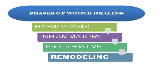
The process of wound healing is intricate and involves overlapped and interconnected processes such as cell migration and proliferation, cellular matrix production, production of growth factors, and cytokines that facilitate the process of wound healing. Because of its intricacy, the process of wound recovery has been classified into four stages: Haemostasis, inflammatory, proliferative, and remodeling phases. The platelets serve as healthcare professionals in healing wounds and repairing the injured blood arteries. In reaction to damage, the blood vessels themselves contract, but this contraction eventually relax. Although the platelets produce chemicals that constrict blood vessels, their primary function is forming a long-lasting clot that seals the injured channel [16,17]
Inflammatory phase: Platelets are activated to initiate the inflammatory phase of the healing process. They produce the substances that cause fibrin clot formation, reestablishing local hemostasis and serving as a temporary extracellular framework for blood cell movement [18]. Concurrently, damaged cells and thrombocytes produce growth factors and cytokines including PDGF (platelet derived growth factor), FGF (fibroblast growth factor), TNF-α (tumor necrosis factor-α), and IL-1β (interleukin-1β) which bring white blood cells to the site of injury. In the beginning neutrophils arrive at the site of injury, where they begin phagocytosing pathogens and removal of dead tissue. Additionally, pro-inflammatory cytokines such as TNF-α, IL-1β, IL-6 (interleukin-6), and IL-8 (interleukin-8) are released by neutrophils, attracting more inflammatory cells to the injured site. Additionally, the neutrophils produce IGF-1 (insulin growth factor-1) and VEGF (vascular endothelial growth factor), that promote the regional growth of vascular endothelial cells, fibroblasts, and keratinocytes [19,20]. A couple of days later, macrophages begin to travel to the area of the wound to carry out removal of dead tissues. Additionally, macrophages encourage the phagocytosis of harmful antigens and release cytokines and growth factors that help regulate the wound healing processes [21].
The proliferative phase: The rapid cell migration, proliferation, and granulation tissue development are characteristics of the proliferative phase of wound healing [22]. FGF, VEGF, EGF (epidermal growth factor), and TGF-β1 (transforming growth factor-β1) are secreted by the injured cells to encourage the growth of fibroblasts, keratinocytes, and endothelial cells. In order to facilitate the movement of cells into the specific region, fibroblasts additionally produce chemicals of the provisional extracellular matrix, such as fibronectin, proteoglycans, and type III collagen. Vascularization near the site of injury starts to reorganize as soon as the damage occurs, but it is most active during the proliferative phase, when it supplies the oxygen and nutrients required for cell migration, proliferation, and the production of extracellular matrix molecules. Endothelial cell growth and vascular system reorganization at the site of injury are stimulated by released mediators such as VEGF and angiopoietins [23]. Re-epithelialization takes place in the proliferative phase to seal the epithelial gap and restore the skin's barrier function [24]. primarily, growth factors trigger the keratinocytes in the outer edges of wounds, which causes the keratinocytes to proliferate and differentiate. This stimulation causes keratinocyte attachment molecules to be lost, which prevents desmosomes and hemidesmosomes from making physical contact and increases the process of migration of these cells across the extracellular matrix. The remodeling phase, the last step of wound healing mechanism in the skin, is dependent on the processes initiated in the earlier stages. Granulation tissue is reduced, transitional extracellular matrix is substituted, and the provisional cells that moved to the new site undergo apoptotic cell death. Because myofibroblast proteins have several sites of attachment to the collagen fibers, TGF-β1 stimulates the fibroblasts to develop into myofibroblasts, which acquire a contractible phenotype and reduce the area that has been injured [25]. Furthermore, MMPs, metal-dependent proteases produced by nearby cells, break down the proteins of the temporary extracellular matrix in order to repair the damaged proteins of the extracellular matrix [26,27]. Therefore, type I collagen, elastin, and other stable extracellular matrix molecules are synthesized by the fibroblasts in remodeling tissue, giving the rejuvenated skin more flexibility and adaptability. below figure (1) shows the phases of wound healing.
Factors That Affect the Wound Healing
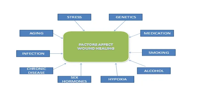
The human being is vulnerable to a wide range of systemic and local diseases that can impair skin healing through a variety of pathways and cause a delay in the process. The next section discusses the main issues that impair the healing of wounds. (Figure 2) [28].
Many vitamins, minerals, fatty acids, carbohydrates, and proteins are necessary for wound repair in order to carry out the proper regeneration processes [29]. Malnutrition hinders repair via increasing inflammation, delaying extracellular matrix reorganization, and reducing angiogenesis, phagocytosis, and fibroblast metabolism [30]. Omega-3 fatty acids (which modulate the arachidonic acid pathway and cell membrane synthesis), vitamin A (which enhances keratinocyte proliferation), vitamin C and carbohydrates (which are in charge of collagen synthesis) are some of the vital nutrients that are crucial for the healing of wounds. Proteins and Amino acids including glutamine, cysteine, arginine and methionine regulate collagen formation and immune cell function. [30] Zinc plays a crucial part in wound cell proliferation and acts as a cofactor for the production of RNA and DNA. A lack of iron hinders the regeneration of extracellular matrix since it functions as a cofactor in the production of collagen. Additionally, as a component of the haemoglobin molecule, iron plays a significant role in ischemia and the transportation of oxygen [31].
Growth Factors Involved in Wound Healing
Over the last several decades, growth factors in recovery of wound have generated a lot of interest in the field of healing. However, platelet-derived growth factor (PDGF) is just one among these treatments that has been shown to enhance recovery in a double-blind, randomized controlled experiment, although the outcomes were very limited [32]. However, given the large number of growth factor and cytokine disruptions found in chronic wounds, it makes sense that correcting all of such problems might be beneficial. For instance, a chronic wound typically expresses a reduced amount of interleukins (IL) 1 and 6 and tumor necrosis factor-α (TNF-α) while exhibiting lower concentrations of vascular endothelial growth factor (VEGF), fibroblast growth factor (FGF), transforming growth factor-β (TGF-β), PDGF, and epidermal growth factor (EGF) [33]. Maybe the main reason for this might be that the chronic wound phenotype cannot be recovered by only replacing one of the many dysregulated and insufficient factors in the environment. On the other hand, the "master regulator" of healing wounds has probably not yet been addressed Anyhow, there are several stories in scientific journals of using a particular growth factor, cytokine, protein, or hormone and getting amazing outcomes in different wound healing models. These include all of the growth-promoting agents described above as well as other targets of interest [34, 35]. But unfortunately, there hasn't been much widespread adoption of these factors, possibly due to their cost. Because many wound healing models are applied and control over the study is sometimes restricted by practical considerations, the data is also frequently challenging to understand. Notably, a number of medical insurance companies have started accepting at least some types of treatment, including Regranex (becaplermin gel, a PDGF). According to an analysis of Blue Cross Blue Shield and United Healthcare policies, under specific circumstances, at least some of these providers will pay for these growth factors. Though apparently exciting treatments like TGF-β supplementation have noticeably failed to yield effects like PDGF, research in the area is undoubtedly still continuing [36]. One of the most important refractive surfaces for sight is the cornea, the transparent front part of the eye made up of five layers Even though it is non-vascular, its position and nature render it vulnerable to a variety of diseases and traumas, such as inflammatory stroma deterioration, chemically caused burns, infectious keratitis, chronic epithelial abnormalities, and mechanical trauma. If left untreated, these disorders can result in vision loss or impaired vision, particularly in low-resource environments wherein the availability of corneal transplants or other cutting-edge treatments is restricted. [37] Since the cornea is an area of the human eye that is continually exposed to the outside world, it is more susceptible to be harmed by a variety of irritants. As a result, corneal wound recovery is a significant medical problem that requires attention in addition to being interesting to fundamental scientists. Healing of corneal wounds is a serious clinical issue. This is because of the growing number of corrective procedures and frequently severe injury to the eye. More than 40,000 corneal transplants are performed each year in the United States alone, and there are around 20 million people who have had LASIK treatment. Despite being typically safe, at least 2% of patients experience issues with LASIK surgery, including ectasia, flap separation, and aberrant wound repair. According to estimates, 20% of people will have eye trauma at some point in their lives. More than one million eye injuries are thought to occur in the United States each year. These steadily rising figures highlight the requirement of improved knowledge of cornea regeneration processes as well as the creation of effective strategies to hasten and enhance wound recovery. The method consists of a series of interconnected activities that are all linked to corneal cells' ability to heal wounds. There are several clear parallels in the ways that endothelial, stromal, as well as epithelial cells perform repair in terms of ECM (extracellular matrix) transformation, growth factor dependency, and migration of cells. The procedure is made more difficult by the well-established relationship that exists between epithelial and stromal cells throughout the course of healing. Simultaneously, the recovery instances of various corneal cells varies significantly. The epidermal layer does not change into other cell types; instead, it repairs itself through the regular migration and maturation of limbal stem cell populations. The healing process of stromal injuries, on the other hand, involves the conversion of stromal keratocytes into fibroblasts and myofibroblasts, as well as the substantial involvement of both local and circulatory cells from the immune system. In contrast to other types of cells, the endothelium of the cornea primarily heals by migration of cells and dispersion. In this process, it can also go through epithelial mesenchymal transition, although cell proliferation is a supporting function. This review's objective is to outline the complicated processes of wound healing by key corneal cell types while also highlighting general features. The use of novel 18 A.V. Ljubimov, M. Saghizadeh / Progress in Retinal and Eye Research 49 (2015) 17e45 biological modulators (such as signalling blockers and microRNA), therapy using genes, stem cells, and nanotechnology to regulate the process of wound healing is highlighted in particularly. While attempting to cite important earlier research in the field of corneal wound recovery, the writers primarily concentrated on recently released data. [38]
FUTURE DIRECTIONS
It should come with no surprise that the area of wound recovery investigation is extremely active given the effects wound repair has on patient care and the economy. Molecular biology and materials science are two apparently unrelated domains that are contributing to emerging studies on wound recovery. Several strategies using creative material design have been used to achieve this goal, with some of them concentrating on new chemicals of relevance in wound healing, such as nitric oxide [39, 40]. Although these techniques might be considered as useful in the healing of wounds and further maybe, therefore, overcome certain limitations of single growth factors or cytokines (particularly with the identification of more widely acting, gregarious molecules that contribute to the wound healing process), The "holy grail" of wound healing continues to remain far off. Embryonic wounds do not scar, as has been known for decades, but it has been considerably harder to comprehend how this happens [41]. Nevertheless, the concept that small injuries may heal with little to no scarring remains fascinating. Although a lot of the earlier debate focused on chronic, non-healing wounds, this is something of a different topic when it comes to wound healing. A wound that occurs in the developing embryo heals via regeneration as opposed to systematic healing [42]. Perhaps this clarifies that the wound healing "switch" activates somewhere in the later trimester of pregnancy, given the comparatively pathogen-free environment observed in utero [43]. Removing the tissue that has been damaged is the first step in healing any injury. Depending on the type of tissue and the extent of the injury, either repair or replacement proceeds next. Last but not least, the process of wound healing must be stopped; otherwise, every damage would result in unregulated growth and multiplication. The way these mechanisms interact depends on the tissue. In the epithelium of the cornea, destroyed tissue is quickly removed, and then new epithelial cells are generated. Endothelial cells are believed to live in a steady-state equilibrium because of contact inhibition from neighboring cells and have a very restricted capacity for regeneration. The profile of gene expression gradually reverts to the preinjury form as corneal restoration takes place over weeks to months, and Matrix metalloproteinases cause the temporary scar matrix to gradually regenerate [44]. Greater awareness of the molecular and cellular alterations which take place while corneal wound recovery will make it possible to develop therapies that target specific stages of the process of healing, producing scars which precisely match the structure of the cornea.
Control of epithelial wound healing
It has been demonstrated that several inhibitory factors can hinder the process of repair with the goal to regulate corneal wound recovery. It has been demonstrated that pro-inflammatory cytokines such as macrophage migration inhibitory factor (MIF) influences angiogenesis, chemokines, and neutrophils. All corneal epithelial and endothelial cells have been shown to contain it, and immunohistochemical studies indicate that it comes out from the wounded eye's epithelium within three hours. On the other hand, the damaged cornea's MIF mRNA expression rises from 6 to 48 hours after injury before reducing. Furthermore, bilateral overexpression of MIF in the aqueous humor is induced by unilaterally damage to the cornea [45]. It was previously demonstrated that platelet-activating factor (PAF) inhibits epithelium cell adherence to extracellular matrix proteins (fibronectin, laminin, and collagen I and IV) by 35–56% while having no apparent impact on epithelium cell migration or multiplication. [46] These findings imply that PAF contributes significantly to the inhibition of corneal wound repair by altering epithelium cell adherence and promoting stromal cell death. Platelet-activating factor receptor (PAF-R) mRNA level is altered by corneal damage; PAF-R mRNA is upregulated after corneal epithelium damage, whereas stroma cells loses the PAF-R gene activity seen in keratocytes. This implies that PAF-R gene expression is stimulated by specific growth factor engagement and increased PAF production following injury, and that these two processes are crucial feedback pathways required to sustain the process of inflammation and control epithelium wound repair. [47] As a result, PAF antagonists may be useful in treating chronic ocular inflammation by preventing the degradation of corneal transparency and visual clarity.
The corneas of humans and mice have high expression of two well-known enzymes: haem-oxygenase (HO), which produces carbon monoxide and antioxidants, and 12/15-lipo-oxygenase (LOX), which produces anti-inflammatory lipid autacoids. It is being demonstrated that Lipoxin A4 (LXA4), an endogenous 12/15-LOX product, is a strong blocker of worsened inflammation and markedly increases re-epithelialization in corneal lesions. In 12/15-LOX+/− mice, in vivo deletion of 12/15-LOX was associated with worsened inflammation and decreased wound healing. The epithelium ablation in mice corneas inhibited the expression of the 12/15-LOX and LXA4 receptor mRNA and inhibited the production of LXA4, which regenerated following the healing process. This implies that LXA4 plays a significant role in the physiological counter-regulatory mechanisms which restrict damage to tissues and encourage inflammatory recovery. [48] Fundamentally, cell division must be controlled. A wound-induced electric field that occurs spontaneously is one environmental cue that has a significant impact on the direction and rate of division of cells in vivo. An indigenous electrical field was demonstrated to govern the pace of wound repair in rat corneas, indicating that it may serve as one regulator of the interaction among cell migration and cell division over the healing process. [49] Nevertheless, the genetic identities of the signaling networks directing electric-field-induced wound recovery and migration of cells in response to electric signals remain unclear [50].
Stromal wound healing
Although solitary stromal damage is uncommon, two instances include the intrastromal wound produced by an intrastromal ring and the microkeratome used in LASIK. [51] To prevent scarring and preserve a favorable refractive result, the stromal tissue must be repaired and replaced as minimally as possible. The majority of studies have detailed keratocyte behavior in vitro.The Fas/Fas ligand system causes keratocytes close to the injury to go through apoptosis, whereas keratocytes farther away become activated fibroblasts and move into the wound, where they start producing new extracellular matrix components and developing scarring in the prevailing impact of the connective tissue growth factors and TGF-β systems. It is believed that corneal wound repair and formation of scars are significantly influenced by connective tissue growth factor (CTGF). Using in situ hybridization, CTGF mRNA has been identified in the activated fibroblasts of the retrocorneal membranes and corneal scars. TGF-β is known to promote fibrosis and is believed to increase the expression of the CTGF gene. (Wunderlich et al. 2000). It was demonstrated that inflammatory cells, keratocytes, or corneal fibroblasts can release IL-1α and/or IL-1β, which may have a paracrine effect in controlling myofibroblast apoptosis. [52] Keratocyte apoptosis is currently proposed as being essential for wound healing after refractive surgery. The amount of damage to the epithelium or stroma determines the degree and extent of apoptosis; It is more noticeable after PRK than after LASIK. It was proposed that pharmacological drugs or changes in surgical methods might induce keratocyte apoptosis [53,54,55]. The plasminogen activator/plasmin system and MMPs coordinate the breakdown and elimination of tissue that is damaged. Plasmin plays a number of functions, which includes as cleaving extracellular matrix proteins, triggering dormant enzymes like procollagenase, and triggering dormant TGF-β. MMPs have a role in angiogenesis and extracellular matrix remodeling. Both of these enzyme systems may be activated and inhibited by growth factors, whose function have been extensively studied. Genes that are differently controlled in corneal wound repair have been found using gene array technology. It was determined that out of 1176 genes, 37 had increased expression and 27 had decreased expression which were five times more in the healing corneas than in the normal, untreated corneas. Excimer laser therapy was discovered to generate thrombospondin-1, laminin-5, and interleukin (IL)-1 in the corneas. Intercellular adhesion molecule (ICAM)-1, macrophage inflammatory proteins, suppressors of cytokine signaling proteins (SOCS), IL-10 receptor, and galectin-7 were among the genes that were elevated. Connexin-31, a gap junction protein; ZO1 and occludin, tight junction proteins; and Smad2, an essential part of the TGF signaling cascade, were among the genes that were down-regulated [56].
REFERENCES


Kamal Singh Bani*, Anil Ahuja, Amit Kumar Sharma, A Comprehensive Review of Wound Healing: Types, Determinants, and Future Directions, Int. J. Med. Pharm. Sci., 2026, 2 (7), 1105-1114. https://doi.org/10.5281/zenodo.21722498
 10.5281/zenodo.21722498