

We use cookies to ensure our website works properly and to personalise your experience. Cookies policy
1Faculty of Medical Science & Research, Sai Nath University, Ranchi, Jharkhand-835219, India.
2Sahyog College of Medical Sciences, Sahyog, Dugdugia, Jharkhand-835210, India.
3Assistant Professor, Faculty of Medical Science & Research, Sai Nath University, Ranchi, Jharkhand-835219, India
Nipah virus (NiV) is a highly risky zoonotic paramyxovirus, which includes the genus of Henipavirus and has emerged as one of the most severe infectious threats of twenty-first century. Since the first identity in Malaysia in 1998–1999, this deadly pathogen has been causing recurrent outbreaks in Bangladesh and India. As of 2024, 435 deaths of 734 confirmed cases have been recorded, resulting in overall case fatality rate (CFR) exceeding 59%. Two main genotypees of the virus have been identified through Molecular virology and comparative genomic evaluation: NiV-Malaysia (NiV-M) and NiV-Bangladesh (NiV-B). Researchers found that their glycoprotein architecture has functionally signaling divergencies, which are mainly responsible for high CFR and sustained human-to-human transmission in South Asian outbreaks. The state of Kerala has witnessed four outbreaks since 2018, which serves as intensively studied template to prevent effective containment or disease in a low- and middle-income health system. According to clinical evidence, therapeutic options are extremely limited. Supportive care as well as empirical ribavirin and compassionate-use monoclonal antibody m102.4. But hope is that some vaccine candidates like ChAdOx1 NiV-B and Hendra virus soluble glycoprotein (HeV-sG) subunit vaccine are now in early clinical evaluation or early trial phases. The implementation of an outbreak preparedness for long-term NiV mitigation and a coordinated One Health Framework is now a demand.
Analysis of the history of Nipah virus (NiV) and its epidemiology, found that it was first solved from the cerebrospinal fluid of a pig farmer in the village of Sungai Nipah in Malaysia in 1999. At the time, an explosive outbreak resulted in 265 people and 105 deaths, indicating that its initial case fatality rate (CFR) was about 40%. In just a few months, the spillover spread among Singapore’s abattoir worker, which proves that the infected pigs were interregional trade [1-3]. Taxonomically this etiological agent is included in the Paramyxoviridae family Henipavirus genus. This has resulted in a close relative of Hendra virus, identified as a bat-borne pathogen in Australia just four years ago. In the last 25 years, NiV has been transformed from an isolated outbreak curiosity to a recurring public health emergency. Since 2001, it has been seen outbreak in Bangladesh, where cumulative mortality rate among confirmed cases has reached about 74%. On the other hand, five discrete outbreaks have been recorded in India so far – especially in the region of Kerala since 2018. India is currently identified as the second most affected nation. The World Health Organization (WHO) has included NiV as a priority pathogen in their Research & Development Blueprint, which ensures the fear of experts that the virus has a full-scale pandemic potential [4-6]. Considering the Public Health Risk, NiV (Nipah Virus) is much more dangerous than other zoonotic agents. Its extraordinarily high case fatality rate (CFR), the power of human-to-human transmission through close-contact, and the presence of the natural reservoir in the natural reservoir in a huge area of South and South East Asia has made the virus a big thread. Moreover, so far, the intense lack of any licensed vaccine and effective antiviral therapy makes its overall risk profile more serious. Besides, Bio-safety Level-4 (BSL-4) containment is required to work on live viruses, which is available in only a few limited facilities around the world. The limitations of this resource naturally slow the speed of research or research [7, 8]. The review paper was originally made based on the progress of various vaccines and therapeutic pipelines from Kerala outbreaks in India between 2018 and 2023, based on the progress of outbreaks in Kerala, India. The main purpose of this study is to present the current knowledge base of NiV—such as virology, epidemiology, clinical medicine, diagnostics and counteraction development. Special attention is given to Indian context and global preparations or global preparations [9].
2. Virology and Molecular Characteristics
2.1 Genome Architecture and Protein Functions
The genetic architecture of Nipah virus (NiV) is mainly developed by an enveloped, negative-sense single-stranded RNA genome, with an estimated length of about 18.2 kilobases (kb). This specific viral genome is mainly responsible for the creation of the overall structure and replication of the virus, which mainly accomplishes the work by encoding six important structural protein. These proteins include nucleocapsid (N) which protects RNA, phosphoprotein (P), and matrix (M) protein in viral assembly. Besides, the very necessary fusion (F) protein and attachment glycoprotein (G), and finally the main catalyst for viral replication of the large RNA-dependent RNA polymerase (L) protein is expressed from this genomic sequence. In addition to this, it creates some non-structural accessory proteins (such as V and W) through the RNA editing process in P gene locus, which plays an important role in hiding host’s interferon antagonism and innate immune evasion or immune resistance [10].
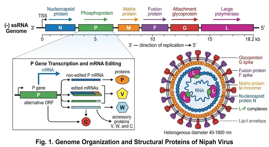
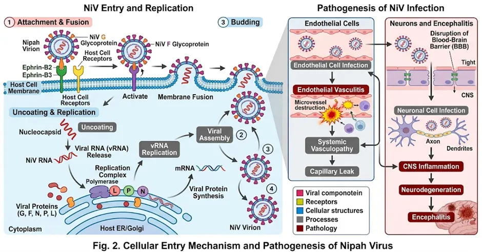
To enter Host cell, its G glycoprotein is associated or mediate with ephrin-B2 and ephrin-B3 receptor that is in the top of cells. These Transmembrane ligands are usually highly conserved and are high-level in neurons, endothelial cells, and smooth muscle; and that is why NiV features such as intense neurotropism and vasculotropism (10). After the receptor binding is completed, F protein activates pH-independent memory fusion, which helps the viral genome to enter host cytoplasm. Evolutionally conserved (evolutionally conserved) and NiV’s dependence on existing ephrin receptors everywhere is its broad mammalian host rate and serious neurological injury [11, 12].
2.2. Genotypic Diversity: NiV-Malaysia versus NiV-Bangladesh
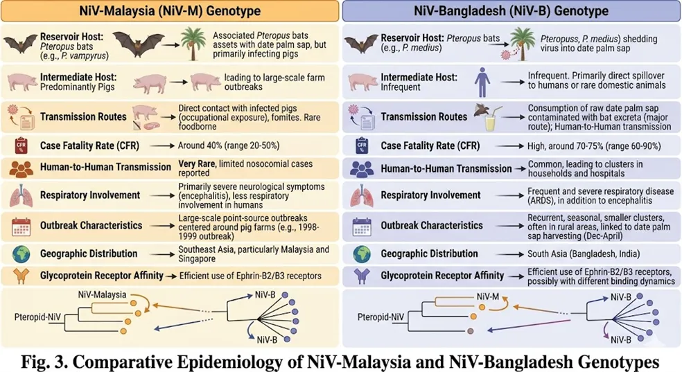
Phylogenetic analysis identifies two main genotype of Nipah virus (NiV) which is completely different from geographic distribution and pathobiological profile. Among these, NiV-Malaysia (NiV-M) genotype is mainly responsible for the outbreak of 1998–1999 and some later sporadic events; the primary bat reservoir of the virus is Pteropus vampyrus and used pigs as an intermediate amplifying host for human infection. On the other hand, NiV-Bangladesh (NiV-B) genotype is responsible for all outbreaks in Bangladesh and India. This linage spread through the bats and has not been founded in the background of spillover or infection in humans [13, 14]. The genomic comparison between two genotype showed that its overall nucleotide identity is about 91.8%. However, their Glycoprotein locus has significant divergence, where amino acid identity decreases to about 92.4%. Different functional studies suggest that it demonstrates a stronger binding affinity with ephrin-B2 and possibly ephrin-B3 reception due to some precise amino acid substitution or changes in NiV-B’s G protein. This special feature may be due to the ability to spread from people to people in Bangladesh Linase (human-to-human transmission capacity) and high mortality (CFR) more. The virus found in Kerala in 2018, has been confirmed by anyone-genome sequencing that it belongs to NiV-B genotype and has more than 99% homology with the strains of Bangladesh. This high genetic match indicates that the virus was not independently produced from the local bats in India, but it is likely a result of a recent cross-border introduction or interborder infection [15].
3. Global Epidemiology and Outbreak Patterns
3.1 Historical Outbreak Timeline
India’s first official NiV outbreak was identified in Siliguri, West Bengal in 2001, where 45 of 66 people died—which caused CFR to 68.2%. Then a second incident occurred in the Rivera district of West Bengal in 2007, where five infected patients die (100% mortality rate), and epidemiologically, as a source of this infection, date palm sap consumption or palm juice is directly linked. Between 2018 and 2023, a severe and concentrated trend of Nipah virus outbreak was observed in Kozhikode district in Kerala. Public health surveillance and scientific investigation confirmed that the presence of specific ecological conditions and fruit bat (Pteropus medius) in the region has served as the main catalyst behind the formation of virus’s recurrent emergence and epidemiological cluster. Its main scientific reason was that the dense Pteropus medius colony existing in the region, which was located close to human settlements. Among all the epidemiolocolations that occurred during this time, the most detailed and thorough study on Kozhikode outbreak of 2018 has been completed. Its index case was a 26-year-old man who probably came in contact with bat-roosting fruit trees [16]. Later, 19 cases of 23 cases were originally spread by nosocomial or family transmission chains (hospital and family infections), one of which was also a healthcare worker who died despite being fully aware of NiV risk. Under this outbreak control, 2640 people were brought under contact tracing and within 45 days of the identification of index case, the situation was fully controlled within 45 days, which received extensive international praise from containment speed. This was followed by 1 case (no death), 1 case (1 death) in Kerala in 2021, and 6 cases (2 deaths) in 2023, which proved that intensive syndromic surveillance and rapid response capacity prepared before, could greatly reduce the secondary spread or secondary social infection [17-20].
Table 1: Molecular Architecture, Genotypic Diversity, and Epidemiological Profiles of Nipah Virus (NiV) Outbreaks
|
Evaluation Category |
Feature / Parameter |
Scientific Specifications & Functional Roles |
|
Genomic Architecture |
Genome Type & Size |
Enveloped, negative-sense, single-stranded RNA genome; approximately 18.2 kb in length. |
|
Structural Proteins |
Nucleocapsid (N): Encapsidates and protects viral RNA.Phosphoprotein (P) & Matrix (M): Directs viral assembly.Fusion (F) Protein: Mediates pH-independent membrane fusion for host cytoplasm entry.Glycoprotein (G): Binds to host transmembrane receptors (ephrin-B2 and ephrin-B3).Large (L) Protein: Functions as the catalytic RNA-dependent RNA polymerase for replication. |
|
|
Non-Structural Proteins |
V and W proteins: Expressed via RNA editing at the P gene locus; mediate host interferon antagonism and innate immune evasion. |
|
|
Tissue Tropism |
Cellular Targets |
High affinity for neurons, endothelial cells, and smooth muscle cells, leading to severe neurotropism and vasculotropism. |
|
Genotypic Diversity |
NiV-Malaysia (NiV-M) |
Associated with the 1998–1999 outbreaks. Natural reservoir: Pteropus vampyrus. Intermediate amplifying host: Pigs. Minimal human-to-human transmission. |
|
NiV-Bangladesh (NiV-B) |
Responsible for outbreaks in Bangladesh and India (including the 2018 Kerala strain, which shares greater than 99% homology). Natural reservoir: Pteropus medius. Driven by direct bat-to-human spillover and exhibits high human-to-human transmission capacity. |
|
|
Sequence Divergence |
Overall nucleotide identity between genotypes is ∼91.8%, dropping to ∼92.4% amino acid identity at the Glycoprotein (G) locus. Precise substitutions in NiV-B increase receptor binding affinity, correlating with higher Case Fatality Rates (CFR). |
|
|
Epidemiological Timeline & Patterns (India) |
Siliguri, West Bengal (2001) |
66 cases, 45 fatalities; 68.2% CFR. First officially identified outbreak in India. |
|
Nadia, West Bengal (2007) |
5 cases, 5 fatalities; 100% CFR. Epidemiologically linked to the consumption of contaminated raw date palm sap. |
|
|
Kozhikode, Kerala (2018–2023) |
Recurrent emergence driven by dense Pteropus medius colonies near human habitats. The 2018 index case originated from bat-roosting trees; 19 of 23 subsequent cases propagated via nosocomial (hospital) and familial transmission chains. Controlled via intensive contact tracing (2,640 individuals monitored). |
4. Transmission Dynamics and Risk Factors
Nipah virus (NiV) infection occurs mainly through three epidemiologically validated transmission pathways, which is responsible for almost every human case. The first and most historically deadly medium of this is the zoonotic spillover from bats to human body. This spillover can occur directly due to contact with contaminated food or bats saliva or red, or indirectly through an intermediate or amplifying animal host (e.g. pigs or pigs in Malaysia). The second important pathway is human-to-human transmission, which is mainly caused by symptomatic patients due to the intensive contact of body fluid such as symptomatic secretions, saliva, urine or blood. Human-to-human transmission acts as the main driving mechanism behind the huge amount of secondary case burden or secondary case burden in outbreaks of Bangladesh and India. Finally, there are nosocomial transmission or hospital-based infections, which are clearly documented in the outbreaks of Siliguri (2001) and Kerala (2018). This particular type of infection indicates the lack of adequate infection prevention and control (infection prevention and control system) during the fight against a strangers in healthcare settings or treatment centres [21-23].
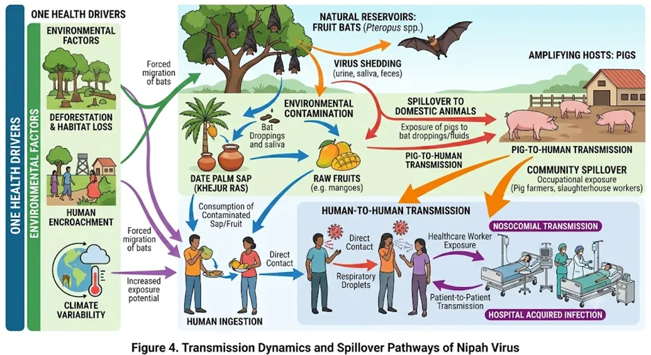
Nipah virus incubation period usually lasts from 4 to 14 days, depending on typeical exposure conditions. However, reviews of various retrospective case analyses have shown that this period may be extended for up to 45 days. A long-prolonged uncertainty windows or uncertainty environments are created due to contact monitoring and quarantine decisions. Considering geographically, India has a huge geographic footprint of potential spillover or infection, as Pteropus bats or bats spreads across 13 states in India, including Kerala, West Bengal and Northeast. Besides this, some specific socio-behavioural risk factors or social-creative risk factors behind infection in India. One of these is to enjoy raw date palm sap in West Bengal, Bihar and Uttar Pradesh in winter, and handling without understanding the partial eating or bats of bats. To change these risky habits, it is necessary to highly urgently targeted community education or specific social awareness activities at the local level [24-26].
5. Clinical Spectrum
NiV infection’s clinical presentation can occur across a very wide spectrum, which can form from complete symptomatic sero-conversion to intense and fatal forms of severe and severe forms. Usually, infection starts with a 3 to 14-day prodromal period in symptomatic diseases, with the main symptoms fever, severe headache, myalgia and vomiting. After that, about 90% of symptoms occur very quickly in neurological features or neurological complications. These Neurological manifestations include various focal brainstem signs, including altered consciousness, seizures, cerebellar ataxia and cranial nerve palsies. These complex symptoms are caused by the fact that the virus has a special predilection or attraction towards neurons and cerebrovascular endothelium. In addition, about 25% of cases of acute respiratory distress syndrome (ARDS) occurs which makes the disease more complicated. This respiratory distress is much more prevalent or prevalent in the current NiV-B infection than the historical NiV-M case, which is essentially one of the main causes of its high mortality or death rates. Another completely different in the medical field and extremely confusing aspects of diagnostically treacherous or diagnostically diagnostic is late-onset or relapsing enphalitis. The patient may appear externally healthy or recovery a few months or years after the appearance. It is believed that this happens due to reactivation of late viral antigen inside the body or residual neuroinflammation, which is caused by a long-term morbidity burden for survivors [27, 28].
6. Diagnostic Approaches
Early and accurate laboratory diagnosis is very important for NiV outbreak containment, which ensures prompt case isolation, targeted contact tracing and proper clinical management. Currently, the Real-time reverse transcription polymerase chain reaction (RT-PCR) test on throat swabs, urine or cerebrospinal fluid offers about 95% sensitivity and is considered the first-line investigation of the first-line [29]. It is possible to get a result within 4–6 hours in BSL-2 certified facilities. Moreover, the WHO-approved RT-PCR protocol is designed primarily by targeting N gene conserved regions, which can provide cross-reactivity between NiV genotypes and Hendra virus. When viral loads decrease in second or third wicks, serological confirmation plays a very effective role as a complement of RT-PCR. In this case, IgM enzyme-linked immunosorbent assay (ELISA) can achieve about 90% sensitivity. On the other hand, autopsy tissues for post-mortem confirmation is used in Immunohistochemical (IHC) analysis, which is about 85% of the reported sensitivity. However, BSL-4 containment is required to virus isolation from clinical specimens and it acts as reference standards for definitive characterisation of novel strains; although its routine deployment is almost impossible due to additional resources demands [30, 31]. Since 2018, NiV’s diagnostic infrastructure has increased significantly. As of 2024, the National Institute of Virology (NIV) Pune served as the national reference laboratory in the country and provides the necessary RT-PCR reagents and quality assurance support to 15 Virus Research and Diagnostic Laboratories (VRDLs) network of high-risk states. Despite this, point-of-care nucleic acid amplification tests still in use in peripheral healthcare facilities, which are large unmet needed in the area primarily in the resource-constrained settings—because most spillover events first appear in these areas [32].
Table 2: Epidemiological Characteristics, Clinical Spectrum, and Diagnostic Modalities of Nipah Virus (NiV) Infection
|
Category |
Parameter |
Scientific Data & Operational Definitions |
|
Epidemiological Pathways |
Primary Transmission Vectors |
2. Human-to-Human: Transmission driven by symptomatic index cases via intensive contact with infectious bodily fluids (secretions, saliva, urine, blood). 3. Nosocomial: Hospital-acquired infections resulting from sub-optimal Infection Prevention and Control (IPC) systems. |
|
Incubation Period |
Standard range: 4–14 days; documented upper limit in retrospective analyses: up to 45 days (prolonging quarantine and contact monitoring windows). |
|
|
Socio-Behavioural Risk Factors |
Consumption of raw date palm sap contaminated by bats; handling/consumption of fruits partially eaten (bat-bitten) by Pteropus species. |
|
|
Clinical Spectrum |
Prodromal Phase |
Duration: 3–14 days. Primary manifestations include pyrexia (fever), debilitating headache, myalgia, and emesis (vomiting). |
|
Neurological Manifestations |
Occurs in ~90% of symptomatic cases; includes altered sensorium, seizures, cerebellar ataxia, and cranial nerve palsies due to viral tropism for neurons and cerebrovascular endothelium. |
|
|
Respiratory & Late Complications |
• Acute Respiratory Distress Syndrome (ARDS): Present in ~25% of cases (highly prevalent in the NiV-B genotype). • Late-Onset/Relapsing Encephalitis: Reactivation of latent viral antigens or residual neuroinflammation occurring months to years post-recovery. |
|
|
Diagnostic Modalities |
Real-Time RT-PCR |
• Target: Conserved regions of the Nucleoprotein (N) gene (provides cross-reactivity with Hendra virus). • Sensitivity & Turnaround: ~95% sensitivity; results within 4–6 hours in BSL-2 facilities. • Specimens: Throat swabs, urine, cerebrospinal fluid (CSF). |
|
Serological & Post-Mortem Assays |
• IgM ELISA: ~90% sensitivity; utilized as a complementary tool during the 2nd and 3rd weeks of illness when viral loads decrease. • Immunohistochemistry (IHC): ~85% sensitivity; utilized on autopsy tissues for post-mortem confirmation. |
|
|
Reference Standards & Infrastructure |
• Definitive Characterization: Viral isolation (requires strict BSL-4 containment). • National Network: Managed by NIV Pune (National Reference Laboratory) supporting 15 Virus Research and Diagnostic Laboratories (VRDLs). |
7. Therapeutic Interventions and Vaccine Development
7.1 Supportive Care and Empirical Therapies
Currently, no antiviral agent licensed or approved for NiV infection treatment. As a result, clinical management of patients with NiV depends largely on intensive supportive care. These supportive care include mechanical ventilation, seizure control and fluid resuscitation. In medical field, Ribavirin, a special nucleoside analogue with broad-spectrum antiviral activity, has been used empirically during the outbreak of Malaysia, Bangladesh and India. Although early in vitro data indicated the effectiveness or effectiveness, no randomised controlled trial has been conducted so far, and the evidence of its clinical benefits remains inconclusive. A possible association was observed in 2018 between ribavirin from a retrospective analysis of Kerala outbreak and improved survival. However, it is difficult to reach a definitive conclusion or final conclusion for confounding due to disease severity. Among other therapeutic approach, murine-derived, humanised monoclonal antibody m102.4 plays an important role. It can target G glycoprotein receptor-binding domain and neutralise both NiV and Hendra virus. It was administrated or applied under compassionate use protocols in 2018 and 2023 in Kerala outbreak. African green monkeys and ferrets are highly promising, and is currently considered for formal accelerated clinical evaluation. However, its use in field settings is currently quite limited due to stockpile limitations and strict cold-chain requirements. The work of manufacturing scale-up and international collaboration is currently ongoing to cut this restriction [33-35].
7.2 Vaccine Pipeline
"CEPI has declared NiV a priority target under their 100-Day Mission framework." There are currently several platforms actively clinical development. Among these is HeV-sG subunit vaccine, which is mainly made from the soluble ectodomain of Hendra virus G glycoprotein and formulated with ISCOM-matrix adjuvant. It has already completed Phase I human safety and immunogenicity trials. By using Shared Conserved Epitopes in Henipaviruses, it has been able to show cross-neutralising antibody responses against both NiV-M and NiV-B [36]. Another important candidate is ChadOx1 NiV-B, a chimpanzee adenovirus-vectored vaccine made in the University of Oxford. It encode the full-length NiV-B G glycoprotein. In 2023, the Phase I/II clinical trials began in Bangladesh, the first evaluation of NiV vaccine on any endemic population. In the preclinical studies done in African green monkey and hamster models, it has displayed robust T-cell and neutralising antibody responses. Besides, Moderna’s mRNA-1215 vaccine encode NiV F and G proteins in lipid nanoparticle formulation. Its preclinical immunogenicity evaluation has been completed and is expected to enter first-in-human studies soon, which is mainly going to use accelerated vaccine development infrastructure during COVID-19 pandemic [37, 38]. According to the WHO R&D Blueprint, if there is no major safety signals during clinical development and global funding commitments, there may be at least one NiV vaccine candidate regulatory approval by 2028. However, endemic population size for traditional field efficacy trials is a big challenge. This is why it is necessary to rely on alternative regulatory pathways, such as those of correlates of protection obtained from challenge models or animal studies.
8. India's Preparedness: Strengths, Gaps and The Kerala Model
Between 2018 and 2023, India’s Nipah response capacity has been significantly improved based on the real experience of four outbreak in Kerala. Kerala Health System has created a state-level NiV contingency plan, which includes a pre-identified isolation facility in the Kozhikode Government Medical College, a standing relationship with NIV Pune for quick diagnostics and community engagement protocols for contact tracing and risk communication. However, there are still some critical gaps or major deficits in the national level. Currently the capacity of BSL-4 laboratory capacity or biological-security level-4 laboratory is limited to only one specific facility—NIV Pune. This creates a large geographic bottleneck or geographic limitations across the country, which slows down sample testing and research pace in emergency situations. In outbreak, in 2023, more than 11,000 contacts were traceed in just three weeks of containment window and 994 sample processes, proved the operational maturity of the system. If concurrent outbreak occurs in multiple states together, there will be additional pressure on this laboratory [39, 40]. Besides, public awareness is still very low in the areas where there is a culture of drinking palm sap in West Bengal, Bihar and Uttar Pradesh. This social habit of drinking raw sap or raw juice in winter harvest season is a big but underappreciated spillover pathway. Besides, training district hospitals in the district hospitals of healthcare worker to handle suspected viral haemorrhagic fever or encephalitis case. This error caused nosocomial spread or hospital-based infections in Siliguri in 2001 and Kozhikar in 2018. Although India’s Integrated Disease Surveillance Programme (IDSP) and Acute Encephalitis Syndrome (AES) surveillance network provides a structural foundation for identification of the disease, NiV’s diagnostic specificity is naturally low in India for multifactorial aetiology or versatile reasons for encephalitis. This is why NiV-specific laboratory testing requires refined clinical algorithms or specific rules that will help to quickly triage. It is important to form a National Nipah Action Plan with a dedicated multi-year funding like the National Action Plan for Antimicrobial Resistance, which will ensure necessary institutional permence to maintain preparatory investments in two outbreaks [41-43].
Table 3: Therapeutic and Preventive Landscape for Nipah Virus
|
Category |
Intervention/Strategy |
Current Status / Key Details |
|
Supportive Care |
Standard Intensive Care |
Mechanical ventilation, seizure control, and fluid resuscitation; remains the primary clinical management. |
|
Empirical Therapy |
Ribavirin |
Used empirically; evidence of clinical benefit remains inconclusive despite some retrospective data suggesting improved survival. |
|
Targeted Therapy |
m102.4 (Monoclonal Antibody) |
Humanised mAb targeting G glycoprotein; used under compassionate use protocols; scale-up/supply-chain challenges remain. |
|
Vaccine Pipeline |
HeV-sG Subunit |
Completed Phase I trials; shows cross-neutralising responses against NiV-M and NiV-B. |
|
Vaccine Pipeline |
ChAdOx1 NiV-B |
Adenovirus-vectored; Phase I/II trials ongoing in Bangladesh; robust T-cell/antibody responses in animal models. |
|
Vaccine Pipeline |
mRNA-1215 |
Lipid nanoparticle formulation; preclinical immunogenicity complete; expected to enter Phase I soon. |
|
India Preparedness |
Kerala Model |
Includes contingency plans, pre-identified isolation facilities, and established diagnostic links with ICMR-NIV Pune. |
|
Systemic Gaps |
BSL-4 Limitations |
Geographic bottleneck exists due to reliance on a single national BSL-4 facility (NIV Pune). |
|
Strategic Framework |
One Health Approach |
Focus on bat-human interface surveillance, land-use management, and genomic surveillance of wildlife. |
9. One Health Framework and Future Directions
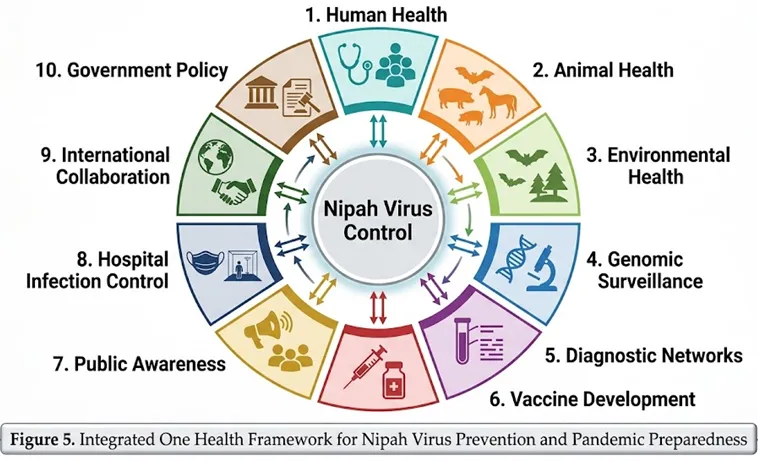
One Health paradigm, recognizing the mutual dependence of human, animal and environmental health, is conceptually indispensable to prevent NiV mitigation. So far all NiV outbreak documented has originated from bat-human interface, influenced by land-use change, deforestation and various cultural practices. These things bring people to contact with Pteropus bat colonies. High-risk districts include the systematic annual surveillance of Pteropus bats—which includes serological and virological screening—to map viral circulation, identify emerging genetic variants and provide early warning signals of elevated spillover risk. The ICMR-NIV Pune bat surveillance programme, which began after 2018, is a commendable start, but P. medius needs expansion in the entire geographic range of 13 endemic states [44, 45]. Live bat caption is an alternative to outbreak-interval monitoring, emerging genomic surveillance technologies (e.g. Environmental RNA sequencing and metagenomic screening of bat guano and ectoparasites) acts as scalable and low-biosafety-demand alternatives. International data-sharing platforms featured by Global Outbreak Alert and Response Network (GOARN) and Global Health Security Agenda (GHSA) and Global Health Security Agenda (GHSA) are highly vital. NiV vaccines require pre-negotiated emergency use authorization frameworks to accelerate development and regulatory approval. Ebola vaccine development and precedents established during COVID-19 pandemic prove that compressed timelines can be achieved if political will and financial resources are properly aligned. In case of high mortality profile-like pathogen, no major outbreak reaction should be the main strategic objective to achieve at least one vaccine platform before outbreak [46]
CONCLUSION
The Global infectious disease risk landscape is a uniquely concerning position. Its high case fatality rate, including wide geographic distribution, bat reservoir, human-to-human and nosocomial transmission, and almost complete absence of licensed countermeasures—together, it has become a pathogen that is most eligible to obtain sustained scientific and public health investment. India’s experience—especially the outbreaks that occurred in Kerala between 2018 and 2023—creating spillover threats—as well as substantive evidence base on how to make effective containment is possible between resources and resources. The molecular divergence between NiV-M and NiV-B, especially in G glycoprotein receptor-binding domain, demanded intensive monitoring of transmission and virulence as a determinant of phenotypic diffrences in transmission and virulence. Therapeutic development requires clinical evaluation of m102.4 and next-generation neutralising antibodies, as well as demonstrating preclinical activity antivirals, it is important to evaluate antivirals. Although promising the vaccine pipeline, it is necessary to create expedited clinical progression and a practgmatic field efficacy assessment framework that is suitable for sparse but geographically concentrated case burden-based pathogen. Finally, it is essential to maintain a sustained commitment to one health principles to prevent a Nipah pandemic. These include continuous bat surveillance, bat-human interface, community behavioural interventions, strong national diagnostic and response infrastructure, and international cooperation in countermeasure development and deployment. The necessary investment is highly modest or little compared to the possible consequences of a large-scale NiV emergence; the education of COVID-19 has made it clear to us how terrible the value of inadequate preparation against zoonotic pandemic threats can be.
REFERENCES

Abhinav Kumar, Parinika Kumari, Rajni Mariam Marandi, Nandani Kumari, Neha Kumari, Md. Sharafat Ansari, Aman Kumar, Shaesta Firdous, Ujjwal Kumar, Romin Kumari, Priya Kumari, Vaibhav Singh, Keshav Kumar, Priyanjali, Anshul Kumar Mahto, Arnab Roy*, Nipah Virus: Epidemiology, Molecular Characteristics, Clinical Dimensions and Pandemic Preparedness — A Comprehensive Scientific Review, Int. J. Med. Pharm. Sci., 2026, 2 (8), 121-134. https://doi.org/10.5281/zenodo.21791228
 10.5281/zenodo.21791228