
View Article

  • Testing for SARS-CoV-2 Serologically Antibodies: Developments and Clinical Significance

  • 1School of Performing and Fine Arts, Om Sterling Global University, Hisar (Haryana), India
    2School of Pharmaceutical Sciences, Om Sterling Global University, Hisar (Haryana), India
     

Abstract

During the COVID-19 pandemic, humoral immune response recording has shown to be a beneficial clinical and/or epidemiological tool. Here, we present some of the most effective uses of antibody challenging in contradiction of the severe acute respiratory syndrome coronavirus 2 (SARS-CoV-2). Anti-nucleocapsid antibodies can aid in differentiating between infection and vaccination, while SARS-CoV-2 neutralizing antibodies and anti-spike circulating IgGs can be used as indicators of illness progression or prophylaxis. Furthermore, we discuss the applicability of the anti-SARS-CoV-2 antibody monitoring post-infection and about vaccination after treatments with well-known anti-CD20 monoclonals, along with the context of different cancers or the autoimmune conditions like multiple sclerosis and rheumatoid arthritis in the age of immunotherapies. Population immunosurveillance is another important use that can be used on the general public or on particular communities, like healthcare professionals. Lastly, we go over how examining antibodies in cerebrospinal fluid might provide information about the neurological issues that are frequently associated with COVID-19.

Keywords

COVID-19, SARS, Antibodies, immunosuppressive medication, Clinical manifestation

Introduction

× Popup Image

The infection associated with SARS-CoV-2 causes both responses of T-cell and B-cell since it targets each viral antigen, includes the protein molecules referred to as spike (S) and nucleocapsid (N). The most widely expressed immunodominant protein is N protein, while S protein is essential for the worm to enter congregation cells. An antibody response is initiated after the first viral encounter and is stimulated by pro-inflammatory cytokines. The existence of these antibodies in livingliquids, for exampleurine, serum, plasma, human milk, saliva and (cerebrospinal fluid) CSF, can be accurately detected by specific antibody assays [1–3]. Different clinical information can be obtained depending on the test's kind, time, and fluid type tested. We will briefly discuss a few clinical uses of testing for anti-SARS-CoV-2 antibodies in this review article, along with an eye toward the ongoing COVID-19 pandemic. Regardless of the severity of the disease, a number of studies show that the majority of immunocompetent individuals experience an adaptive immune reaction after coming into interaction with the worm. Starting as soon as 1-3 weeks post infection, antibodies against N and S proteins, includes those of the IgA, IgM, and IgG classes, can be seen in the serum; however, whereas IgM degrades quickly, IgG and IgA antibodies might last for long time. However, the precise length and the titer of anti-SARS-CoV-2 antibody persistent in the bloodstream following infection clearance vary and are probably donor-specific, in addition to being dependent on the severity of the disease [4,5]. More precisely, individuals with more severe COVID-19 have longer persistence and higher titers [6]. Antibody titers typically correspond with the severity of the illness [7-10]. The antibody titer and how well it binds to particular SARS-CoV-2 antigens (such as S and N proteins) can be measured using a variety of tests, and their specific neutralizing efficacy can also be ascertained. There are two main kinds of binding testing. Point-of-care tests are mainly horizontal movement strategies that identify antibodies in a plasma droplet and are conducted in any type of location, such as a nursing home, hospital ward, or place of employment. Specialized staff are needed for laboratory tests, which involve techniques like chemiluminescence assays (CIA/CLIA) and ELISA, which recognize antibodies from CSF, dried blood spots, plasma, or serum. The FDA (https://www.fda. gov/medical-devices/coronavirus-disease-2019-covid-19-emergency-use-authorizations-medical-devices/in-vitro-diagnostics-euas-serology-and-other-adaptive-immune-response-tests-sars-cov-2) states that a total of 85 tests have gotten EUAs for serology. With this approval, laboratories are categorized into three groups: H, which are settings for patient care; M, It is for fulfilling the requirements to conduct tests with a moderate level of complexity; and W is for fulfilling the requirements to conduct tests with a high level of complexity. The capacity of anti-S antibodies to block the attachment of virus to its specific receptor, the angiotensin converting enzyme 2 (ACE2), and consequently entrance into human cells, is indicated by neutralizing assays (Fig. 1). This category of assays includes competitive neutralization, virus neutralization, and pseudo-virus neutralization [11]. The latter type (in the plate format) is on a commercial basis accessible and simple to set up as well as carry out in a typical wet lab, whereas the first two take more time, specialist personnel, and equipment for handling pathogens. According to this link (https://www.fda.gov/medical-devices/coronavirus-disease-2019-covid-19 emergency-use-authorizations-medical-devices/in-vitro-diagnostics-euas-serology-and-other-adaptive-immune-response-tests-sars-cov-2), only two tests have gained FDA approval as of yet. Serological tests that be able to show humoral immune reactions beside SARS-CoV-2 are essential for informing public health professionals and guiding policy and decision-making in the medical field. At the moment, the majority of antibody testing are carried out in the central clinical laboratory, which restricts widespread entry for a variety of people. Furthermore, it's critical to have extremely sensitive assays which can distinguish among vaccination and SARS-CoV-2 infection. In order to achieve this, a number of cutting-edge techniques are currently being developed, such as a microfluidic cartridge-based device and a biosensor (multiplexed nanoplasmonic) [12, 13]. These might be used as PoC (point-of-care) antibody monitoring techniques, even in the non-specialised settings like a nursing home or a workplace.
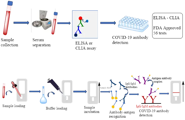
Figure 1. Tests and procedures approved by the FDA. Higher specificity and sensitivity are obtained from testing using laboratory techniques, such as ELISA or CLIA, but point-of-care testing, such as using lateral flow methods, is very helpful in specialized environments, such nursing homes or workplaces. The idea behind neutralizing antibody detection techniques is that antibodies have the ability to obstruct the RBD-ACE2 interaction.

2. Clinical applications

After considering the biological fluid employed and the analyte assessed (kind of antigen or type of Ig), multiple applications in clinical can be guided by tests for antibodies.

2.1. The identification of acute SARS-CoV-2 contamination would not be made with antibody testing

Molecular testing, such as PCR identification of SARS-CoV-2 genes, is more sensitive and specific than antibody testing against any viral antigen. However, among groups that have had vaccinations, antibody testing may be helpful, for example, in identifying infected individuals. In order to achieve this, specific testing is being created to identify antibodies in the blood or saliva that are neutralizing the S, RBD, and N proteins at the same time [14]. Saliva contains SARS-CoV-2 antibodies, which are the initial line of defense against the infection. After an infection has healed, they are found in the mucosa, or more specifically, in saliva. It has been shown that even up to 1.2 yearslaterminor COVID-19, antibody persistence was still present in saliva and plasma [15]. Interestingly, compared to patients with severe COVID-19 symptoms, salivary IgG and IgA antibodies might be found earlier in milder cases [16]. Severe COVID-19 infection, however, resulted with higher blood and salivary antibody titers than mild or asymptomatic infections [17]. In moderate cases, salivary IgA titers rapidly dropped after six weeks, but in cases with severe COVID-19, they were still detectable at least week ten. To sum up, tests for both IgG and IgA exhibit higher levels of sensitivity as well as specificity when used to assess the IgG and IgA immune response as well as confirm recent or ongoing SARS-CoV-2 infections [18-21].

SARS-CoV-2 IgG FcγR ELISAs are another cutting-edge technique for seroprevalence studies. This method carefully combines antigen-antibody binding in solution with isotype-specific immune complex detection, enabling the long-term identification of anti-SARS-CoV-2 IgG antibody responses within populations with a challenging immunological background and/or where S-protein-based vaccination regimens have been introduced [22]. Additionally, antibody testing may be helpful for the poorly defined clinical condition known as long-COVID detection. The findings highlight the difficulty in diagnosing protracted COVID-19 infections and how this difficulty is exacerbated in a large number of patients who were not examined during their acute sickness and/or do not have SARS-CoV-2 antibodies [23]. It's been demonstrated, specifically, that 42%–53% of people with lengthy COVID exhibited detectable SARS-CoV-2 specific T cell responses even though they did not have detectable SARS-CoV-2 antibodies. Furthermore, using techniques from machine learning that the input parameters like serum pro-inflammatory, anti-inflammatory, antiviral, as well as antiviral cytokine measurements as well as anti-SARS-CoV-2 antibody measurements, patients admitted to hospitals can be divided into both low and high-risk clinical categories with different cytokine and antibodies profiles that can guide personalised therapy [24]. In light of the global anti-COVID-19 immunization effort that is currently underway in several nations, tracking breakthrough infections is also quite important. As the primary sero-surveillance target to date, the S protein, is targeted by COVID-19 vaccines to elicit immune responses, other targets are being investigated to differentiate vaccination from illness. In the first two months after the start of symptoms, the compassion of N seropositivity for theminor COVID-19 was 85%; however, in those who did not exhibit symptoms, the sensitivity was lower at 67%. Serological reactions to the N may be useful in diagnosing of infections by SARS-CoV-2 after vaccination, as N-specific IgG concentration was not changed by immunization in the infection-naïve individuals [25]. Similarly, the attainment of N antibody detection was examined in the Canadian cohort of adult paramedics in order to determine prior infections by COVID-19 and compare variations between vaccinated volunteers and untreated volunteers. It was discovered that, in order to attain the best outcomes from the tests, the vaccinated and untreated groups need distinct criteria, particularly when it comes to identifying SARS-CoV-2 infections during the previous nine months [26].

2.2. Antibody analysis to gauge the severity of an illness and provide personal defense against occurrence and recurrence

Several studies use anti-SARS-CoV-2 serologic reactions to prophesy the rigorousness of COVID-19 in persons who have not received vaccinations. In order to do this, evaluations have been made of the serological response kinetics and the relationship among antibody titers and disease prognosis. It has been shown that for patients who need additional oxygen, antibody titers increased gradually over a period of up to three weeks after the onset of symptoms. Antibody titers were significantly greater for individuals who required invasive ventilation [13]. Serology worked well in anticipating the need for invasive ventilation, and antibody titers at admission were also significantly higher in critically sick patients [27]. High IgG levels against S were shown to be negatively connected with function of pulmonary and the degree of lung CT abnormalities, but favorably correlated with the biomarkers of immunological inflammation and activation, according to a different study that produced similar results. Hence, it was suggested that S-specific IgG levels serve as a useful immunological surrogate marker to recognise people who may need close monitoring following COVID-19 and who are at risk for chronic lung injury [28]. In the context of protection, vaccine recipients were evaluated in the coronavirus efficacy (COVE) phase 3 clinical trial for their ability to neutralize and bind circulating antibodies as indicators of protection and risk for the COVID-19 disease. Antibodies were measured four weeks after the first immunization and at that time as well. The study revealed that individuals who had post-vaccination 50% neutralization titers of the projected vaccine efficacies for 10, 100, and 1000 were 78%, 91%, and 96%, respectively. This indicates the neutralizing activity measurement is a reliable indicator of efficacy of vaccine and can assist in developing vaccination regimens [29]. Many projects are in motion to offer lateral flow PoC (Point of Care) assays with the ability to quantify RBD-ACE2 NAb (neutralizing antibody) levels by whole blood, with results which can be seen or measured quantifiably on a tiny device [30]. The community is operating under the premise which neutralization serves as a stand-in for real protection.  The finding that these tests can exhibit a strong association with traditional neutralization testing is also noteworthy [31]. Compared to their peers who were not vaccinated, recovering entities who had improved from COVID-19 previously had stronger immune responses following vaccination (hybrid immunity); however, it is still unknown how breakthrough infections after immunization will impact the humoral immunological response and expected protective levels. 104 vaccinated people, including those with breakthrough infections, hybrid immunity, and no history of infections, had their neutralizing antibody responses evaluated. in a study that addressed this. It was demonstrated that vaccination following a natural infection and immunological sera following a breakthrough infection both widely neutralise variations of SARS-CoV-2 to a comparable extent. These findings imply the fact, whether or not vaccination occurs before or after, increased being in contact with antigens originating via spontaneous infection significantly increases the amount, the extent and character of the humoral immune response [32]. However, more research on the molecular aspects of hybrid immunity is necessary in the future. Omicron variants differ from the original SARS-CoV-2 mutations in a few key ways. As a result, individuals infected during the initial COVID-19 wave were protected against reinfection with omicron by 18.0% to 69.2%, depending on their particular characteristics [33]. According to studies along the same lines, vaccination in conjunction with the Omicron/BA.1 infection Probably resistant to Delta and other variations is hybrid immunity. This is because it shows how much protection against the formerly dominant Delta variant is elicited by omicron infection, regardless of past immunisation status. On the other hand, even with considerable increase, infection via Omicron/BA.1 alone provided only a restricted cross-VoCs safety [34]. Remarkably, extended investigations (to a maximum 18 months) have demonstrated circulating antibodies even in cases of minor illness, suggesting that the more research is needed to understand the specific immunological processes governing persistence [35]. Furthermore, patients with more severe forms of the condition have been observed to have larger titers of antibodies, as previously mentioned. In contrast to asymptomatic patients, same high titers were also found in deceased patients, suggesting that a fatal infection is not linked to a compromised humoral response [36].

3. Antibody analysis to evaluate immunosuppressive medication properties and adjust treatment regimens to provide the best possible vaccination response in susceptible populations

Although this response is frequently age- and gender-specific and diminishes over time, healthy individuals react to full vaccinations satisfactorily [37–40]. Nonetheless, following immunization, binding or deactivating antibody reactions in healthy individuals are diminished against certain mutations, particularly omicron; for this reason, giving a booster dose is beneficial [41–44]. Numerous research have been carried out to far to deal with the matter of vaccination in response in people using immune-stimulating medications. The primary metric employed in the investigations that follow, with the exception of a few that we specifically mention using a neutralizing assay, is binding titers against S protein. Given that both types of procedures have FDA approval, using either ELISA or electrochemiluminescence has equivalent value from a methodological standpoint. Patients with cancer, multiple sclerosis, and rheumatic disorders are included in studies after immunosuppression. It has been shown that patients with inflammatory bowel disease who were using routinely used immunosuppressive medications had different COVID-19 vaccine-induced antibody responses. More precisely, participants who were healthy controls were recruited from nine UK sites, together with individuals on one of six different immunosuppressive therapy regimens: tofacitinib, vedolizumab, ustekinumab, infliximab, or thiopurines with infliximab. Antibodies were tested 53–92 days after the second dosage of the COVID-19 vaccine, which was administered to eligible participants in two doses. Depending on the medication, COVID-19 vaccines differed in their immunogenicity and were reduced in those taking tofacitinib, infliximab, or thiopurines with infliximab [45]. Another prospective observational multicenter study revealed a considerably reduced reaction from neutralising antibodies in individuals as opposed to control. 478 patients with Cryoglobulinemic vasculitis and systematic lupus erythematosus (SSc), rheumatoid arthritis (RA), as well as 13 other systemic vasculitis patients were included in the study. A higher frequency of vaccination non-response was linked to patients receiving rituximab, mycophenolate-mofetil, or glucocorticoids [46]. Furthermore, it has been demonstrated that methotrexate (MTX) therapy for autoimmune rheumatic illnesses decreases the immunogenicity of the SARS-CoV-2 immunization in a manner that is age-dependent. It has been proposed that in patients older than 60, retaining MTX for a minimum of 10 days following immunization greatly enhances the antibody response [47]. Another research of patients receiving continuous rituximab treatment indicates that only 36% of the patients experienced seroconversion following immunization, indicating that the anti-CD 20 monoclonal antibody rituximab reduces the effectiveness of vaccinations [48]. Interestingly, B-cell depleted patients in ANCA-associated vasculitis patients exhibited a comparable vaccination-induced antigen-specific T-cell response, even in the absence of a discernible humoral immunological response, in both B-cell cured patients and normal controls [49]. In an upcoming cohort study, participants receiving rituximab for RA were given a third dose of the vaccination if their serological responses to the first two were insufficient. This was done to examine whether giving more doses of the vaccine could affect this outcome. Although a strong serological response was not elicited by this third dosage given between six and nine months following a rituximab infusion, it was thought to enhance the immunological response of cellular T-cells [50]. The 6-month gap between therapy and immunization has to be reevaluated in light of these rituximab-treated patient studies [51].  In a similar vein, the amount of antibodies in individuals with MS (multiple sclerosis) receiving anti-CD20 medication (ocrelizumab or rituximab) were evaluated. After the first, second, and third vaccinations, it was discovered that 14.0%, 37.7%, and 33.3% were seropositive; however, there was no difference in antibody levels following the third and second doses. These results point to the necessity of developing treatment plans that provide B cell reconstitution prior to increasing immunization [52]. Patients using anti-CD20 monoclonal antibodies had the lowest seroconversion in a multicenter prospective trial of MS patients, with patients using sphingosine-1-phosphate receptor modulators coming in second [53]. Two additional studies [54] independently confirmed this finding. One of these studies was a sizable one from Israel, which not only demonstrated that patients treated either fingolimod or ocrelizumab showed decreased humoral responses. However, fingolimod also inhibited cellular immune responses, despite no improvement seen after the third boosting dosage. Even yet, vaccination after more than five months from the date of ocrelizumab infusion was linked to improved seropositivity [55], and CD4+ and CD8+ T-cell responds were maintained [56, 57]. After receiving two doses of the mRNA-1273 or BNT162b2 vaccination, 130 patients receiving haemodialysis, and 13 patients receiving peritoneal dialysis had their plasma samples evaluated. Although low-level or undetected IgG antibodies against S were present in 35% of patients, 49% and 77% of patients had low or undetectable neutralising antibodies against both vaccine-matched SARS-CoV-2 and Delta, respectively. In these situations, it's important to regularly track antibody responses in order to determine the most effective preventative and/or therapeutic approach [58]. Vaccination efficacy may be compromised in patients following transplantation of allogeneic stem cells, contingent upon the degree of restoration of the immune system. It's been demonstrated that the majority of patients 138 out of 182 individuals, or 75.8% did generate a high antibody titer; this actual world investigation demonstrated that the majority of patients respond to mRNA vaccinations in a satisfactory way, despite the fact that patients having transplanting allogeneic stem cells has not been included in the first registration studies [59]. Conversely, following a routine two-dose immunization, recipients of seroconverted kidney transplants demonstrated poor neutralization against developing variants of concern [60]. IgG levels rose in response to each S epitope under investigation considerably in liver transplant recipients in contrast to those who received kidney transplants after immunisation, according to a comparison of kidney and liver transplant patients. It appears that patients of liver transplants respond more strongly to immunization than recipients of kidney transplants to vaccination; this difference in immunosuppression may not fully account for this occurrence [61]. Tests were conducted to determine if circulating antibodies bound to RBD one month following the first and second doses of the COVID-19 immunisation, and again three months following the second dosage, in people living with HIV (PLWH) who are on suppressive antiretroviral treatment. It was demonstrated that after receiving simultaneous COVID-19 immunization, PLWH with healthy CD4+ T-cell numbers and well-managed viral loads typically developed robust first humoral responses [62].

COVID-19-positive cancer patients generally have lower survival rates. The rates of seroconversion were reduced or even negligible in patients with haematological malignancies., whereas the majority of Cancer sufferers possess a nearly 100% rate of seroconversion following immunisation against SARS-CoV-2 infection. After two vaccine shots, 90.3% of people were seropositive in 515 cancer patients, according to a Florida study [63]. However, in patients with solid tumours (98.1%) was higher than in those with haematologic cancer (84.7%), and in patients via lymphoid cancer (70.0%), it was lower still. considerably, compared to individuals 53.3 percent had received treatment six to 24 months earlier. or those who (94.2%) never had anti-CD20 medication, patients who received immunization within 6 months of anti-CD20 monoclonal antibody treatment had a considerably lower seroconversion (6.3%). The concept of immunogenicity was evaluated in 85 patients Immunocheckpoint inhibitors (ICIs): an array of well-established malignancies in another prospective observational trial. The second dose resulted in a significant increase in seroconversion rates despite the relatively weak responses after the priming dose, the delivery of the third booster dose significantly enhanced the responses of antibodies [64]. Comparably, 76.3% of patients with hematologic malignancies established humoral immunity, and 79% of them had a cellular response in a cohort of patients. An inferior humoral response was linked to hypogammaglobulinemia, lymphopenia, active hematologic treatment, and anti-CD20 therapy within the preceding six months. Patients receiving anti-CD20 therapy showed a considerable distinction between the cellular and humoral reactions; in these cases, the cellular response was 71.1% and the humoral response was 17.5%. T-cell numbers were maintained in these patients despite the confirmation of B-cell aplasia [65]. Lastly, following two doses of the BNT162b2 mRNA vaccine, considerably reduced anti-S antibodies against the strain of Wuhan were observed in a group of individuals who had either received CAR-T cells or a bone marrow transplant, with correspondingly reduced cross-recognition between the proteins, Beta, and Delta Omicron S-RBD and Omicron S-RBD. The Omicron variant was not neutralized by either cohort, whereas the Delta variations and the wildtype WA1 were [66] each batch rendered the wildtype ineffective. Following immunization, Additionally, the neutralising antibody titers were measured in patients suffering from Waldenström macroglobulinemia (WM) or multiple myeloma (MM). Even after two booster shots, patients with MM, particularly those receiving anti-CD38 or anti-BCMA therapy, had reduced levels of neutralizing antibodies against SARS-CoV-2 [67–70]. Race, illness, vaccine, and treatment features all have an impact on vaccine-mediated antibody production in multiple myeloma (MM) [71]. According to the research, patients with WM who received either a single dose of the AZD1222 vaccine or two doses of the BNT162b2 vaccine produced fewer neutralizing antibodies than controls. Furthermore, even after a booster dose of the vaccine, active treatment with rituximab or Bruton's tyrosine kinase inhibitors has been shown to be an independent predictor of inadequate antibody response following immunization [72, 73]. The rates of seroconversion were 100% and 94.7%, respectively, in patients with myeloid malignancy, which included 23 patients with myelodysplastic syndrome (MDS) and 46 patients with acute myeloctic leukemia (AML), with no discernible variation from healthy controls. However, compared to healthy controls or AML patients, patients with MDS had a noticeably reduced antibody titer. According to this study, individuals with myeloid cancers may respond better to vaccinations than those with lymphoid cancers [74]. Evidence supports the administration of a third dose of the vaccination in patients with multiple myeloma, B cell non-Hodgkin lymphoma, or chronic lymphocytic leukemia, even though some of these individuals may still exhibit vaccine failure [75]. Lastly, in a different cohort of patients with lymphoma who have received full vaccination against CD20 treatment, their CD8+ T-cell responses attain comparable frequencies and magnitudes to those of controls, even though these patients do not exhibit humoral immune responses [76]. Overall, it is evident that a poor immune response to vaccination may be predicted by the subtype of hematologic malignancy and B-cell depleting medication. Monoclonal antibodies and antiviral medications have recently become accessible for the early treatment of COVID-19 as well as for prevention prior to or following exposure. Patients with hematologic malignancies who are at high risk for severe COVID-19 and vaccination non-responders should be offered these medications [77, 78]. Research indicates that those undergoing immunosuppressive therapies or suffering from hematologic malignancies might require extra doses of vaccinations [63]. Clearly, recommendations are needed to differentiate patients with hematological malignancies from those in an immunocompromised state, and to guide vaccination regimens and preventative measures in oncology patients [79].

4. Public health policy at the population level or in specific situations can be informed by antibody testing to evaluate seroprevalence in the community following infection and/or immunization

To acquire precise epidemiological data regarding the COVID-19 pandemic, surveillance testing is necessary for making public health decisions. Random sampling of a population can be used in surveillance tests to ascertain incidence and prevalence. Therefore, tests must be able to distinguish between immunity resulting from vaccination and immunity resulting from current illness. The accurate assessment of infection rates at the population level might provide valuable insights into the efficacy of pandemic containment strategies, such as social distance and vaccination. Similar to this, significant locations like hospitals, nursing homes, crucial businesses, and universities can conduct these serological surveys [80-82]. In terms of methodology, these studies use FDA-approved assays for anti-S or anti-RBD determination, unless otherwise indicated.

4.1. Research on populations

Interesting population-level statistics came from Australia. The first six months of the pandemic saw the only COVID-19 epidemic to affect the entire country of Australia as of mid-2021. The largest national SARS-CoV-2 seroprevalence survey conducted in Australia found that, out of 11,317 specimens, only 71 were positive for SARS-CoV-2-specific antibodies, and none of the seropositive specimens had neutralizing antibodies. This finding underscored the population's lack of exposure to the virus and the urgent need for vaccine protection [83]. In a different nationwide study conducted in Mexico using 9640 blood samples, seroprevalence was calculated based on demographic and socioeconomic factors. The seroprevalence in the country was 24.9%, with a lower rate for those over 60. Urban and metropolitan locations, poor socioeconomic level, low education, and workers all showed higher seroprevalence. 67.3% of seropositive individuals had no symptoms. These findings indicated that social distance, lockdown procedures, and immunization campaigns should take into account the fact that vulnerable populations are more likely to contract the virus [84]. Following each significant wave of the COVID-19 pandemic, a prospective cross-sectional investigation was carried out to determine the frequency of unidentified SARS-CoV-2 infection in Hong Kong's general population. There were 4198 participants in the study. Anti-SARS-CoV-2 IgG positivity was confirmed in just six patients; the adjusted frequency of undetected infection was 0.15%. When these results were extrapolated to the entire population, they showed that for every confirmed case that was reported, there were less than 1.9 unknown infections, and the overall prevalence of SARS-CoV-2 infection in Hong Kong prior to the immunization program was estimated to be less than 0.45% [85]. A total of 110,000 adults (16 years of age or older) were randomly chosen between November and December 2020 (before to the launch of the vaccination) for a population-based cross-sectional study in Norway. They were asked to fill out a questionnaire and donate a dried blood spot sample. The adjusted and weighted seroprevalence for the country was 0.9%. Seroprevalence in this paradigm was equivalent to instances identified by virology [86]. A serosurvey was carried out in Greece from March to December 2020. It was intended to be a cross-sectional survey that was conducted once a month. 705 (1.26%) of the 55,947 serum samples that were obtained tested positive for antibodies; in December 2020, a greater seroprevalence of 9.09% was noted. Elevated seroprevalence levels were seen in highly populated urban regions in comparison to the national average [87]. It's interesting to note that ethnic minority groups have been diagnosed with SARS-CoV-2 more frequently in high-income nations, according to surveillance data. The cumulative incidence of SARS-CoV-2 was assessed in six ethnic groups in Amsterdam, the Netherlands, in order to test this further. The cumulative SARS-CoV-2 incidence was higher in participants with South Asian, African, Turkish, Moroccan, and Ghanaian backgrounds than in persons with Dutch origins (159%). Additionally, the main ethnic minority groups in Amsterdam had greater incidence of SARS-CoV-2, especially during the second wave. The seroprevalence of SARS-CoV-2 antibodies might potentially provide important epidemiological data regarding the dynamics of population infection [88]. In order to evaluate the changing SARS-CoV-2 seroprevalence associated with Belgium's first national lockdown, a statewide seroprevalence research was carried out across seven periods, utilizing 3000–4000 leftover samples. The three-week lockdown saw a rise in seroprevalence from 1.8% to 5.3% (lockdown to begin in mid-March 2020). Seroprevalence then leveled off. According to this, throughout the lockdown, a tiny but growing percentage of Belgians had serologically detectable symptoms of SARS-CoV-2 exposure. When confinement restrictions were loosened and the entire lockdown was released, this fraction did not grow any more [89]. A cross-sectional study of seroprevalence across the federal states of Germany, called SaarCoPS, was conducted. They calculated that the adult infection rate, including senior persons, was 1.02%, the underreporting rate was 2.68 times higher, and the infection fatality rate was 2.09%. These kinds of research are crucial because they can offer a useful starting point for assessing the dynamics of upcoming pandemics and the influence of public health initiatives on the spread of viruses and public health [90]. Along with other investigations, these have also been reported from Malawi and Cyprus [91, 92]. These kinds of studies can assist in determining how to plan booster doses and build COVID-19 vaccine regimens that take past SARS-CoV-2 infection and time since immunization into account [93].

4.2. Studies in specialized settings, hospitals, and medical personnel

Healthcare workers (HCW) in two Irish hospitals had a seroprevalence of SARS-CoV-2 in October 2020 of 15 and 4. 1%, respectively. Six months later, when anti-nucleocapsid and anti-spike antibodies were measured in the same HCW population, seroprevalence rose to 21 and 13%, respectively; 26% of infections remained undetected. Of the participants who received all vaccinations, 23 out of 4111 (0.6%) suffered breakthrough infection; all of them had anti-S antibodies [94]. In Sweden, healthcare workers who provide care to patients with COVID-19 have a greater incidence of SARS-CoV-2 infection than blood donors. This Swedish study suggested using multiple serological targets to confirm previous infection because it found significant difference between different IgG assays. According to their findings, CD4+ T-cell reactivity is not a good indicator of prior infection and does not always mean that naive people are immune to infection [95]. In Switzerland, healthcare workers who had direct contact with COVID-19 patients showed a marginally elevated absolute risk of seropositivity in contrast to those who did not, indicating the efficacy of personal protective equipment (PPE) and other interventions aimed at lowering nosocomial viral transmission. The analysis revealed that the highest likelihood of seropositivity was associated with home contact with known COVID-19 individuals [96].  Research using comparable designs have been reported from several locations worldwide. True infection rates can be ascertained by referring to a study [97] conducted on hospital personnel in Colombia, where seroprevalence was shown to be greater than the detectable prevalence of acute infections. These studies have also been utilized to identify the subset of healthcare providers most at risk for infection, with results indicating that individuals working in acute medical units and closely interacting with COVID-19 patients were more vulnerable to infection [98]. This also applied to a hospital in Belgium, where seroprevalence was higher in individuals who handled respiratory specimens, were in contact with patients or COVID-19 confirmed subjects, or who reported having an immunodeficiency or an ongoing hematological malignancy [99]. Similar results were observed in a Greek tertiary hospital, where physicians who interacted with patients were naturally more exposed to infection [100]. In order to ascertain the method of nosocomial infection, 685 healthcare workers were enlisted at a Japanese hospital prior to receiving the anti-COVID-19 vaccine. Compared to HCWs working in non-COVID-19 wards, positive rates for those employed in COVID-19 wards were noticeably higher. It was calculated that the rate of total silent infection was 6.0% by deducting the positive rates of PCR from the IgG (RBD) rates [101]. In a cross-sectional investigation conducted by medical staff at a University Hospital in Munich, Germany, 2.4% of 4554 patients had an overall seroprevalence of SARS-CoV-2-IgG. Those who provide direct patient care, such as those employed in COVID-19 units, are equally likely to test positive for antibodies as those who do not deal directly with patients. A higher likelihood of infection was noted in employees who reported contact with SARS-CoV-2-positive colleagues, personal acquaintances, or interaction with COVID-19 patients without the proper personal protection equipment [102]. Six to eight months after the second dose, 535 vaccinated healthcare workers from Israel with known prior infection status had their SARS-CoV-2 anti-S-IgG levels measured. It was found that, when interpreted in conjunction with vaccination timing, anti-S serological assays could confirm or rule out prior infections within the preceding three months [103]. Using a different methodology, infection rates were determined in fixed cohorts using >90,000 app-based dataset and 1% of the local population tested for antibodies using PCR. A 300,000-person catchment region was surveyed for the study. Seropositivity risk was shown to be higher in a number of high-exposure categories, including nurses. The greatest risk factor was, as would be predicted, contact with a COVID-19-affected person; infection rates were unaffected by using public transit, having children enrolled in school, or traveling [104]. Health professionals at five public hospitals spread across several Ethiopian areas had their seroprevalence evaluated. There were 1997 sera gathered in all. 39.6% was the total seroprevalence. Of the 821 HCWs who tested positive for COVID-19, 224 had a history of symptoms that were compatible with the virus, whereas 436 had neither a history of COVID-19-like symptoms nor contact with COVID-19 cases. These results underscore the noteworthy amount of asymptomatic infection load in Ethiopian hospitals and could be indicative of the extent of transmission in the broader populace [105]. The canton of Fribourg tested the seroprevalence of children under the age of six in another vulnerable demographic. There were 871 kids in all, with a 33-month median age; 412 (47%) of them were girls. Intotal, 180 (21%) of children were seropositive. The primary exposure risk was the number of family members who tested positive for SARS-CoV-2 (PCR test); family size did not correlate with an increased risk of infection. Neither the number of contacts in extra-familial care nor extra-familial care itself raises a child's likelihood of being SARS-CoV-2 seropositive [106]. This strategy may be helpful for vulnerable groups, such as those who are homeless (PEH). According to a Danish study, PEH and related shelter workers had a prevalence of SARS-CoV-2 antibodies that was more than twice as high as the general population. The phase at which PEH should be eligible for a vaccine could be determined by taking these results into account [107]. Nursing facilities are a vulnerable population as well. In a study conducted in Flanders, Belgium, seroprevalence was found among staff and residents who were chosen at random from 20 nursing facilities spread out geographically. Throughout the 20 assisted living facilities, the seroprevalence ranged from 0.0% to 45.0%. According to this study, SARS-CoV-2 affects nursing homes more frequently than the general public. The observed wide range indicates that epidemiological data in specialized settings should be evaluated cautiously and raises the possibility that some risk factors for the spread among residents and staff may be related to the nursing home itself [108]. In a different Danish study, the seroprevalence of SARS-CoV-2 was three times greater in residents of low-socioeconomic social housing areas than in the Danish community as a whole. Males had a substantially greater seroprevalence, which grew marginally with age. Being seropositive was strongly associated with living in a household with more than four people or in a household with several generations [109].

5. Using inflammatory indicators and anti-SARS-CoV-2 antibodies in CSF as a substitute for long- and neurological-COVID

Assessing neurological illness linked to COVID-19 is another enlightening clinical use of anti-SARS-CoV-2 antibody testing. Antibodies in the CSF were evaluated in order to look into the pathophysiological cause of encephalopathy and extended comatose or stuporous state in many sick COVID-19 patients. Anti-SARS-CoV-2 antibodies were detected in the CSF of all eight patients analyzed, and four of the eight had high titers, which were equivalent to high serum values. This suggested a breakdown of the blood-brain barrier (BBB), which probably made it easier for cytokines and other inflammatory mediators to enter the central nervous system (CNS) and increased neuroinflammation and neurodegeneration [3]. In a related investigation, 16 individuals with neurological complaints had their serum and CSF samples tested for COVID-19 antibody responses. In serum, 81% of patients had S-specific IgG, while in CSF, 56% of patients had it. Interestingly, anti-SARS-CoV-2 antibodies were present in the CSF of all patients with elevated markers of CNS damage; furthermore, anti-SARS-CoV-2 CSF antibodies had the highest predictive value for neuronal damage compared to all tested clinical variables and biomarkers [110]. Levels of IgGs in both serum and CSF were also associated with the severity of the disease. Pro-inflammatory cytokines (IL-6, TNFα, IL-12p70) and IL-10 were only elevated in the CSF of stroke COVID-19 subjects in a cross-sectional study of CSF neuro-inflammatory profiles from 18 COVID-19 subjects with neurological complications (stroke, encephalopathy, headache); a similar increase was also noted in non-COVID-19 stroke subjects. While there was no evidence of SARS-CoV-2 viral RNA, anti-SARS-CoV-2 antibodies were found in the CSF of 77% of COVID-19 patients with severe disease, and CSF-CRP was found in all subjects with critical stages of COVID-19 (7/18) but only in 1/82 controls [111]. In a different investigation, anti-neuronal and anti-glial autoantibodies were examined in blood and CSF samples from 11 extremely sick COVID-19 patients who had unexplained neurological symptoms like myoclonus, oculomotor dysfunction, delirium, dystonia, and epileptic seizures. Anti-neuronal autoantibodies were detected in serum or CSF from all patients; the antigens included unique, yet-to-be-identified epitopes as well as recognized intracellular and neuronal surface antigens. It was discovered that these antigens localized in the olfactory bulb, hippocampal, neuropil, and vascular endothelium of basal ganglia. Confirmation is still needed for the theory that some COVID-19-induced autoantibodies might be hiding in plain sight because of possible molecular mimicry of SARS-CoV-2 proteins with human polypeptides. Certain features of multi-organ disease in COVID-19 may be explained by any kind of autoantibody [112]. Upon isolating CSF-derived monoclonal antibodies from a patient afflicted with severe COVID-19, it was shown that these antibodies specifically targeted neural and antiviral antigens, with one clone reacting to both spike protein and neural tissue [113]. Interestingly, no autoantibodies were found in a separate group of sixty prospective individuals with severe COVID-19 and encephalopathy. The cytokines IL-18, IL-6, and IL-8 were higher in both the serum and CSF of these neuro-COVID-19 patients, however only the CSF showed an increase in MCP1, and the serum alone showed an increase in IL-10, IL-1RA, IP-10, MIG, and NfL. During the next 18 months, there was a strong correlation between the degree of neurologic dysfunction in everyday activities and the levels of 14-3-3 and NfL in CSF [114]. When taken as a whole, these data do not support either particular neuroinflammation or direct SARS-CoV-2 infection of the central nervous system as the pathophysiology of neurological problems in COVID-19 [115]. Therefore, it is still unclear what the function and potential neurological cross-reactivity of CSF anti-SARS-CoV2 IgG antibodies are. When intrathecal inflammation is absent, evidence from CSF profile in COVID-19 patients with neurological symptoms primarily points to BBB breakdown, which is consistent with cerebrospinal endotheliopathy. Since both acute symptoms and long-term COVID-19 symptoms may be influenced by ongoing BBB failure and elevated cytokine levels in that setting [116, 117], measuring anti-SARS-CoV-2 circulating IgGs in the CSF probably offers a trustworthy biomarker for the onset of long-term COVID-19 symptoms.

Table 3: Research on neutralizing monoclonal antibodies for Active and passive immunization against SARS

Antibody type and production process

Viral challenge, animal, and dosage of passive immunization

References

IgG1 is a human monoclonal antibody. Epstein-Barr virus immortalization of SARS patients' memory B cell repertoire enhanced by CpG2006.

BALB/c mice. Intraperitoneal administration of a monoclonal antibody two days before the intranasal challenge with 104 TCID50 SARS-CoV.


IgG1 is a human monoclonal antibody. Naive antibody phage display library IgG1 is screened.

Ferret. Twenty-four hours prior to intratracheal challenge with 104 TCID50 of virus, 10 mg/kg of monoclonal antibody was given.


6. SUMMARY

Anti-SARS-CoV-2 antibody analysis(testing) will continue to be a vital tool as the pandemic progresses and new strains appear. As previously mentioned, useful information on disease prediction, preclusion, epidemiology, and care for immunocompromised individuals and sensitive groups (e.g., the hospitalized or elderly) can be collected with caution and precision through testing. Furthermore, as variants of concern are likely to continue to emerge, identifying distinct serotypes based on the humoral unaffected response provides additional tools for the ongoing worldwide endeavor. Certain antibody tests are less sensitive against Omicron because of antigenic traits that clearly differentiate it from earlier SARS-CoV-2 variations, according to the paradigm (and substantial evolutionary leap) of Omicron strain with its subvariants. While monitoring humoral immune responses in individual patient’s aids in illness prediction, testing updates as needed will ensure that we preserve the ability to track SARS-CoV-2 seroprevalence in the population post-infection and/or vaccination. It is important to note, however, that the titers of circulating antibodies cannot completely predict an individual's level of protection against reinfection with a current or new strain due to the acquisition of memory (B- and T-) immune cells following infection and/or vaccination. Furthermore, it is important to remember that tissues, particularly the mucosa, rather than blood, are the primary locations of mounted immunological responses following microbial or viral infection. As such, it is not appropriate to overestimate the role of circulating antibodies in forecasting general immunity and defense against illnesses in the future.

Consent for Publication

Not applicable.

Availability of Data and Materials

All the data is available in the manuscript.

Funding

None.

ACKNOWLEDGEMENT

None.

Conflict of Interest

The authors declare that there is no conflict of interest.

Ethical Approval

Not applicable.

REFERENCES

  1. Doba˜no C, Alonso S, Vidal M, Jim´enez A, Rubio R, Santano R, et al. Multiplex antibody analysis of IgM, IgA and IgG to SARS-CoV-2 in saliva and serum from infected children and their close contacts. Front Immunol 2022;13:751705.
  2. Scaggs Huang F. Are SARS-CoV-2 antibodies detectable in human milk after vaccination against COVID-19? J Pediatric Infect Dis Soc 2022;11(4):126.
  3. Alexopoulos H, Magira E, Bitzogli K, Kafasi N, Vlachoyiannopoulos P, Tzioufas A, et al. Anti–SARS-CoV-2 antibodies in the CSF, blood-brain barrier dysfunction, and neurological outcome: studies in 8 stuporous and comatose patients. NeurolNeuroimmunolNeuroinflamm 2020;7(6): e893.
  4. Cagigi A, Yu M, ¨Osterberg B, Svensson J, Falck-Jones S, Vangeti S, et al. Airway antibodies emerge according to COVID-19 severity and wane rapidly but reappear after SARS-CoV-2 vaccination. JCI Insight 2021;6(22).
  5. Terpos E, Mentis A, Dimopoulos MA. Loss of anti-SARS-CoV-2 antibodies in mild Covid-19. N Engl J Med 2020;383(17):1695.
  6. Takita M, Yoshida T, Tsuchida T, Nakagama Y, Kido Y, Suzuki S, et al. Low SARS- CoV-2 antibody titers may be associated with poor clinical outcomes for patients with severe COVID-19. Sci Rep 2022;12(1):9147.
  7. Casadevall A, Focosi D, Pirofski LA, Shoham S. Single monoclonal antibodies should not be used for COVID-19 therapy: a call for antiviral stewardship. Clinical Infectious Diseases. 2024 Aug 8:ciae408.https://doi.org/10.1093/cid/ciae408.
  8. Declercq J, Gerlo S, Van Nevel S, De Ruyck N, Holtappels G, Delesie L, Tobback E, Lammens I, Gerebtsov N, Sedeyn K, Saelens X. Repeated COVID-19 mRNA-based vaccination contributes to SARS-CoV-2 neutralizing antibody responses in the mucosa. Science Translational Medicine. 2024 Oct 23;16(770):eadn2364.DOI: 10.1126/scitranslmed.adn2364.
  9. Zhang Q, Jiao L, Chen Q, Bulstra CA, Geldsetzer P, de Oliveira T, Yang J, Wang C, Bärnighausen T, Chen S. COVID-19 antibody responses in individuals with natural immunity and with vaccination-induced immunity: a systematic review and meta-analysis. Systematic Reviews. 2024 Jul 19;13(1):189.https://doi.org/10.1186/s13643-024-02597-y.
  10. Katzmarzyk M, Naughton R, Sitaras I, Jacobsen H, Higdon MM, Deloria Knoll M. Evaluating the Quality of Studies Assessing COVID-19 Vaccine Neutralizing Antibody Immunogenicity. Vaccines. 2024 Oct 30;12(11):1238.  https://doi.org/10.3390/vaccines12111238.
  11. Chmielewska AM, Czarnota A, Bie´nkowska-Szewczyk K, Grzyb K. Immune response against SARS-CoV-2 variants: the role of neutralization assays. NPJ Vaccines 2021;6(1):142. Nov 29.
  12. Adi W, Biswas D, Shelef MA, Yesilkoy F. Multiplexed COVID-19 antibody quantification from human sera using label-free nanoplasmonic biosensors. Biomed Opt Express 2022;13(4):2130–43. Apr 1.
  13. Moeller ME, Engsig FN, Bade M, Fock J, Pah P, Soerensen AL, et al. Rapid quantitative point-of-care diagnostic test for post COVID-19 vaccination antibody monitoring. MicrobiolSpectr 2022;10(2):e0039622.
  14. Colwill K, Galipeau Y, Stuible M, Gervais C, Arnold C, Rathod B, et al. A scalable serology solution for profiling humoral immune responses to SARS-CoV-2 infection and vaccination. ClinTransl Immunology 2022;11(3): e1380.
  15.  Pinilla YT, Heinzel C, Caminada LF, Consolaro D, Esen M, Kremsner PG, et al. SARS-CoV-2 antibodies are persisting in saliva for more than 15 months after infection and become strongly boosted after vaccination. Front Immunol 2021;12: 798859.
  16.  Costantini VP, Nguyen K, Lyski Z, Novosad S, Bardossy AC, Lyons AK, et al. Development and validation of an enzyme immunoassay for detection and quantification of SARS-CoV-2 salivary IgA and IgG. J Immunol 2022;208(6): 1500–8.
  17. Trougakos IP, Terpos E, Zirou C, Sklirou AD, Apostolakou F, Gumeni S, et al. Comparative kinetics of SARS-CoV-2 anti-spike protein RBD IgGs and neutralizing antibodies in convalescent and naïve recipients of the BNT162b2 mRNA vaccine versus COVID-19 patients. BMC Med 2021;19(1):208.
  18. Lapuente D, Winkler TH, Tenbusch M. B-cell and antibody responses to SARS-CoV-2: infection, vaccination, and hybrid immunity. Cellular & Molecular Immunology. 2024 Feb;21(2):144-58. https://doi.org/10.1038/s41423-023-01095-w.
  19. Röltgen K, Boyd SD. Antibody and B cell responses to SARS-CoV-2 infection and vaccination: the end of the beginning. Annual Review of Pathology: Mechanisms of Disease. 2024 Jan 24;19(1):69-97. https://doi.org/10.1146/annurev-pathmechdis-031521-042754.
  20. Srivastava K, Carreño JM, Gleason C, Monahan B, Singh G, Abbad A, Tcheou J, Raskin A, Kleiner G, van Bakel H, Sordillo EM. SARS-CoV-2-infection-and vaccine-induced antibody responses are long lasting with an initial waning phase followed by a stabilization phase. Immunity. 2024 Mar 12;57(3):587-99.
  21. Movsisyan M, Truzyan N, Kasparova I, Chopikyan A, Sawaqed RE, Bedross A, Sukiasyan M, Dilbaryan K, Shariff S, Kantawala B, Hakobjanyan G. Tracking the evolution of anti-SARS-CoV-2 antibodies and long-term humoral immunity within 2 years after COVID-19 infection. Scientific Reports. 2024 Jun 11;14(1):13417. https://doi.org/10.1038/s41598-024-64414-9.
  22. Deschermeier C, Ehmen C, von Possel R, Murawski C, Rushton B, Amuasi J, et al. Fcγ-receptor-based enzyme-linked immunosorbent assays for sensitive, specific, and persistent detection of anti-SARS-CoV-2 nucleocapsid protein IgG antibodies in human sera. J ClinMicrobiol 2022;60(6): e0007522.
  23. Krishna BA, Lim EY, Mactavous L, Lyons PA, Doffinger R, Bradley JR, et al. Evidence of previous SARS-CoV-2 infection in seronegative patients with long COVID. EBioMedicine 2022; 81:104129.
  24. Mueller YM, Schrama TJ, Ruijten R, Schreurs MWJ, Grashof DGB, van de Werken HJG, et al. Stratification of hospitalized COVID-19 patients into clinical severity progression groups by immuno-phenotyping and machine learning. Nat Commun 2022;13(1):915.
  25. van den Hoogen LL, Smits G, van Hagen CCE, Wong D, Vos ERA, van Boven M, et al. Seropositivity to nucleoprotein to detect mild and asymptomatic SARS-CoV- 2 infections: a complementary tool to detect breakthrough infections after COVID-19 vaccination? Vaccine 2022;40(15):2251–7.
  26. Asamoah-Boaheng M, Goldfarb DM, Barakauskas V, Kirkham TL, Demers PA, Karim ME, et al. Evaluation of the performance of a multiplexed serological assay in the detection of SARS-CoV-2 infections in a predominantly vaccinated population. MicrobiolSpectr 2022;10(1):e0145421.
  27. Kritikos A, Gabellon S, Pagani JL, Monti M, Bochud PY, Manuel O, et al. Anti- SARS-CoV-2 titers predict the severity of COVID-19. Viruses 2022;14(5).
  28. Nairz M, Sahanic S, Pizzini A, B¨ohm A, Tymoszuk P, Mitterstiller AM, et al. Quantity of IgG response to SARS-CoV-2 spike glycoprotein predicts pulmonary recovery from COVID-19. Sci Rep 2022;12(1):3677.
  29. Gilbert PB, Montefiori DC, McDermott AB, Fong Y, Benkeser D, Deng W, et al. Immune correlates analysis of the mRNA-1273 COVID-19 vaccine efficacy clinical trial. Science 2022;375(6576):43–50. Jan 7.
  30. Chan MMH, Leung KY, Zhang RRQ, Liu D, Fan Y, Khong MKW, et al. Diagnostic value of a SARS-CoV-2 rapid test kit for detection of neutralizing antibodies as a point-of-care surveillance test. MicrobiolSpectr 2022;10(2): e0099321.
  31. Fulford TS, Van H, Gherardin NA, Zheng S, Ciula M, Drummer HE, et al. A point- of-care lateral flow assay for neutralising antibodies against SARS-CoV-2. EBioMedicine 2021; 74:103729.
  32. Bates TA, McBride SK, Leier HC, Guzman G, Lyski ZL, Schoen D, et al. Vaccination before or after SARS-CoV-2 infection leads to robust humoral response and antibodies that effectively neutralize variants. Sci Immunol 2022;7(68): eabn8014.
  33. Rothberg MB, Kim P, Shrestha NK, Kojima L, Tereshchenko LG. Protection against the omicron variant offered by previous SARS-CoV-2 infection: a retrospective cohort study. Clin Infect Dis 2022: ciac604.
  34.  Khan K, Karim F, Cele S, Reedoy K, San JE, Lustig G, et al. Omicron infection enhances Delta antibody immunity in vaccinated persons. Nature 2022.
  35. Choe PG, Hong J, Park J, Chang E, Kang CK, Kim NJ, et al. Persistent antibody responses up to 18 months after mild SARS-CoV-2 infection. J Infect Dis 2022.
  36.  Choteau M, Scohy A, Messe S, Luyckx M, Dechamps M, Montiel V, et al. Development of SARS-CoV2 humoral response including neutralizing antibodies is not sufficient to protect patients against fatal infection. Sci Rep 2022;12(1): 2077.
  37. Ali K, Berman G, Zhou H, Deng W, Faughnan V, Coronado-Voges M, et al. Evaluation of mRNA-1273 SARS-CoV-2 vaccine in adolescents. N Engl J Med 2021;385(24):2241–51.
  38. Terpos E, Trougakos IP, Apostolakou F, Charitaki I, Sklirou AD, Mavrianou N, et al. Age-dependent and gender-dependent antibody responses against SARS- CoV-2 in health workers and octogenarians after vaccination with the BNT162b2 mRNA vaccine. Am J Hematol 2021;96(7): E257–9.
  39. Terpos E, Karalis V, Sklirou AD, Apostolakou F, Ntanasis-Stathopoulos I, Bagratuni T, et al. Third dose of the BNT162b2 vaccine results in very high levels of neutralizing antibodies against SARS-CoV-2: results of a prospective study in 150 health professionals in Greece. Am J Hematol 2022.
  40. Ntanasis-Stathopoulos I, Karalis V, Sklirou AD, Gavriatopoulou M, Alexopoulos H, Malandrakis P, et al. Third dose of the BNT162b2 vaccine results in sustained high levels of neutralizing antibodies against SARS-CoV-2 at 6 months following vaccination in healthy individuals. Hemasphere 2022;6(7): e747.
  41. Furukawa K, Tjan LH, Kurahashi Y, Sutandhio S, Nishimura M, Arii J, et al. Assessment of neutralizing antibody response against SARS-CoV-2 variants after 2 to 3 doses of the BNT162b2 mRNA COVID-19 vaccine. JAMA Netw Open 2022;5 (5): e2210780.
  42. Rosati M, Terpos E, Agarwal M, Karalis V, Bear J, Burns R, et al. Distinct neutralization profile of spike variants by antibodies induced upon SARS-CoV-2 infection or vaccination. Am J Hematol 2022;97(1): E3–7.
  43. Madhi SA, Kwatra G, Myers JE, Jassat W, Dhar N, Mukendi CK, et al. Population immunity and Covid-19 severity with omicron variant in South Africa. N Engl J Med 2022;386(14):1314–26.
  44. Wu M, Wall EC, Carr EJ, Harvey R, Townsley H, Mears HV, et al. Three-dose vaccination elicits neutralising antibodies against omicron. Lancet 2022;399 (10326):715–7.
  45.  Alexander JL, Kennedy NA, Ibraheim H, Anandabaskaran S, Saifuddin A, Castro Seoane R, et al. COVID-19 vaccine-induced antibody responses in immunosuppressed patients with inflammatory bowel disease (VIP): a multicentre, prospective, case-control study. Lancet Gastroenterol Hepatol 2022; 7(4):342–52.
  46. Ferri C, Ursini F, Gragnani L, Raimondo V, Giuggioli D, Foti R, et al. Impaired immunogenicity to COVID-19 vaccines in autoimmune systemic diseases. High prevalence of non-response in different patients’ subgroups. J Autoimmun 2021; 125:102744.
  47. Arumahandi de Silva AN, Frommert LM, Albach FN, Klotsche J, Scholz V, Jeworowski LM, et al. Pausing methotrexate improves immunogenicity of COVID- 19 vaccination in elderly patients with rheumatic diseases. Ann Rheum Dis 2022; 81(6):881–8.
  48. Magliulo D, Wade SD, Kyttaris VC. Immunogenicity of SARS-CoV-2 vaccination in rituximab-treated patients: effect of timing and immunologic parameters. Clin Immunol 2022; 234:108897.
  49. Marty PK, Van Keulen VP, Erskine CL, Shah M, Hummel A, Stachowitz M, et al. Antigen specific humoral and cellular immunity following SARS-CoV-2 vaccination in ANCA-associated vasculitis patients receiving B-cell depleting therapy. Front Immunol 2022; 13:834981.
  50. Jyssum I, Kared H, Tran TT, Tveter AT, Provan SA, Sexton J, et al. Humoral and cellular immune responses to two and three doses of SARS-CoV-2 vaccines in rituximab-treated patients with rheumatoid arthritis: a prospective, cohort study. Lancet Rheumatol 2022;4(3): e177–87.
  51. Troldborg A, Thomsen MK, Bartels LE, Andersen JB, Vils SR, Mistegaard CE, et al. Time since rituximab treatment is essential for developing a humoral response to COVID-19 mRNA vaccines in patients with rheumatic diseases. J Rheumatol 2022;49(6):644–9.
  52. Bajwa HM, Novak F, Nilsson AC, Nielsen C, Holm DK, Østergaard K, et al. Persistently reduced humoral and sustained cellular immune response from first to third SARS-CoV-2 mRNA vaccination in anti-CD20-treated multiple sclerosis patients. MultSclerRelatDisord 2022; 60:103729.
  53. Bsteh G, Hegen H, Traxler G, Krajnc N, Leutmezer F, Di Pauli F, et al. Comparing humoral immune response to SARS-CoV2 vaccines in people with multiple sclerosis and healthy controls: an Austrian prospective multicenter cohort study. Eur J Neurol 2022;29(5):1538–44.
  54. Capuano R, Bisecco A, Conte M, Donnarumma G, Altieri M, Grimaldi E, et al. Six- month humoral response to mRNA SARS-CoV-2 vaccination in patients with multiple sclerosis treated with ocrelizumab and fingolimod. MultSclerRelatDisord 2022; 60:103724.
  55. Milo R, Staun-Ram E, Karussis D, Karni A, Hellmann MA, Bar-Haim E, et al. Humoral and cellular immune responses to SARS-CoV-2 mRNA vaccination in patients with multiple sclerosis: an Israeli multi-center experience following 3 vaccine doses. Front Immunol 2022; 13:868915.
  56. R¨auber S, Korsen M, Huntemann N, Rolfes L, Müntefering T, Dobelmann V, et al. Immune response to SARS-CoV-2 vaccination in relation to peripheral immune cell profiles among patients with multiple sclerosis receiving ocrelizumab. J Neurol Neurosurg Psychiatry 2022:328197.
  57.  Sabatino JJJ, Mittl K, Rowles WM, McPolin K, Rajan JV, Laurie MT, et al. Multiple sclerosis therapies differentially affect SARS-CoV-2 vaccine-induced antibody and T cell immunity and function. JCI Insight 2022;7(4).
  58. Bassi J, Giannini O, Silacci-Fregni C, Pertusini L, Hitz P, Terrot T, et al. Poor neutralization and rapid decay of antibodies to SARS-CoV-2 variants in vaccinated dialysis patients. PLoS ONE 2022;17(2): e0263328.
  59. Beerlage A, Leuzinger K, Valore L, Mathew R, Junker T, Drexler B, et al. Antibody response to mRNA SARS-CoV-2 vaccination in 182 patients after allogeneic hematopoietic cell transplantation. Transpl Infect Dis 2022: e13828.
  60.  Benning L, Morath C, Bartenschlager M, Nusshag C, K¨alble F, Buylaert M, et al. Neutralization of SARS-CoV-2 variants of concern in kidney transplant recipients after standard COVID-19 vaccination. Clin J Am Soc Nephrol 2022;17(1):98–106.
  61.  Furian L, Russo FP, Zaza G, Burra P, Hartzell S, Bizzaro D, et al. Differences in humoral and cellular vaccine responses to SARS-CoV-2 in kidney and liver transplant recipients. Front Immunol 2022; 13:853682.
  62. Brumme ZL, Mwimanzi F, Lapointe HR, Cheung PK, Sang Y, Duncan MC, et al. Humoral immune responses to COVID-19 vaccination in people living with HIV receiving suppressive antiretroviral therapy. NPJ Vaccines 2022;7(1):28.
  63. Giuliano AR, Lancet JE, Pilon-Thomas S, Dong N, Jain AG, Tan E, et al. Evaluation of antibody response to SARS-CoV-2 mRNA-1273 vaccination in patients with cancer in Florida. JAMA Oncol 2022;8(5):748–54.
  64. Terpos E, Liontos M, Fiste O, Zagouri F, Briasoulis A, Sklirou AD, et al. SARS-CoV- 2 neutralizing antibodies kinetics postvaccination in cancer patients under treatment with immune checkpoint inhibition. Cancers (Basel) 2022;14(11).
  65. Jim´enez M, Rold´an E, Fern´andez-Naval C, Villacampa G, Martinez-Gallo M, Medina-Gil D, et al. Cellular and humoral immunogenicity of the mRNA-1273 SARS-CoV-2 vaccine in patients with hematologic malignancies. Blood Adv 2022; 6(3):774–84.
  66. Bergamaschi C, Pagoni M, Rosati M, Angel M, Tzannou I, Vlachou M, et al. Reduced antibodies and innate cytokine changes in SARS-CoV-2 BNT162b2 mRNA vaccinated transplant patients with hematological malignancies. Front Immunol 2022; 13:899972.
  67. Terpos E, Trougakos IP, Gavriatopoulou M, Papassotiriou I, Sklirou AD, Ntanasis- Stathopoulos I, et al. Low neutralizing antibody responses against SARS-CoV-2 in older patients with myeloma after the first BNT162b2 vaccine dose. Blood 2021; 137(26):3674–6.
  68. Terpos E, Gavriatopoulou M, Ntanasis-Stathopoulos I, Briasoulis A, Gumeni S, Malandrakis P, et al. The neutralizing antibody response post COVID-19 vaccination in patients with myeloma is highly dependent on the type of anti- myeloma treatment. Blood Cancer J 2021;11(8):138.
  69. Terpos E, Gavriatopoulou M, Ntanasis-Stathopoulos I, Briasoulis A, Gumeni S, Malandrakis P, et al. Booster BNT162b2 optimizes SARS-CoV-2 humoral response in patients with myeloma: the negative effect of anti-BCMA therapy. Blood 2022; 139(9):1409–12.
  70. Ntanasis-Stathopoulos I, Karalis V, Gavriatopoulou M, Malandrakis P, Sklirou AD, Eleutherakis-Papaiakovou E, et al. Second booster BNT162b2 restores SARS-CoV- 2 humoral response in patients with multiple myeloma, excluding those under anti-BCMA therapy. HemaSphere 2022.
  71. Nooka AK, Shanmugasundaram U, Cheedarla N, Verkerke H, Edara VV, Valanparambil R, et al. Determinants of neutralizing antibody response after SARS CoV-2 vaccination in patients with myeloma. J Clin Oncol 2022: JCO2102257.
  72. Gavriatopoulou M, Terpos E, Ntanasis-Stathopoulos I, Briasoulis A, Gumeni S, Malandrakis P, et al. Poor neutralizing antibody responses in 106 patients with WM after vaccination against SARS-CoV-2: a prospective study. Blood Adv 2021;5 (21):4398–405.
  73. Terpos E, Fotiou D, Karalis V, Ntanasis-Stathopoulos I, Sklirou AD, Gavriatopoulou M, et al. SARS-CoV-2 humoral responses following booster BNT162b2 vaccination in patients with B-cell malignancies. Am J Hematol 2022.
  74. Mori A, Onozawa M, Tsukamoto S, Ishio T, Yokoyama E, Izumiyama K, et al. Humoral response to mRNA-based COVID-19 vaccine in patients with myeloid malignancies. Br J Haematol 2022.
  75. Re D, Seitz-Polski B, Brglez V, Carles M, Graça D, Benzaken S, et al. Humoral and cellular responses after a third dose of SARS-CoV-2 BNT162b2 vaccine in patients with lymphoid malignancies. Nat Commun 2022;13(1):864.
  76.  Riise J, Meyer S, Blaas I, Chopra A, Tran TT, Delic-Sarac M, et al. Rituximab- treated patients with lymphoma develop strong CD8 T-cell responses following COVID-19 vaccination. Br J Haematol 2022.
  77. Ludwig H, Sonneveld P, Facon T, San-Miguel J, Avet-Loiseau H, Mohty M, et al. COVID-19 vaccination in patients with multiple myeloma: a consensus of the European Myeloma Network. Lancet Haematol 2021;8(12):e934–46.
  78. Langerbeins P, Hallek M. COVID-19 in patients with hematologic malignancy. Blood 2022. May 11blood.2021012251.
  79.  Seebacher NA. The antibody response of haematological malignancies to COVID- 19 infection and vaccination. Br J Cancer 2022;126(5):691–2.
  80. Lee CC, Segaloff HE, Cole D, Rosenblum HG, Morgan CN, Somers T, et al. A cohort study measuring SARS-CoV-2 seroconversion and serial viral testing in university students. BMC Infect Dis 2022;22(1):314.
  81. Shanmugam SN, Byeon H. Comprehending symmetry in epidemiology: A review of analytical methods and insights from models of COVID-19, Ebola, Dengue, and Monkeypox. Medicine. 2024 Oct 11;103(41): e40063.DOI: 10.1097/MD.0000000000040063.
  82. Gashema P, Musafiri T, Ndahimana F, Iradukunda H, Saramba E, Nyakatswau ST, Gahamanyi N, Iradukunda PG, Ahmed A, Dzinamarira T, Muvunyi CM. Mpox in East Africa: Learning from COVID-19 and Ebola to Strengthen Public Health Responses. Viruses. 2024 Oct 8;16(10): 1578.https://doi.org/10.3390/v16101578.
  83. Vette KM, Machalek DA, Gidding HF, Nicholson S, O’Sullivan MVN, Carlin JB, et al. Seroprevalence of severe acute respiratory syndrome coronavirus 2-specific antibodies in Australia after the first epidemic wave in 2020: a national survey. Open Forum Infect Dis 2022;9(3). Marofac002.
  84. Basto-Abreu A, Carnalla M, Torres-Ibarra L, Romero-Martínez M, Martínez- Barnetche J, L´opez-Martínez I, et al. Nationally representative SARS-CoV-2 antibody prevalence estimates after the first epidemic wave in Mexico. Nat Commun 2022;13(1):589.
  85. Boon SS, Wong MCS, Ng RWY, Leung DTM, Chen Z, Lai CKC, et al. Seroprevalence of unidentified SARS-CoV-2 infection in Hong Kong during 3 pandemic waves. JAMA Netw Open 2021;4(11):e2132923.
  86. Anda EE, Braaten T, Borch KB, Nøst TH, Chen SLF, Lukic M, et al. Seroprevalence of antibodies against SARS-CoV-2 in the adult population during the pre- vaccination period, Norway, winter 2020/21. Euro Surveill 2022;27(13).
  87. Koureas M, Bogogiannidou Z, Vontas A, Kyritsi MA, Mouchtouri VA, Dadouli K, et al. SARS-CoV-2 sero-surveillance in Greece: evolution over time and epidemiological attributes during the pre-vaccination pandemic era. Diagnostics (Basel) 2022;12(2).
  88.  Coyer L, Boyd A, Schinkel J, Agyemang C, Galenkamp H, Koopman ADM, et al. Differences in SARS-CoV-2 infections during the first and second wave of SARS- CoV-2 between six ethnic groups in Amsterdam, the Netherlands: a population- based longitudinal serological study. Lancet Reg Health Eur 2022; 13:100284.
  89. Herzog SA, De Bie J, Abrams S, Wouters I, Ekinci E, Patteet L, et al. Seroprevalence of IgG antibodies against SARS-CoV-2 - a serial prospective cross- sectional nationwide study of residual samples, Belgium, March to October 2020. Euro Surveill 2022;27(9).
  90. Lohse S, Sternjakob-Marthaler A, Lagemann P, Sch¨ope J, Rissland J, Seiwert N, et al. German federal-state-wide seroprevalence study of 1(st) SARS-CoV-2 pandemic wave shows importance of long-term antibody test performance. Commun Med (Lond) 2022; 2:52.
  91. Mamais I, Malatras A, Papagregoriou G, Giallourou N, Kakouri AC, Karayiannis P, et al. Circulating IgG levels in SARS-CoV-2 convalescent individuals in cyprus. J Clin Med 2021;10(24).
  92. Mandolo J, Msefula J, Henrion MYR, Brown C, Moyo B, Samon A, et al. SARS- CoV-2 exposure in Malawian blood donors: an analysis of seroprevalence and variant dynamics between January 2020 and July 2021. BMC Med 2021;19(1): 303.
  93. Decarreaux D, Pouquet M, Souty C, Vilcu AM, Pr´evot-Monsacre P, Fouri´e T, et al. Seroprevalence of SARS-CoV-2 IgG antibodies and factors associated with SARS- CoV-2 IgG neutralizing activity among primary health care workers 6 months after vaccination rollout in France. Viruses 2022;14(5).
  94. Allen N, Brady M, Ni Riain U, Conlon N, Domegan L, Carrion Martin AI, et al. Prevalence of antibodies to SARS-CoV-2 following natural infection and vaccination in Irish hospital healthcare workers: changing epidemiology as the pandemic progresses. Front Med (Lausanne) 2021; 8:758118.
  95. Marklund E, Leach S, Nystr¨om K, Lundgren A, Liljeqvist JÅ, Nilsson S, et al. Longitudinal follow up of immune responses to SARS-CoV-2 in health care workers in Sweden with several different commercial IgG-assays, measurement of neutralizing antibodies and CD4(+) T-cell responses. Front Immunol 2021;12: 750448.
  96. Piccoli L, Ferrari P, Piumatti G, Jovic S, Rodriguez BF, Mele F, et al. Risk assessment and seroprevalence of SARS-CoV-2 infection in healthcare workers of COVID-19 and non-COVID-19 hospitals in Southern Switzerland. Lancet Reg Health Eur 2021; 1:100013.
  97. Caballero N, Nieto MA, Suarez-Zamora DA, Moreno S, Remolina CI, Dur´an D, et al. Prevalence of SARS-CoV-2 infection and SARS-CoV-2-specific antibody detection among healthcare workers and hospital staff of a university hospital in Colombia. IJID Reg 2022; 3:150–6.
  98. Colton H, Hodgson D, Hornsby H, Brown R, Mckenzie J, Bradley KL, et al. Risk factors for SARS-CoV-2 seroprevalence following the first pandemic wave in UK healthcare workers in a large NHS Foundation Trust. Wellcome Open Res 2021;6: 220.
  99. Gr´egoire C, Huynen P, Gofflot S, Seidel L, Maes N, Vranken L, et al. Predictive factors for the presence and long-term persistence of SARS-CoV-2 antibodies in healthcare and university workers. Sci Rep 2022;12(1):9790.
  100. Vlachoyiannopoulos P, Alexopoulos H, Apostolidi I, Bitzogli K, Barba C, Athanasopoulou E, et al. Anti-SARS-CoV-2 antibody detection in healthcare workers of two tertiary hospitals in Athens, Greece. Clinical Immunol 2020;221: 108619.
  101. Nishimura M, Sugawa S, Ota S, Suematsu E, Shinoda M, Shinkai M. Detection of silent infection of severe acute respiratory syndrome coronavirus 2 by serological tests. PLoS ONE 2022;17(5): e0267566.
  102.  Erber J, Kappler V, Haller B, Mijoˇcevi´c H, Galhoz A, Prazeres da Costa C, et al. Infection control measures and prevalence of SARS-CoV-2 IgG among 4,554 university hospital employees, Munich, Germany. Emerg Infect Dis 2022;28(3): 572–81.
  103. Abu Jabal K, Edelstein M. Using SARS-CoV-2 anti-S IgG levels as a marker of previous infection: example from an Israeli healthcare worker cohort. Int J Infect Dis 2022; 120:22–4.
  104.  Klein C, Borsche M, Balck A, F¨oh B, Rahm¨oller J, Peters E, et al. One-year surveillance of SARS-CoV-2 transmission of the ELISA cohort: a model for population-based monitoring of infection risk. Sci Adv 2022;8(15): eabm5016.
  105. Gelanew T, Seyoum B, Mulu A, Mihret A, Abebe M, Wassie L, et al. High seroprevalence of anti-SARS-CoV-2 antibodies among Ethiopian healthcare workers. BMC Infect Dis 2022;22(1):261.
  106. Rouge Elton PA, Schmitt PA, Faouzi M, Zimmermann P, Ritter Schenk C. Seroprevalence of SARS-CoV-2 antibodies and associated risk factors in children less than 6 years of age in the canton of Fribourg, Switzerland (COVPED study): a population-based cross-sectional study. Swiss Med Wkly 2022;152: w30173.
  107. Eriksen ARR, Fogh K, Hasselbalch RB, Bundgaard H, Nielsen SD, Jørgensen CS, et al. SARS-CoV-2 antibody prevalence among homeless people and shelter workers in Denmark: a nationwide cross-sectional study. BMC Public Health 2022;22(1):1261.
  108. Janssens H, Heytens S, Meyers E, De Schepper E, De Sutter A, Devleesschauwer B, et al. Pre-vaccination SARS-CoV-2 seroprevalence among staff and residents of nursing homes in Flanders (Belgium) in fall 2020. Epidemiol Infect 2022;150: 1–25.
  109. Fogh K, Eriksen ARR, Hasselbalch RB, Kristensen ES, Bundgaard H, Nielsen SD, et al. Seroprevalence of SARS-CoV-2 antibodies in social housing areas in Denmark. BMC Infect Dis 2022;22(1):143.
  110. Cunningham JL, Virhammar J, R¨onnberg B, Dopico XC, Kolstad L, Albinsson B, et al. Antibody responses to severe acute respiratory syndrome coronavirus 2 in the serum and cerebrospinal fluid of patients with coronavirus disease 2019 and neurological symptoms. J Infect Dis 2022;225(6):965–70.
  111. Garcia MA, Barreras PV, Lewis A, Pinilla G, Sokoll LJ, Kickler T, et al. Cerebrospinal fluid in COVID-19 neurological complications: neuroaxonal damage, anti-SARS-Cov2 antibodies but no evidence of cytokine storm. J Neurol Sci 2021; 427:117517.
  112. Franke C, Ferse C, Kreye J, Reincke SM, Sanchez-Sendin E, Rocco A, et al. High frequency of cerebrospinal fluid autoantibodies in COVID-19 patients with neurological symptoms. Brain BehavImmun 2021; 93:415–9.
  113. Song E, Bartley CM, Chow RD, Ngo TT, Jiang R, Zamecnik CR, et al. Divergent and self-reactive immune responses in the CNS of COVID-19 patients with neurological symptoms. Cell Rep Med 2021;2(5):100288.
  114. Guasp M, Mu˜noz-S´anchez G, Martínez-Hern´andez E, Santana D, Carbayo ´A, Naranjo L, et al. CSF biomarkers in COVID-19 associated encephalopathy and encephalitis predict long-term outcome. Front Immunol 2022; 13:866153.
  115. Lewis A, Frontera J, Placantonakis DG, Lighter J, Galetta S, Balcer L, et al. Cerebrospinal fluid in COVID-19: a systematic review of the literature. J Neurol Sci 2021; 421:117316.
  116. Jarius S, Pache F, K¨ortvelyessy P, Jelˇ ci´c I, Stettner M, Franciotta D, et al. Cerebrospinal fluid findings in COVID-19: a multicenter study of 150 lumbar punctures in 127 patients. J Neuroinflammation 2022;19(1):19.
  117. Alexopoulos H, Trougakos IP, Dimopoulos MA, Terpos E. Clinical usefulness of testing for severe acute respiratory syndrome coronavirus 2 antibodies. European Journal of Internal Medicine. 2023 Jan 1; 107:7-16. https://doi.org/10.1016/j.ejim.2022.11.009.

Reference

  1. Doba˜no C, Alonso S, Vidal M, Jim´enez A, Rubio R, Santano R, et al. Multiplex antibody analysis of IgM, IgA and IgG to SARS-CoV-2 in saliva and serum from infected children and their close contacts. Front Immunol 2022;13:751705.
  2. Scaggs Huang F. Are SARS-CoV-2 antibodies detectable in human milk after vaccination against COVID-19? J Pediatric Infect Dis Soc 2022;11(4):126.
  3. Alexopoulos H, Magira E, Bitzogli K, Kafasi N, Vlachoyiannopoulos P, Tzioufas A, et al. Anti–SARS-CoV-2 antibodies in the CSF, blood-brain barrier dysfunction, and neurological outcome: studies in 8 stuporous and comatose patients. NeurolNeuroimmunolNeuroinflamm 2020;7(6): e893.
  4. Cagigi A, Yu M, ¨Osterberg B, Svensson J, Falck-Jones S, Vangeti S, et al. Airway antibodies emerge according to COVID-19 severity and wane rapidly but reappear after SARS-CoV-2 vaccination. JCI Insight 2021;6(22).
  5. Terpos E, Mentis A, Dimopoulos MA. Loss of anti-SARS-CoV-2 antibodies in mild Covid-19. N Engl J Med 2020;383(17):1695.
  6. Takita M, Yoshida T, Tsuchida T, Nakagama Y, Kido Y, Suzuki S, et al. Low SARS- CoV-2 antibody titers may be associated with poor clinical outcomes for patients with severe COVID-19. Sci Rep 2022;12(1):9147.
  7. Casadevall A, Focosi D, Pirofski LA, Shoham S. Single monoclonal antibodies should not be used for COVID-19 therapy: a call for antiviral stewardship. Clinical Infectious Diseases. 2024 Aug 8:ciae408.https://doi.org/10.1093/cid/ciae408.
  8. Declercq J, Gerlo S, Van Nevel S, De Ruyck N, Holtappels G, Delesie L, Tobback E, Lammens I, Gerebtsov N, Sedeyn K, Saelens X. Repeated COVID-19 mRNA-based vaccination contributes to SARS-CoV-2 neutralizing antibody responses in the mucosa. Science Translational Medicine. 2024 Oct 23;16(770):eadn2364.DOI: 10.1126/scitranslmed.adn2364.
  9. Zhang Q, Jiao L, Chen Q, Bulstra CA, Geldsetzer P, de Oliveira T, Yang J, Wang C, Bärnighausen T, Chen S. COVID-19 antibody responses in individuals with natural immunity and with vaccination-induced immunity: a systematic review and meta-analysis. Systematic Reviews. 2024 Jul 19;13(1):189.https://doi.org/10.1186/s13643-024-02597-y.
  10. Katzmarzyk M, Naughton R, Sitaras I, Jacobsen H, Higdon MM, Deloria Knoll M. Evaluating the Quality of Studies Assessing COVID-19 Vaccine Neutralizing Antibody Immunogenicity. Vaccines. 2024 Oct 30;12(11):1238.  https://doi.org/10.3390/vaccines12111238.
  11. Chmielewska AM, Czarnota A, Bie´nkowska-Szewczyk K, Grzyb K. Immune response against SARS-CoV-2 variants: the role of neutralization assays. NPJ Vaccines 2021;6(1):142. Nov 29.
  12. Adi W, Biswas D, Shelef MA, Yesilkoy F. Multiplexed COVID-19 antibody quantification from human sera using label-free nanoplasmonic biosensors. Biomed Opt Express 2022;13(4):2130–43. Apr 1.
  13. Moeller ME, Engsig FN, Bade M, Fock J, Pah P, Soerensen AL, et al. Rapid quantitative point-of-care diagnostic test for post COVID-19 vaccination antibody monitoring. MicrobiolSpectr 2022;10(2):e0039622.
  14. Colwill K, Galipeau Y, Stuible M, Gervais C, Arnold C, Rathod B, et al. A scalable serology solution for profiling humoral immune responses to SARS-CoV-2 infection and vaccination. ClinTransl Immunology 2022;11(3): e1380.
  15.  Pinilla YT, Heinzel C, Caminada LF, Consolaro D, Esen M, Kremsner PG, et al. SARS-CoV-2 antibodies are persisting in saliva for more than 15 months after infection and become strongly boosted after vaccination. Front Immunol 2021;12: 798859.
  16.  Costantini VP, Nguyen K, Lyski Z, Novosad S, Bardossy AC, Lyons AK, et al. Development and validation of an enzyme immunoassay for detection and quantification of SARS-CoV-2 salivary IgA and IgG. J Immunol 2022;208(6): 1500–8.
  17. Trougakos IP, Terpos E, Zirou C, Sklirou AD, Apostolakou F, Gumeni S, et al. Comparative kinetics of SARS-CoV-2 anti-spike protein RBD IgGs and neutralizing antibodies in convalescent and naïve recipients of the BNT162b2 mRNA vaccine versus COVID-19 patients. BMC Med 2021;19(1):208.
  18. Lapuente D, Winkler TH, Tenbusch M. B-cell and antibody responses to SARS-CoV-2: infection, vaccination, and hybrid immunity. Cellular & Molecular Immunology. 2024 Feb;21(2):144-58. https://doi.org/10.1038/s41423-023-01095-w.
  19. Röltgen K, Boyd SD. Antibody and B cell responses to SARS-CoV-2 infection and vaccination: the end of the beginning. Annual Review of Pathology: Mechanisms of Disease. 2024 Jan 24;19(1):69-97. https://doi.org/10.1146/annurev-pathmechdis-031521-042754.
  20. Srivastava K, Carreño JM, Gleason C, Monahan B, Singh G, Abbad A, Tcheou J, Raskin A, Kleiner G, van Bakel H, Sordillo EM. SARS-CoV-2-infection-and vaccine-induced antibody responses are long lasting with an initial waning phase followed by a stabilization phase. Immunity. 2024 Mar 12;57(3):587-99.
  21. Movsisyan M, Truzyan N, Kasparova I, Chopikyan A, Sawaqed RE, Bedross A, Sukiasyan M, Dilbaryan K, Shariff S, Kantawala B, Hakobjanyan G. Tracking the evolution of anti-SARS-CoV-2 antibodies and long-term humoral immunity within 2 years after COVID-19 infection. Scientific Reports. 2024 Jun 11;14(1):13417. https://doi.org/10.1038/s41598-024-64414-9.
  22. Deschermeier C, Ehmen C, von Possel R, Murawski C, Rushton B, Amuasi J, et al. Fcγ-receptor-based enzyme-linked immunosorbent assays for sensitive, specific, and persistent detection of anti-SARS-CoV-2 nucleocapsid protein IgG antibodies in human sera. J ClinMicrobiol 2022;60(6): e0007522.
  23. Krishna BA, Lim EY, Mactavous L, Lyons PA, Doffinger R, Bradley JR, et al. Evidence of previous SARS-CoV-2 infection in seronegative patients with long COVID. EBioMedicine 2022; 81:104129.
  24. Mueller YM, Schrama TJ, Ruijten R, Schreurs MWJ, Grashof DGB, van de Werken HJG, et al. Stratification of hospitalized COVID-19 patients into clinical severity progression groups by immuno-phenotyping and machine learning. Nat Commun 2022;13(1):915.
  25. van den Hoogen LL, Smits G, van Hagen CCE, Wong D, Vos ERA, van Boven M, et al. Seropositivity to nucleoprotein to detect mild and asymptomatic SARS-CoV- 2 infections: a complementary tool to detect breakthrough infections after COVID-19 vaccination? Vaccine 2022;40(15):2251–7.
  26. Asamoah-Boaheng M, Goldfarb DM, Barakauskas V, Kirkham TL, Demers PA, Karim ME, et al. Evaluation of the performance of a multiplexed serological assay in the detection of SARS-CoV-2 infections in a predominantly vaccinated population. MicrobiolSpectr 2022;10(1):e0145421.
  27. Kritikos A, Gabellon S, Pagani JL, Monti M, Bochud PY, Manuel O, et al. Anti- SARS-CoV-2 titers predict the severity of COVID-19. Viruses 2022;14(5).
  28. Nairz M, Sahanic S, Pizzini A, B¨ohm A, Tymoszuk P, Mitterstiller AM, et al. Quantity of IgG response to SARS-CoV-2 spike glycoprotein predicts pulmonary recovery from COVID-19. Sci Rep 2022;12(1):3677.
  29. Gilbert PB, Montefiori DC, McDermott AB, Fong Y, Benkeser D, Deng W, et al. Immune correlates analysis of the mRNA-1273 COVID-19 vaccine efficacy clinical trial. Science 2022;375(6576):43–50. Jan 7.
  30. Chan MMH, Leung KY, Zhang RRQ, Liu D, Fan Y, Khong MKW, et al. Diagnostic value of a SARS-CoV-2 rapid test kit for detection of neutralizing antibodies as a point-of-care surveillance test. MicrobiolSpectr 2022;10(2): e0099321.
  31. Fulford TS, Van H, Gherardin NA, Zheng S, Ciula M, Drummer HE, et al. A point- of-care lateral flow assay for neutralising antibodies against SARS-CoV-2. EBioMedicine 2021; 74:103729.
  32. Bates TA, McBride SK, Leier HC, Guzman G, Lyski ZL, Schoen D, et al. Vaccination before or after SARS-CoV-2 infection leads to robust humoral response and antibodies that effectively neutralize variants. Sci Immunol 2022;7(68): eabn8014.
  33. Rothberg MB, Kim P, Shrestha NK, Kojima L, Tereshchenko LG. Protection against the omicron variant offered by previous SARS-CoV-2 infection: a retrospective cohort study. Clin Infect Dis 2022: ciac604.
  34.  Khan K, Karim F, Cele S, Reedoy K, San JE, Lustig G, et al. Omicron infection enhances Delta antibody immunity in vaccinated persons. Nature 2022.
  35. Choe PG, Hong J, Park J, Chang E, Kang CK, Kim NJ, et al. Persistent antibody responses up to 18 months after mild SARS-CoV-2 infection. J Infect Dis 2022.
  36.  Choteau M, Scohy A, Messe S, Luyckx M, Dechamps M, Montiel V, et al. Development of SARS-CoV2 humoral response including neutralizing antibodies is not sufficient to protect patients against fatal infection. Sci Rep 2022;12(1): 2077.
  37. Ali K, Berman G, Zhou H, Deng W, Faughnan V, Coronado-Voges M, et al. Evaluation of mRNA-1273 SARS-CoV-2 vaccine in adolescents. N Engl J Med 2021;385(24):2241–51.
  38. Terpos E, Trougakos IP, Apostolakou F, Charitaki I, Sklirou AD, Mavrianou N, et al. Age-dependent and gender-dependent antibody responses against SARS- CoV-2 in health workers and octogenarians after vaccination with the BNT162b2 mRNA vaccine. Am J Hematol 2021;96(7): E257–9.
  39. Terpos E, Karalis V, Sklirou AD, Apostolakou F, Ntanasis-Stathopoulos I, Bagratuni T, et al. Third dose of the BNT162b2 vaccine results in very high levels of neutralizing antibodies against SARS-CoV-2: results of a prospective study in 150 health professionals in Greece. Am J Hematol 2022.
  40. Ntanasis-Stathopoulos I, Karalis V, Sklirou AD, Gavriatopoulou M, Alexopoulos H, Malandrakis P, et al. Third dose of the BNT162b2 vaccine results in sustained high levels of neutralizing antibodies against SARS-CoV-2 at 6 months following vaccination in healthy individuals. Hemasphere 2022;6(7): e747.
  41. Furukawa K, Tjan LH, Kurahashi Y, Sutandhio S, Nishimura M, Arii J, et al. Assessment of neutralizing antibody response against SARS-CoV-2 variants after 2 to 3 doses of the BNT162b2 mRNA COVID-19 vaccine. JAMA Netw Open 2022;5 (5): e2210780.
  42. Rosati M, Terpos E, Agarwal M, Karalis V, Bear J, Burns R, et al. Distinct neutralization profile of spike variants by antibodies induced upon SARS-CoV-2 infection or vaccination. Am J Hematol 2022;97(1): E3–7.
  43. Madhi SA, Kwatra G, Myers JE, Jassat W, Dhar N, Mukendi CK, et al. Population immunity and Covid-19 severity with omicron variant in South Africa. N Engl J Med 2022;386(14):1314–26.
  44. Wu M, Wall EC, Carr EJ, Harvey R, Townsley H, Mears HV, et al. Three-dose vaccination elicits neutralising antibodies against omicron. Lancet 2022;399 (10326):715–7.
  45.  Alexander JL, Kennedy NA, Ibraheim H, Anandabaskaran S, Saifuddin A, Castro Seoane R, et al. COVID-19 vaccine-induced antibody responses in immunosuppressed patients with inflammatory bowel disease (VIP): a multicentre, prospective, case-control study. Lancet Gastroenterol Hepatol 2022; 7(4):342–52.
  46. Ferri C, Ursini F, Gragnani L, Raimondo V, Giuggioli D, Foti R, et al. Impaired immunogenicity to COVID-19 vaccines in autoimmune systemic diseases. High prevalence of non-response in different patients’ subgroups. J Autoimmun 2021; 125:102744.
  47. Arumahandi de Silva AN, Frommert LM, Albach FN, Klotsche J, Scholz V, Jeworowski LM, et al. Pausing methotrexate improves immunogenicity of COVID- 19 vaccination in elderly patients with rheumatic diseases. Ann Rheum Dis 2022; 81(6):881–8.
  48. Magliulo D, Wade SD, Kyttaris VC. Immunogenicity of SARS-CoV-2 vaccination in rituximab-treated patients: effect of timing and immunologic parameters. Clin Immunol 2022; 234:108897.
  49. Marty PK, Van Keulen VP, Erskine CL, Shah M, Hummel A, Stachowitz M, et al. Antigen specific humoral and cellular immunity following SARS-CoV-2 vaccination in ANCA-associated vasculitis patients receiving B-cell depleting therapy. Front Immunol 2022; 13:834981.
  50. Jyssum I, Kared H, Tran TT, Tveter AT, Provan SA, Sexton J, et al. Humoral and cellular immune responses to two and three doses of SARS-CoV-2 vaccines in rituximab-treated patients with rheumatoid arthritis: a prospective, cohort study. Lancet Rheumatol 2022;4(3): e177–87.
  51. Troldborg A, Thomsen MK, Bartels LE, Andersen JB, Vils SR, Mistegaard CE, et al. Time since rituximab treatment is essential for developing a humoral response to COVID-19 mRNA vaccines in patients with rheumatic diseases. J Rheumatol 2022;49(6):644–9.
  52. Bajwa HM, Novak F, Nilsson AC, Nielsen C, Holm DK, Østergaard K, et al. Persistently reduced humoral and sustained cellular immune response from first to third SARS-CoV-2 mRNA vaccination in anti-CD20-treated multiple sclerosis patients. MultSclerRelatDisord 2022; 60:103729.
  53. Bsteh G, Hegen H, Traxler G, Krajnc N, Leutmezer F, Di Pauli F, et al. Comparing humoral immune response to SARS-CoV2 vaccines in people with multiple sclerosis and healthy controls: an Austrian prospective multicenter cohort study. Eur J Neurol 2022;29(5):1538–44.
  54. Capuano R, Bisecco A, Conte M, Donnarumma G, Altieri M, Grimaldi E, et al. Six- month humoral response to mRNA SARS-CoV-2 vaccination in patients with multiple sclerosis treated with ocrelizumab and fingolimod. MultSclerRelatDisord 2022; 60:103724.
  55. Milo R, Staun-Ram E, Karussis D, Karni A, Hellmann MA, Bar-Haim E, et al. Humoral and cellular immune responses to SARS-CoV-2 mRNA vaccination in patients with multiple sclerosis: an Israeli multi-center experience following 3 vaccine doses. Front Immunol 2022; 13:868915.
  56. R¨auber S, Korsen M, Huntemann N, Rolfes L, Müntefering T, Dobelmann V, et al. Immune response to SARS-CoV-2 vaccination in relation to peripheral immune cell profiles among patients with multiple sclerosis receiving ocrelizumab. J Neurol Neurosurg Psychiatry 2022:328197.
  57.  Sabatino JJJ, Mittl K, Rowles WM, McPolin K, Rajan JV, Laurie MT, et al. Multiple sclerosis therapies differentially affect SARS-CoV-2 vaccine-induced antibody and T cell immunity and function. JCI Insight 2022;7(4).
  58. Bassi J, Giannini O, Silacci-Fregni C, Pertusini L, Hitz P, Terrot T, et al. Poor neutralization and rapid decay of antibodies to SARS-CoV-2 variants in vaccinated dialysis patients. PLoS ONE 2022;17(2): e0263328.
  59. Beerlage A, Leuzinger K, Valore L, Mathew R, Junker T, Drexler B, et al. Antibody response to mRNA SARS-CoV-2 vaccination in 182 patients after allogeneic hematopoietic cell transplantation. Transpl Infect Dis 2022: e13828.
  60.  Benning L, Morath C, Bartenschlager M, Nusshag C, K¨alble F, Buylaert M, et al. Neutralization of SARS-CoV-2 variants of concern in kidney transplant recipients after standard COVID-19 vaccination. Clin J Am Soc Nephrol 2022;17(1):98–106.
  61.  Furian L, Russo FP, Zaza G, Burra P, Hartzell S, Bizzaro D, et al. Differences in humoral and cellular vaccine responses to SARS-CoV-2 in kidney and liver transplant recipients. Front Immunol 2022; 13:853682.
  62. Brumme ZL, Mwimanzi F, Lapointe HR, Cheung PK, Sang Y, Duncan MC, et al. Humoral immune responses to COVID-19 vaccination in people living with HIV receiving suppressive antiretroviral therapy. NPJ Vaccines 2022;7(1):28.
  63. Giuliano AR, Lancet JE, Pilon-Thomas S, Dong N, Jain AG, Tan E, et al. Evaluation of antibody response to SARS-CoV-2 mRNA-1273 vaccination in patients with cancer in Florida. JAMA Oncol 2022;8(5):748–54.
  64. Terpos E, Liontos M, Fiste O, Zagouri F, Briasoulis A, Sklirou AD, et al. SARS-CoV- 2 neutralizing antibodies kinetics postvaccination in cancer patients under treatment with immune checkpoint inhibition. Cancers (Basel) 2022;14(11).
  65. Jim´enez M, Rold´an E, Fern´andez-Naval C, Villacampa G, Martinez-Gallo M, Medina-Gil D, et al. Cellular and humoral immunogenicity of the mRNA-1273 SARS-CoV-2 vaccine in patients with hematologic malignancies. Blood Adv 2022; 6(3):774–84.
  66. Bergamaschi C, Pagoni M, Rosati M, Angel M, Tzannou I, Vlachou M, et al. Reduced antibodies and innate cytokine changes in SARS-CoV-2 BNT162b2 mRNA vaccinated transplant patients with hematological malignancies. Front Immunol 2022; 13:899972.
  67. Terpos E, Trougakos IP, Gavriatopoulou M, Papassotiriou I, Sklirou AD, Ntanasis- Stathopoulos I, et al. Low neutralizing antibody responses against SARS-CoV-2 in older patients with myeloma after the first BNT162b2 vaccine dose. Blood 2021; 137(26):3674–6.
  68. Terpos E, Gavriatopoulou M, Ntanasis-Stathopoulos I, Briasoulis A, Gumeni S, Malandrakis P, et al. The neutralizing antibody response post COVID-19 vaccination in patients with myeloma is highly dependent on the type of anti- myeloma treatment. Blood Cancer J 2021;11(8):138.
  69. Terpos E, Gavriatopoulou M, Ntanasis-Stathopoulos I, Briasoulis A, Gumeni S, Malandrakis P, et al. Booster BNT162b2 optimizes SARS-CoV-2 humoral response in patients with myeloma: the negative effect of anti-BCMA therapy. Blood 2022; 139(9):1409–12.
  70. Ntanasis-Stathopoulos I, Karalis V, Gavriatopoulou M, Malandrakis P, Sklirou AD, Eleutherakis-Papaiakovou E, et al. Second booster BNT162b2 restores SARS-CoV- 2 humoral response in patients with multiple myeloma, excluding those under anti-BCMA therapy. HemaSphere 2022.
  71. Nooka AK, Shanmugasundaram U, Cheedarla N, Verkerke H, Edara VV, Valanparambil R, et al. Determinants of neutralizing antibody response after SARS CoV-2 vaccination in patients with myeloma. J Clin Oncol 2022: JCO2102257.
  72. Gavriatopoulou M, Terpos E, Ntanasis-Stathopoulos I, Briasoulis A, Gumeni S, Malandrakis P, et al. Poor neutralizing antibody responses in 106 patients with WM after vaccination against SARS-CoV-2: a prospective study. Blood Adv 2021;5 (21):4398–405.
  73. Terpos E, Fotiou D, Karalis V, Ntanasis-Stathopoulos I, Sklirou AD, Gavriatopoulou M, et al. SARS-CoV-2 humoral responses following booster BNT162b2 vaccination in patients with B-cell malignancies. Am J Hematol 2022.
  74. Mori A, Onozawa M, Tsukamoto S, Ishio T, Yokoyama E, Izumiyama K, et al. Humoral response to mRNA-based COVID-19 vaccine in patients with myeloid malignancies. Br J Haematol 2022.
  75. Re D, Seitz-Polski B, Brglez V, Carles M, Graça D, Benzaken S, et al. Humoral and cellular responses after a third dose of SARS-CoV-2 BNT162b2 vaccine in patients with lymphoid malignancies. Nat Commun 2022;13(1):864.
  76.  Riise J, Meyer S, Blaas I, Chopra A, Tran TT, Delic-Sarac M, et al. Rituximab- treated patients with lymphoma develop strong CD8 T-cell responses following COVID-19 vaccination. Br J Haematol 2022.
  77. Ludwig H, Sonneveld P, Facon T, San-Miguel J, Avet-Loiseau H, Mohty M, et al. COVID-19 vaccination in patients with multiple myeloma: a consensus of the European Myeloma Network. Lancet Haematol 2021;8(12):e934–46.
  78. Langerbeins P, Hallek M. COVID-19 in patients with hematologic malignancy. Blood 2022. May 11blood.2021012251.
  79.  Seebacher NA. The antibody response of haematological malignancies to COVID- 19 infection and vaccination. Br J Cancer 2022;126(5):691–2.
  80. Lee CC, Segaloff HE, Cole D, Rosenblum HG, Morgan CN, Somers T, et al. A cohort study measuring SARS-CoV-2 seroconversion and serial viral testing in university students. BMC Infect Dis 2022;22(1):314.
  81. Shanmugam SN, Byeon H. Comprehending symmetry in epidemiology: A review of analytical methods and insights from models of COVID-19, Ebola, Dengue, and Monkeypox. Medicine. 2024 Oct 11;103(41): e40063.DOI: 10.1097/MD.0000000000040063.
  82. Gashema P, Musafiri T, Ndahimana F, Iradukunda H, Saramba E, Nyakatswau ST, Gahamanyi N, Iradukunda PG, Ahmed A, Dzinamarira T, Muvunyi CM. Mpox in East Africa: Learning from COVID-19 and Ebola to Strengthen Public Health Responses. Viruses. 2024 Oct 8;16(10): 1578.https://doi.org/10.3390/v16101578.
  83. Vette KM, Machalek DA, Gidding HF, Nicholson S, O’Sullivan MVN, Carlin JB, et al. Seroprevalence of severe acute respiratory syndrome coronavirus 2-specific antibodies in Australia after the first epidemic wave in 2020: a national survey. Open Forum Infect Dis 2022;9(3). Marofac002.
  84. Basto-Abreu A, Carnalla M, Torres-Ibarra L, Romero-Martínez M, Martínez- Barnetche J, L´opez-Martínez I, et al. Nationally representative SARS-CoV-2 antibody prevalence estimates after the first epidemic wave in Mexico. Nat Commun 2022;13(1):589.
  85. Boon SS, Wong MCS, Ng RWY, Leung DTM, Chen Z, Lai CKC, et al. Seroprevalence of unidentified SARS-CoV-2 infection in Hong Kong during 3 pandemic waves. JAMA Netw Open 2021;4(11):e2132923.
  86. Anda EE, Braaten T, Borch KB, Nøst TH, Chen SLF, Lukic M, et al. Seroprevalence of antibodies against SARS-CoV-2 in the adult population during the pre- vaccination period, Norway, winter 2020/21. Euro Surveill 2022;27(13).
  87. Koureas M, Bogogiannidou Z, Vontas A, Kyritsi MA, Mouchtouri VA, Dadouli K, et al. SARS-CoV-2 sero-surveillance in Greece: evolution over time and epidemiological attributes during the pre-vaccination pandemic era. Diagnostics (Basel) 2022;12(2).
  88.  Coyer L, Boyd A, Schinkel J, Agyemang C, Galenkamp H, Koopman ADM, et al. Differences in SARS-CoV-2 infections during the first and second wave of SARS- CoV-2 between six ethnic groups in Amsterdam, the Netherlands: a population- based longitudinal serological study. Lancet Reg Health Eur 2022; 13:100284.
  89. Herzog SA, De Bie J, Abrams S, Wouters I, Ekinci E, Patteet L, et al. Seroprevalence of IgG antibodies against SARS-CoV-2 - a serial prospective cross- sectional nationwide study of residual samples, Belgium, March to October 2020. Euro Surveill 2022;27(9).
  90. Lohse S, Sternjakob-Marthaler A, Lagemann P, Sch¨ope J, Rissland J, Seiwert N, et al. German federal-state-wide seroprevalence study of 1(st) SARS-CoV-2 pandemic wave shows importance of long-term antibody test performance. Commun Med (Lond) 2022; 2:52.
  91. Mamais I, Malatras A, Papagregoriou G, Giallourou N, Kakouri AC, Karayiannis P, et al. Circulating IgG levels in SARS-CoV-2 convalescent individuals in cyprus. J Clin Med 2021;10(24).
  92. Mandolo J, Msefula J, Henrion MYR, Brown C, Moyo B, Samon A, et al. SARS- CoV-2 exposure in Malawian blood donors: an analysis of seroprevalence and variant dynamics between January 2020 and July 2021. BMC Med 2021;19(1): 303.
  93. Decarreaux D, Pouquet M, Souty C, Vilcu AM, Pr´evot-Monsacre P, Fouri´e T, et al. Seroprevalence of SARS-CoV-2 IgG antibodies and factors associated with SARS- CoV-2 IgG neutralizing activity among primary health care workers 6 months after vaccination rollout in France. Viruses 2022;14(5).
  94. Allen N, Brady M, Ni Riain U, Conlon N, Domegan L, Carrion Martin AI, et al. Prevalence of antibodies to SARS-CoV-2 following natural infection and vaccination in Irish hospital healthcare workers: changing epidemiology as the pandemic progresses. Front Med (Lausanne) 2021; 8:758118.
  95. Marklund E, Leach S, Nystr¨om K, Lundgren A, Liljeqvist JÅ, Nilsson S, et al. Longitudinal follow up of immune responses to SARS-CoV-2 in health care workers in Sweden with several different commercial IgG-assays, measurement of neutralizing antibodies and CD4(+) T-cell responses. Front Immunol 2021;12: 750448.
  96. Piccoli L, Ferrari P, Piumatti G, Jovic S, Rodriguez BF, Mele F, et al. Risk assessment and seroprevalence of SARS-CoV-2 infection in healthcare workers of COVID-19 and non-COVID-19 hospitals in Southern Switzerland. Lancet Reg Health Eur 2021; 1:100013.
  97. Caballero N, Nieto MA, Suarez-Zamora DA, Moreno S, Remolina CI, Dur´an D, et al. Prevalence of SARS-CoV-2 infection and SARS-CoV-2-specific antibody detection among healthcare workers and hospital staff of a university hospital in Colombia. IJID Reg 2022; 3:150–6.
  98. Colton H, Hodgson D, Hornsby H, Brown R, Mckenzie J, Bradley KL, et al. Risk factors for SARS-CoV-2 seroprevalence following the first pandemic wave in UK healthcare workers in a large NHS Foundation Trust. Wellcome Open Res 2021;6: 220.
  99. Gr´egoire C, Huynen P, Gofflot S, Seidel L, Maes N, Vranken L, et al. Predictive factors for the presence and long-term persistence of SARS-CoV-2 antibodies in healthcare and university workers. Sci Rep 2022;12(1):9790.
  100. Vlachoyiannopoulos P, Alexopoulos H, Apostolidi I, Bitzogli K, Barba C, Athanasopoulou E, et al. Anti-SARS-CoV-2 antibody detection in healthcare workers of two tertiary hospitals in Athens, Greece. Clinical Immunol 2020;221: 108619.
  101. Nishimura M, Sugawa S, Ota S, Suematsu E, Shinoda M, Shinkai M. Detection of silent infection of severe acute respiratory syndrome coronavirus 2 by serological tests. PLoS ONE 2022;17(5): e0267566.
  102.  Erber J, Kappler V, Haller B, Mijoˇcevi´c H, Galhoz A, Prazeres da Costa C, et al. Infection control measures and prevalence of SARS-CoV-2 IgG among 4,554 university hospital employees, Munich, Germany. Emerg Infect Dis 2022;28(3): 572–81.
  103. Abu Jabal K, Edelstein M. Using SARS-CoV-2 anti-S IgG levels as a marker of previous infection: example from an Israeli healthcare worker cohort. Int J Infect Dis 2022; 120:22–4.
  104.  Klein C, Borsche M, Balck A, F¨oh B, Rahm¨oller J, Peters E, et al. One-year surveillance of SARS-CoV-2 transmission of the ELISA cohort: a model for population-based monitoring of infection risk. Sci Adv 2022;8(15): eabm5016.
  105. Gelanew T, Seyoum B, Mulu A, Mihret A, Abebe M, Wassie L, et al. High seroprevalence of anti-SARS-CoV-2 antibodies among Ethiopian healthcare workers. BMC Infect Dis 2022;22(1):261.
  106. Rouge Elton PA, Schmitt PA, Faouzi M, Zimmermann P, Ritter Schenk C. Seroprevalence of SARS-CoV-2 antibodies and associated risk factors in children less than 6 years of age in the canton of Fribourg, Switzerland (COVPED study): a population-based cross-sectional study. Swiss Med Wkly 2022;152: w30173.
  107. Eriksen ARR, Fogh K, Hasselbalch RB, Bundgaard H, Nielsen SD, Jørgensen CS, et al. SARS-CoV-2 antibody prevalence among homeless people and shelter workers in Denmark: a nationwide cross-sectional study. BMC Public Health 2022;22(1):1261.
  108. Janssens H, Heytens S, Meyers E, De Schepper E, De Sutter A, Devleesschauwer B, et al. Pre-vaccination SARS-CoV-2 seroprevalence among staff and residents of nursing homes in Flanders (Belgium) in fall 2020. Epidemiol Infect 2022;150: 1–25.
  109. Fogh K, Eriksen ARR, Hasselbalch RB, Kristensen ES, Bundgaard H, Nielsen SD, et al. Seroprevalence of SARS-CoV-2 antibodies in social housing areas in Denmark. BMC Infect Dis 2022;22(1):143.
  110. Cunningham JL, Virhammar J, R¨onnberg B, Dopico XC, Kolstad L, Albinsson B, et al. Antibody responses to severe acute respiratory syndrome coronavirus 2 in the serum and cerebrospinal fluid of patients with coronavirus disease 2019 and neurological symptoms. J Infect Dis 2022;225(6):965–70.
  111. Garcia MA, Barreras PV, Lewis A, Pinilla G, Sokoll LJ, Kickler T, et al. Cerebrospinal fluid in COVID-19 neurological complications: neuroaxonal damage, anti-SARS-Cov2 antibodies but no evidence of cytokine storm. J Neurol Sci 2021; 427:117517.
  112. Franke C, Ferse C, Kreye J, Reincke SM, Sanchez-Sendin E, Rocco A, et al. High frequency of cerebrospinal fluid autoantibodies in COVID-19 patients with neurological symptoms. Brain BehavImmun 2021; 93:415–9.
  113. Song E, Bartley CM, Chow RD, Ngo TT, Jiang R, Zamecnik CR, et al. Divergent and self-reactive immune responses in the CNS of COVID-19 patients with neurological symptoms. Cell Rep Med 2021;2(5):100288.
  114. Guasp M, Mu˜noz-S´anchez G, Martínez-Hern´andez E, Santana D, Carbayo ´A, Naranjo L, et al. CSF biomarkers in COVID-19 associated encephalopathy and encephalitis predict long-term outcome. Front Immunol 2022; 13:866153.
  115. Lewis A, Frontera J, Placantonakis DG, Lighter J, Galetta S, Balcer L, et al. Cerebrospinal fluid in COVID-19: a systematic review of the literature. J Neurol Sci 2021; 421:117316.
  116. Jarius S, Pache F, K¨ortvelyessy P, Jelˇ ci´c I, Stettner M, Franciotta D, et al. Cerebrospinal fluid findings in COVID-19: a multicenter study of 150 lumbar punctures in 127 patients. J Neuroinflammation 2022;19(1):19.
  117. Alexopoulos H, Trougakos IP, Dimopoulos MA, Terpos E. Clinical usefulness of testing for severe acute respiratory syndrome coronavirus 2 antibodies. European Journal of Internal Medicine. 2023 Jan 1; 107:7-16. https://doi.org/10.1016/j.ejim.2022.11.009.

Photo
Sunita Devi
Corresponding author

School of Pharmaceutical Sciences, Om Sterling Global University, Hisar (Haryana), India

Photo
Saroj Kumari
Co-author

School of Performing and Fine Arts, Om Sterling Global University, Hisar (Haryana), India

Photo
Gayatari Sharma
Co-author

School of Performing and Fine Arts, Om Sterling Global University, Hisar (Haryana), India

Saroj Kumari, Gayatari Sharma, Sunita Devi*, Testing for SARS-CoV-2 Serologically Antibodies: Developments and Clinical Significance, Int. J. Med. Pharm. Sci., 2026, 2 (7), 122-140. https://doi.org/10.5281/zenodo.21128768

More related articles
mRNA Technology in Pharmacy from Vaccine to Therap...
Anjali Mane, P. M. Mohite, Harshali Tambvekar, D. R. Jadge...
Preparation and Evaluation of Herbal Hand Sanitize...
Ravi Ahirwar, Prabhakar Kaushik, Pooja Vishwakarma, Piyush Verma,...
Forced Degradation Studies and Stability-Indicating RP-HPLC Methods: A Comprehen...
Shubham Harihar, Vishal Babar, Sudarshan Nagrale, Amit Pondkule...
Idiopathic Interstitial Pneumonia: The Diagnostic Challenges with Case Presentat...
Muthurajan Paramasivam, Pranav Aravind, Brindha Chakravarthy, Aravind Shanmugam, Akash Sabapathy...
Related Articles
An Updated Review on COVID-19: Variants, Long COVID, Diagnostics, and Therapeuti...
Narendra Umale, Tanishka Ramteke, Swaraj Yewale, Sumedh Tajane, Nutan Ingale, Nisha Mate, Nehal Khan...
Immunoglobulin E (IgE): From Discovery and Structure to Molecular Regulation, Ge...
Dinesh P., S. Parthiban, S. Nandhini, P. Krishnakumar, N. Nandhakumar, M. Madhumithra, G. Rathinavel...
Review on Process Analytical Technology in Pharmaceutical Manufacturing...
Roja K., Sarumathi B., Saminathan C., Akshaya A....
AI-Driven Stability Testing in Pharmaceutical Industry...
Yashraj Agarkar , Santosh Jain, Prachi Udapurkar , Chanchal Chavan, Akshay Kadam...
mRNA Technology in Pharmacy from Vaccine to Therapeutic Applications...
Anjali Mane, P. M. Mohite, Harshali Tambvekar, D. R. Jadge...
More related articles
mRNA Technology in Pharmacy from Vaccine to Therapeutic Applications...
Anjali Mane, P. M. Mohite, Harshali Tambvekar, D. R. Jadge...
Preparation and Evaluation of Herbal Hand Sanitizer...
Ravi Ahirwar, Prabhakar Kaushik, Pooja Vishwakarma, Piyush Verma, Pankaj Kumar Pandey, Nikhil Singh,...
mRNA Technology in Pharmacy from Vaccine to Therapeutic Applications...
Anjali Mane, P. M. Mohite, Harshali Tambvekar, D. R. Jadge...
Preparation and Evaluation of Herbal Hand Sanitizer...
Ravi Ahirwar, Prabhakar Kaushik, Pooja Vishwakarma, Piyush Verma, Pankaj Kumar Pandey, Nikhil Singh,...