

We use cookies to ensure our website works properly and to personalise your experience. Cookies policy
1Department of Phytochemistry, Nalanda Institute of Pharmaceutical Sciences, Siddharth Nagar, Kantepudi(V), Sattenapalli(M), Guntur (Dist)- 522438, AP, India
2Department of Pharmaceutics, Nalanda Institute of Pharmaceutical Sciences, Siddharth Nagar, Kantepudi (V), Sattenapalli (M), Guntur (Dist)-522438, AP, India
3Nalanda Institute of Pharmaceutical Sciences, Siddhardh Nagar, Kantepudi (v), Sattenapalli(M), Guntur (DIST)-522438, AP, India
Asthma is a chronic and multifaceted respiratory condition that affects over 300 million individuals across the globe. It is characterized by persistent inflammation of the airways, which leads to episodes of wheezing, breathlessness, chest tightness, and coughing. The most prevalent form of asthma is classified as Type 2 or T2-high asthma. In this variant, the immune response is heavily driven by eosinophils, mast cells, and T-helper 2 cells. These components release a cascade of cytokines, including interleuki, interleukin-5 and interleukin-13 This release promotes several processes: the production of immunoglobulin E , which is integral to allergic responses; the recruitment of eosinophils—white blood cells that contribute to inflammation and tissue damage. Conversely, non-Type 2 or T2-low asthma is typically associated with a different inflammatory profile characterized by neutrophilic inflammation. This type of asthma is driven by T-helper and T-helper 17 immune responses, which are often present in older adults, smokers, and those suffering from more severe manifestations of the disease. Among asthmatic patients, approximately 80–85% of cases are classified as T2-high asthma, while only 15–20% are T2-low asthma. Treatment of asthma focuses heavily on controlling inflammation. Inhaled corticosteroids remain the cornerstone therapy for managing T2-high asthma. For more severe or treatment-resistant cases, biologic therapies targeting specific inflammatory pathways, such as anti-IgE (omalizumab), anti-IL-5 (mepolizumab, benralizumab), and anti-IL-4/IL-13 (dupilumab), have shown great promise. For T2-low asthma, macrolide antibiotics like azithromycin and other novel therapies are being explored. This article reviews the safety, efficacy, and indications of the currently approved biologics and discusses potential novel biologics for asthma.
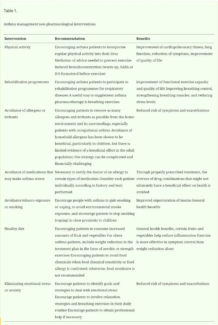
Asthma is an incurable disease, but with medical treatment, patients can achieve a full and high quality of life comparable to healthy individuals. Asthma control is divided into two domains: symptom control and risk of adverse outcomes. Neglected symptom control often leads to worsening health and the return of recurrent exacerbations. In the short term, symptom control can be provided through the Asthma Control Test (ACT) and Asthma Control Questionnaire (ACQ), use of short-acting inhaled β2 agonist bronchodilator (SABA) releasers for complications, and avoidance of contact with triggers. One aspect of treatment is to take into account the severity of asthma. Severe asthma remains uncontrolled despite optimal treatment, but it must be distinguished from intractable asthma. Moderate asthma is well controlled with a low or medium dose of inhaled corticosteroids (ICSs) (plus SABA as needed). Mild asthma is well controlled with low-intensity treatment. The management of bronchial asthma can be divided into pharmacological and non-pharmacological. According to Šperková (2018), prevention of symptoms of already diagnosed asthma is possible by avoiding contact with the allergen or other trigger. Non-pharmacological strategies in asthma management include certain interventions eg.physical activity, attending rehabilitation programmes, avoiding allergens, medication, food, tobacco or irritants, healthy eating, eliminating emotional stress or anxiety. Pharmacological treatment consists of maintenance treatment, controllers and relievers, anti-inflammatory reliever (AIR), and maintenance and reliever therapy (MART). Maintenance treatment is prescribed for everyday use, even though a person does not have asthma symptoms, as the medication is intended to be used continuously. They include ICS-containing medications (ICS, ICS-LABA, ICS-LABA-LAMA), leukotriene receptor antagonist [LTRA], and biologic therapy. Controllers can be defined as basic anti-inflammatory drugs administered regularly, which are intended for daily long-term use. They provide asthma control through an anti-inflammatory effect and target both domains of asthma control (symptoms and future risk). Another solution is antileukotriens in tablet form. The onset of action of the drugs is not immediate. Relievers are taken only when there is difficulty (difficulty breathing, wheezing, chest heaviness). They have a bronchodilating effect and remove the symptoms of the disease. Relief comes quickly after taking relievers, but they do not cure chronic inflammation. Increased use of relievers is a signal of worsening asthma symptoms. Treatment of asthma involves a combination of medications – controllers and relievers The relievers consist of the anti-inflammatory component ICS-formoterol and the ICS-SABA and SABA combinatio They are often recommended as a short-term treatment when bronchoconstriction occurs. The use of relievers via inhalation technique is considered the fastest way to bring relief from breathing difficulties, worsening asthma, or exacerbations, because it provides targeted delivery of the drug, is faster acting, requires a small dose, and is easy to take. Very important is how much of the drug reaches the target organ. This depends on the correct inhalation technique, the characteristics of the inhalation system, and the size of the emitted particles. Oral steroids have more side effects and do not have superiority over ICSs in the management of asthma. However, they are more beneficial in the management of severe acute exacerbations. An AIR is an inhaler that contains a low dose of ICS and a rapid-acting bronchodilator. Patients can also use it before exercise, other physical activity, or exposure to allergens as a prevention against bronchoconstriction or worsening of symptoms. Maintenance and reliever therapy (MART) is used only with combination ICS-formoterol inhalers such as budesonide-formoterol and beclomethasone-formoterol. MART treatment means that the patient takes ICS-formoterol every day (as a maintenance dose) and, if necessary, a releaser.
Table 1; asthama management non-pharmacological interventions
Treatment with antihistamines can be administered in adult patients by inhalation, orally or parenterally (subcutaneously – administered subcutaneously, intramuscularly – administered into the muscle, or intravenously – administered into a vein). Inhaled antihistamines come in the form of metered-dose aerosol inhalers, which can be triggered by pressure or inhalation, powder inhalers, fine particle inhalers, and nebulizers. They vary depending on the type, drug molecule, aerosol cloud velocity, and the degree of ease of handling the inhaler When using a releaser, proper inhalation technique is key. There are two basic types of inhalation devices: pressurized metered-dose inhalers (pMDIs) and dry powder inhalers (DPIs). The inhalation technique is similar in both cases; we first remove the cap of the inhaler. In the case of pressurized dose inhalers, we shake the inhaler before use. For DPIs, we make sure that the device is filled. Next, exhale gently and insert the mouthpiece between the lips, wrapping it tightly around the lips. Activate the inhaler and inhale deeply. Hold your breath for five to ten seconds, then slowly exhale. Repeat the process if necessary.Histamine Receptors in the LungIt has been elucidated that four types of histamine receptors such as H1, H2, H3, and H4 exist in the airway and pulmonary tissue. The bronchoconstriction of smooth muscle mediated via H1 receptors is one of the most well-known biological actions of histamine in the respiratory system. It was reported long before that histamine evoked a contraction of human bronchi, and bronchoconstriction was recognized first as one of the biological actions of histamine. While histamine contracts bronchial smooth muscles as strongly as muscarinic M1 receptor agonists, histamine contracts pulmonary peripheral tissue samples more strongly than M1 receptor agonists. This result seemed to suggest the higher sensitivity of peripheral airways to histamine, although it was possible that the contraction of vascular smooth muscles was involved in the contraction of the pulmonary peripheral tissue samples because the sample contained vessels. The response of pulmonary arteries to histamine is biphasic induced by vascular contraction via H1 receptors and vascular dilation via H2 receptors. Histamine induces plasma leakage from postcapillary venules by affecting the bronchial microcirculatory system. Histamine increases the secretion of mucous glycoprotein from the human airway in vitro. This action is inhibited by the H2 receptor antagonist (H2RA), cimetidine, not H1 receptor antagonists (H1RAs). Histamine also accelerates the chloride ion transport of airway epithelial cells, which is closely associated with water transfer in the airway.
Histamine and Mast Cells in Asthma
Histamine has been a well-known chemical mediator released from mast cells in the immediate allergic reaction for a long time and has been thought to have a critical role in the asthma pathophysiology. Histamine is released into the surface of the airway by inhaled allergens and direct contact with a bronchoscope, and is recovered in bronchoalveolar lavage fluid (BALF). Under the same condition, other chemical mediators are released from mast cells, such as 9α, 11β-prostaglandin F2-α, and tryptase, and are also recovered in BALF. In fact, it was reported that the histamine concentration in the BALF of patients with asthma was significantly higher than that from patients with allergic rhinitis. Furthermore, Tomioka et al. estimated that the number of mast cells in BALF of asthmatic patients was greater than that of control subjects (Figure 1).
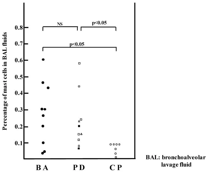
Figure1: bronchoalveolar lavage fluid
Percentage of mast cells in BAL fluids in asthmatics (BA: ●), patients with other pulmonary diseases (PD): interstitial pneumonia (□), sarcoidosis (△), and chronic obstructive pulmonary diseases (■), and control patients (CP: ○). (Bars indicate SEM). The figure was modified from reference with permission: Tomioka et al., Mast cells in bronchoalveolar lumen of patients with bronchial asthma. Am. Rev. Respir. Dis. Histamine is a representative bioamine that exerts strong and diverse biological actions, and was the first chemical mediator historically found to evoke bronchial smooth muscle contraction in asthma. Dale and Laidlaw first reported the action of bronchoconstriction by histamine. They reported that the bronchoconstriction of guinea pigs induced by venous injection of histamine is similar to that observed by allergen challenge to sensitized guinea pigs with antigen. Therefore, they suggested that histamine was an important chemical mediator that evokes bronchoconstriction in allergen challenge and might be involved in airway obstruction. Since this study suggested that the airways of guinea pigs were the most sensitive to histamine in terms of undergoing airway smooth muscle contractions, the guinea pigs were used most frequently throughout the twentieth century as an animal model of asthma induced by allergen challenge. In addition, almost all histamine in the skin is localized in mast cells as in the mast cells that exist in the airways. They determined that histamine released from mast cells was involved in the airway response after allergen challenge. Furthermore, since IgE and high-affinity IgE receptors on the surface of mast cells were identified, the concepts of allergen sensitization and the release of chemical mediators such as histamine from mast cells were combined. Based on this historical background, it was clearly recognized that histamine was an important chemical mediator involved in the bronchoconstriction induced by an immediate reaction in bronchial asthma
Histamine in the Pathophysiology of Asthma
Curry reported a historical finding concerning asthma pathophysiology, showing histamine induced bronchoconstriction in asthmatics by injection or inhalation at a low dose of histamine that had no effect in normal subjects. Based on these results, the concept of airway hyperresponsiveness to histamine as a physical characteristic of asthmatics was proposed. Afterward, since the airways of asthmatics were hyperresponsive to many airway smooth-muscle-contracting agents, it has been recognized as nonspecific airway hyperresponsiveness showing airway abnormality. Terfenadine, a strong H1RA, induced bronchodilation in asthmatics the same as β2-stimulant. This result suggested that the continuous release of histamine from mast cells in the lung of asthmatics was causing contractive tension of the airway smooth muscle. These lines of evidence suggested that histamine was deeply involved in the asthma pathophysiology. Furthermore, terfenadine reversed completely the decrease in the forced expiratory volume in one second (FEV1) as an index of airway obstruction induced by histamine. On the other hand, terfenadinereversed partially the decrease of FEV1 induced by allergen inhalation)
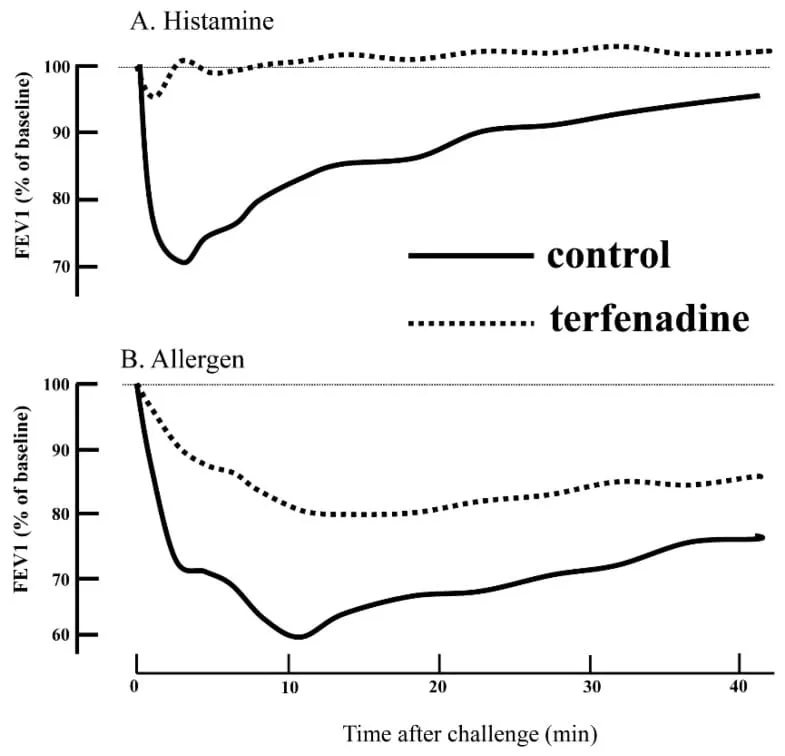
Time after challenge(min)
The effects of terfenadine on bronchoconstriction induced by (A) Histamine and (B) Allergen. Terfenadine (dotted curve) significantly attenuated bronchoconstriction induced by allergic challenge. (Solid curves indicate pretreatment with terfenadine. Dotted curves indicate pretreatment with placebo. The figure was modified from reference with permission: Rafferty et al., The contribution of histamine to immediate bronchoconstriction provoked by inhaled allergen and adenosine 5’ monophosphate in atopic asthma. Am. Rev. Respir. Dis. It was reported that the histamine concentration in the BALF of patients with asthma was significantly higher than that of normal subjects as shown. Furthermore, mast cells and basophils were increased in the BALF of asthmatics during exacerbations. The mRNA level of l-histidine decarboxylase, a histamine synthase, was markedly elevated in the pulmonary tissue of patients with asthmatic death. Histamine N-methyl transferase (HMT) is the principal enzyme that metabolizes histamine in the airway. HMT activity was measured in human trachea and bronchi. In addition, the contractile response of isolated human bronchi to histamine was potentiated in the presence of an HMT inhibitor, SKF 91488. These results suggest that HMT plays an important role in degrading histamine and in regulating the airway response to histamine (Figure 3). In addition, it was reported that polymorphisms of H1R and HMT gene in the patients with allergic asthma were significantly different from those of nonallergic asthma, suggesting that polymorphisms of H1R and HMT gene were involved in the pathogenesis of allergic asthma.
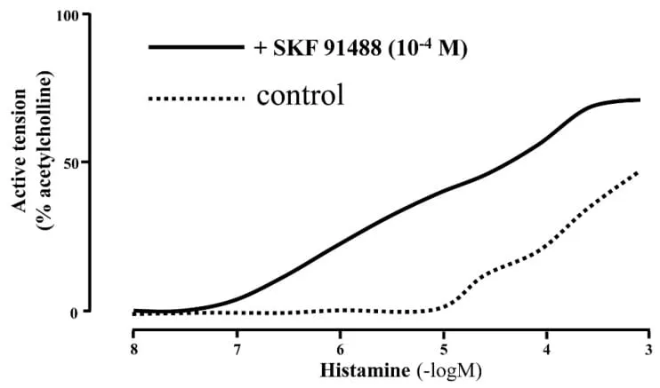
Figure2: Histamine (-logM)
Effect of SKF 91488 and aminoguanidine on contractile response of human bronchi to histamine. Dose–response curves to histamine in SKF 91488 (10−4 M) and control are analyzed. Data are expressed as percentage of response to acetylcholine (10−3 M). Histamine N-methyltransferase (HMT) regulates contraction of airway smooth muscle by histamine. HMT in epithelium degrades histamine actively. The figure was modified from reference with permission: Yamauchi et al., Structure and function of human histamine N-methyltransferase: critical enzyme in histamine metabolism in airway. Am. J. Physiol. 1994, 267, L342–L349.
Histamine Transport in the Pathophysiology of Asthma
Histamine is synthesized and stored in the vesicles of mast cells and basophils. Upon immunological stimulation of mast cells and basophils, histamine is released from storage vesicles into the extracellular space activating G-protein-coupled receptors H1, H2, H3, and H4 . However, to terminate the effects of histamine via histamine receptors on targets cells such as bronchial smooth muscle cells, the histamine concentration in the extracellular space should be regulated by the degradation of histamine. The degradation enzyme, HMT, is critical in metabolizing histamine into inactive forms of the metabolite and was documented to be significant for the relationship between airway responsiveness and HMT activities when using the HMT inhibitor, SFK91488, in animal models. Biochemical analysis suggested that the HMT enzyme is primarily localized in cytoplasmic space while histamine is unable to easily enter the intracellular space because at physiological pH, histamine exists as an organic cation. Therefore, transport machineries of histamine are required to enter the intracellular space and to obtain access to the HMT enzyme. We hypothesized the two potential mechanisms: the HMT enzyme translocates to the plasma membrane upon some stimulation and possibly achieves access to histamine (membrane translocation hypothesis) and other molecules assist the transport of histamine into cells (transporter hypothesis). In terms of the first hypothesis (membrane translocation hypothesis), we clarified the translocation of HMT to the plasma membrane upon stimulation of adrenergic receptor with isoproterenol. In regard to the second hypothesis (transporter hypothesis), supportive evidence was reported for comparatively high-capacity transporters (solute carrier cation transporters 22As; SLC22As) in the reuptake of endogenous substrates, such as norepinephrine, dopamine, serotonin, and histamine. Among these transporters, organic cation transporter (OCT)-2 (SLC22A2) and -3 (SLC22A3) revealed the ability to transport histamine into cells via a potential-sensitive mode. OCT-2 was exclusively expressed in the kidney, while OCT-3 was ubiquitously expressed. In order to clarify the molecular mechanisms of how the histamine transport system is associated with HMT inactivation, we prepared three types of cells stably expressing HMT alone, plasma membrane targeting form of HMT due to N-terminal myristylation, and HMT plus OCT-2 for in vivo/in vitro assays followed by measuring the concentration of extracellular as well as intracellular histamine. Cells expressing HMT with OCT-2 indicated significantly increased intracellular histamine contents compared with cells expressing HMT with myristylation modification, suggesting that the transporter is critically required for histamine metabolism. Based on previous studies on the distribution and capability of histamine transport, organic cation transporter-3 (OCT-3) is presumed to be the most important transporter in allergic asthma, although the functional characterizations of all transporters identified to date have not been fully completed in regard to histamine transporter activities. In general, both membrane drug transporters and metabolic enzymes are involved in the clearance of drug. Likewise, both OCT-3 and metabolic enzyme HMT are intimately interconnected and operate in the reduction of the extracellular histamine concentration. Therefore, alterations in the histamine transport activities in the respiratory tissues would critically influence the pharmacokinetics of histamine. Oct3/Slc22a3 (mouse homologue of human OCT-3)-deficient mice were generated and exhibited increased accumulation of histamine in the brain as well as in the peripheral plasma. As far as we know to date, there are 11 studies on functional and phenotypic alterations suspected to be related to human OCT-3 polymorphisms. Yamauchi et al. reported that four major genetic polymorphisms (S116A, R120R, I140T, and A411A) of the OCT-3 gene including synonymous alterations were investigated concerning the relationship between the allele frequency and asthma severities, and were classified into two groups (mild and moderate/severe) on the basis of the Japanese asthma severity guidelines. Patients with moderate and/or severe symptoms exhibited significantly lower frequency of the C allele compared to those showing mild symptoms, suggesting that a genetic alteration of the OCT-3 gene may have relevance to the asthma severity of asthma
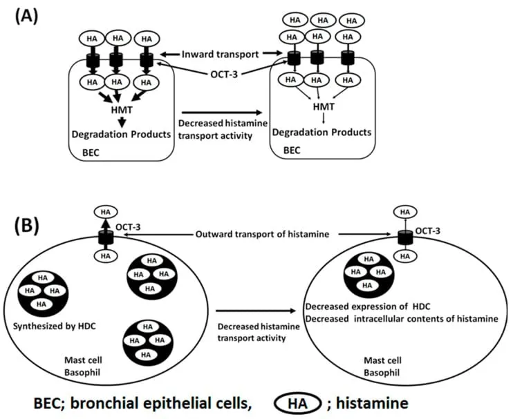
Figure3: bronchial epithelial cells
Scheme of histamine transport system through OCT-3. (A) Inward transport of histamine in bronchial epithelial cells. Genetic polymorphisms of OCT-3 affect the inward transport activity of histamine, resulting in the increased amounts of histamine in extracellular space and possibly confer stronger stimulation on H1R and induce contraction of bronchial smooth muscle cells. (B) Outward transport system of histamine through OCT-3 in professional histamine-producing cells (mast cells and basophils). Genetic polymorphisms of OCT-3 affect the outward transport activity of histamine, resulting in the decreased amounts of histamine contents in the vesicles through homeostatic
A New Insight on the Immunological Pathway in Asthma
Asthma is an inflammatory disorder of the conducting airways that has a strong association with allergic sensitization. The disease is characterized by a polarized Th-2 (T-helper-2)-type T-cell response and the levels of IL-4, IL-5, and IL-13 are upregulated in the airways of patients with bronchial asthma. After an exposure to allergens, specific IgE on the high-affinity IgE receptors of mast cells conjugates with allergens, IgE and allergen complex activate mast cells through IgE receptors and release chemical mediators including histamine, leukotrienes, prostaglandins, and so forth. This immunological event causes an immediate reaction in the airways of asthmatics. shift from a Th2 cell-dependent, IgE-mediated disease to a more complicated heterogeneous disease. Recent studies clearly show that not only Th2 cytokines but also other T-cell-related cytokines such as IL-17A and IL-22 as well as epithelial cell cytokines such as IL-25, IL-33, and thymic stromal lymphopoietin (TSLP) are involved in the pathogenesis of asthma. Recently, type 2 innate lymphoid cells (ILC2) were found to represent a critical innate source of type 2 cytokines. In the classical immunological pathway, the role of mast cells is critical in the immediate reaction. However, over the past decade, the understanding of asthma pathogenesis has made a significant On the other hand, recent advances in the immunoreactions of asthma revealed that in the innate immune response, epithelial cells release innate immune molecules such as IL-33, TSLA, and so forth in response to foreign stimulants such as microorganisms, dust, smoke, and so forth. Consequently, TSLP or IL-33 activates ICL2 to release various Th2 type cytokines such as IL-5 and IL-13, inducing eosinophilic inflammation in the airways. This pathway of innate immunity explains well the pathogenesis of asthmatic acute exacerbations caused by infection or inhalation of dusty air. However, mast cell activation is not involved in the pathway of the innate immunity of asthma pathogenesis. A New Aspect of H1RA and H4RA in Asthma Therapy For three decades, the primary therapies for asthma included ICS and LABA, LAMA, theophylline, and LTRA, combined with ICS depending on the severity. As described above, the clinical trials of H1RA for asthma have not been successful and recent advances in immunology revealed IgE-independent immunological pathways, suggesting a critical immune pathway without mast cells in the asthma pathogenesis The therapeutic use of H1RAs to the comorbidity of asthma and allergic rhinitis. More than 60% of asthmatics represented the comorbidity with allergic rhinitis. These lines of evidence suggest that ICS may be enough for asthma therapy and that the significance of mast cells in asthma is small. However, Omalizmab, an anti-IgE antibody, demonstrated excellent clinical effects on severe allergic asthmatics. This clinical effect reminded us that mast cells are still playing critical roles in the asthma pathophysiology. Since asthma is a disease with heterogenous immunological pathways including IgE-independent pathways, H1RAs would not be effective in the general population of asthmatics. In other words, H1RAs can be expected to be clinically effective in some subtypes of asthma. Bronchial asthma and allergic rhinitis (AR) are thought to share a common pathogenesis. More than 60 percent of patients with adult asthma showed symptoms of AR and were diagnosed with AR. In addition, asthma and AR symptoms often co-occur in patients with asthma and AR (Table 2) [64]. H1RAs are recommended for AR in clinical guidelines. In this regard, asthmatics with AR may represent a promising subtype in which H1RAs may be very effective on both asthmatic and AR symptoms (Figure 7). Allergic asthmatics are thought to represent more than 70% of total asthmatics. Therefore, asthmatics with AR are a large subtype.
Table2: comorbidity of asthama with allergic Rhinitis
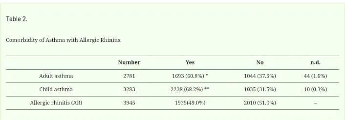
Adult asthma vs AR p < 0.001; ** Child asthma vs. AR p < 0.001 (x2 analysis). Reference, Yamauchi et al. Allergol.
CONCLUSION:
In recent decades, the treatment of bronchial asthma has advanced significantly, not only due to a better understanding of the pathophysiology of the disease, but also to the discovery of new therapeutic strategies. Management based on an individualized approach in the control of the patient’s condition is key. ICSs remain the mainstay of long-term treatment, while their combination with long-acting β2-agonists (LABAs) significantly improves clinical outcomes. Biological therapies are coming to the fore, bringing significant advances in treatment for patients with severe eosinophilic or allergic asthma. Monoclonal antibodies such as anti-IL-E (omalizumab), anti-IL-5 (mepolizumab, reslizumab), or anti-IL-4/IL-13 (dupilumab) allow a personalized approach to therapy, thus achieving better disease control and reducing the need for systemic corticosteroids. Within the nephromacological form of disease management, an individualized approach to the patient is important, including patient education, elimination of triggers, and incorporation of physical activity into the patient’s daily routine. However, the challenge remains the identification of disease phenotypes and the development of therapeutic approaches targeting inflammatory processes occurring at the molecular level.
REFERENCES


D. Rama Brahma Reddy*, K. Malleswari, A. Gopi Raju, A Review on Autocoder Inflammation Antihistimines Bronchial Asthma, Int. J. Med. Pharm. Sci., 2026, 2 (8), 630-639. https://doi.org/10.5281/zenodo.22006056
 10.5281/zenodo.22006056