

We use cookies to ensure our website works properly and to personalise your experience. Cookies policy
1Department of Pharmacology, Dr. Shivajirao Kadam College of Pharmacy, Kasabe Digraj, Sangli, Maharashtra, India
2Department of Pharmaceutical Analysis, Vikas College of Pharmacy, Jangoan, Maharashtra, India
3Department of Pharmacology, Appasaheb Birnale College of Pharmacy, Sangli, Maharashtra, India
Cardiovascular diseases are a leading cause of global mortality, and oxidative stress plays a central role in the development of myocardial injury. The present study investigated the cardioprotective effect of the ethanolic extract of Rotula aquatica Lour. against isoproterenol-induced cardiotoxicity in Wistar rats. Animals were allocated to normal, isoproterenol control, propranolol-treated, and extract-treated groups. Cardioprotective activity was evaluated by electrocardiography, serum cardiac biomarkers, lipid profile, antioxidant parameters, inflammatory markers, and histopathological examination of myocardial tissue. Administration of Rotula aquatica extract attenuated isoproterenol-induced myocardial damage by improving cardiac biochemical indices, restoring endogenous antioxidant defence, reducing inflammatory responses, normalizing electrocardiographic abnormalities, and preserving myocardial architecture. The protective effect was comparable to the standard treatment and appeared to be dose dependent. These findings suggest that Rotula aquatica possesses significant cardioprotective potential, possibly through antioxidant and anti-inflammatory mechanisms, and may serve as a promising natural therapeutic agent against experimental myocardial injury.
Cardiovascular diseases (CVDs) remain the leading cause of morbidity and mortality worldwide, accounting for a substantial proportion of global deaths. Among these disorders, myocardial infarction (MI) is one of the most serious manifestations, resulting from prolonged interruption of coronary blood flow that causes irreversible myocardial injury and progressive impairment of cardiac function. The pathogenesis of MI involves complex mechanisms, including oxidative stress, inflammatory responses, calcium overload, metabolic disturbances, and structural remodelling of the myocardium, all of which contribute to cardiomyocyte death and cardiac dysfunction. [1–3,19,20] Experimental animal models are essential for understanding the mechanisms of myocardial injury and for evaluating the efficacy of potential cardioprotective agents before clinical application. Among the available models, isoproterenol-induced myocardial infarction is widely employed because it is simple, reproducible, economical, and closely mimics the biochemical, electrocardiographic, and histopathological alterations observed in human myocardial infarction. [4–6,22] Isoproterenol is a synthetic non-selective β-adrenergic agonist that produces myocardial injury when administered at high doses. Excessive β-adrenergic stimulation increases heart rate, myocardial contractility, and oxygen demand, leading to relative myocardial ischaemia and hypoxia. In addition, auto-oxidation of isoproterenol generates reactive oxygen species (ROS), resulting in oxidative stress, calcium overload, lipid peroxidation, mitochondrial dysfunction, and inflammatory responses that culminate in myocardial necrosis. These pathological changes are associated with elevated serum cardiac biomarkers, depletion of endogenous antioxidant enzymes, electrocardiographic abnormalities, and histological damage resembling human myocardial infarction. [5–10,22] Oxidative stress is considered one of the major contributors to myocardial injury. Excessive production of ROS, including superoxide anions, hydrogen peroxide, and hydroxyl radicals, damages membrane lipids, proteins, nucleic acids, and mitochondria. Lipid peroxidation increases membrane permeability, causing leakage of intracellular enzymes such as creatine kinase-MB (CK-MB), lactate dehydrogenase (LDH), and aspartate aminotransferase (AST) into the circulation. Simultaneously, endogenous antioxidant defence systems, including superoxide dismutase (SOD), catalase (CAT), glutathione peroxidase (GPx), and reduced glutathione (GSH), become depleted, thereby aggravating myocardial damage. Restoration of the antioxidant defence system is therefore considered an important therapeutic strategy for preventing myocardial injury. [8–10,21] Current pharmacological management of cardiovascular diseases includes β-blockers, angiotensin-converting enzyme inhibitors, calcium channel blockers, antiplatelet agents, statins, and diuretics. Although these drugs have significantly improved patient outcomes, prolonged treatment is often associated with adverse effects, increased treatment costs, and reduced patient compliance. Consequently, considerable attention has been directed towards identifying natural products with improved safety profiles and cardioprotective potential. [12] Medicinal plants are rich sources of biologically active phytochemicals, including flavonoids, alkaloids, phenolic compounds, glycosides, tannins, and triterpenoids, which exhibit antioxidant, anti-inflammatory, antihyperlipidaemic, and membrane-stabilising properties. Previous studies have demonstrated that these phytoconstituents protect the myocardium by scavenging free radicals, reducing lipid peroxidation, restoring endogenous antioxidant enzymes, improving lipid metabolism, and suppressing inflammatory responses. [10,13–15]
Rotula aquatica Lour. (Family: Boraginaceae) is a medicinal plant widely distributed in the Western Ghats of India and has long been used in traditional Ayurvedic medicine for the treatment of urolithiasis and other ailments. Phytochemical investigations have identified several bioactive constituents, including alkaloids, steroids, bauerenol, and rhabdiol, which are responsible for its diverse pharmacological activities. Previous investigations have reported antioxidant, anti-inflammatory, antibacterial, antidiabetic, hypolipidaemic, analgesic, and antiurolithiatic properties of the plant, suggesting its potential role in protecting against oxidative stress-mediated diseases. [16–18] Despite its extensive traditional use and documented pharmacological properties, scientific evidence supporting the cardioprotective efficacy of Rotula aquatica against isoproterenol-induced myocardial injury remains limited. Therefore, the present study was undertaken to evaluate the cardioprotective effect of the ethanolic extract of Rotula aquatica Lour. against isoproterenol-induced cardiotoxicity in Wistar rats by assessing electrocardiographic alterations, serum cardiac biomarkers, antioxidant status, lipid profile, inflammatory mediators, and histopathological changes. The findings of this study may provide experimental evidence supporting the therapeutic potential of Rotula aquatica as a natural cardioprotective agent. [5,6,10,18]
MATERIALS AND METHODS
Chemicals, Reagents and Instruments
All chemicals and reagents used in the present study were of analytical grade. Isoproterenol and Propranolol were used for induction and standard treatment, respectively. Isoproterenol hydrochloride (ISO) was used for induction of myocardial injury, whereas propranolol hydrochloride was used as the standard cardioprotective drug.(standard cardioprotective drug), Methanol, ethanol, petroleum ether, chloroform Quercetin (standard antioxidant compound), Gallic acid 1,1-diphenyl-2-picrylhydrazyl (DPPH), Folin–Ciocalteu reagent, Sodium carbonate Trichloroacetic acid (TCA), Thiobarbituric acid (TBA), Phosphate buffer solution (PBS), Normal saline (0.9% NaCl), Formalin solution (10%).
Instruments
UV–Visible spectrophotometer, rotary evaporator, Soxhlet apparatus, centrifuge, ECG recording system, and biochemical analyser.
Induction of myocardial infarction
Myocardial injury was induced by subcutaneous administration of isoproterenol hydrochloride (5 mg/kg body weight) dissolved in normal saline for 7 consecutive days. [19]
Plant material
Rotula aquatica Lour. roots were collected from Kerala, India, during the study period. The plant material was authenticated by a qualified taxonomist in the Department of Botany, Dr. Patangrao Kadam Mahavidyalaya, Sangli, Maharashtra, India. A voucher specimen was deposited in the departmental herbarium for future reference. The collected roots were thoroughly washed with distilled water to remove adhering soil and other impurities, shade-dried at room temperature until a constant weight was obtained, and coarsely powdered using a mechanical grinder. The powdered material was passed through a suitable sieve and stored in airtight containers until extraction.
Extraction conditions
The powdered root material (100 g) was extracted with methanol using a Soxhlet extraction apparatus. The extraction was continued for approximately 6–8 h until the solvent in the siphon tube became colourless, indicating complete extraction of soluble constituents. The obtained extract was filtered and concentrated under reduced pressure using a rotary vacuum evaporator to remove the solvent. The concentrated extract was transferred to airtight containers and stored at 4°C until further phytochemical and pharmacological investigations. [20]
Quantitative phytochemical analysis
Total flavonoid content (TFC) of the methanolic extract of Rotula aquatica roots was determined using the aluminium chloride colorimetric method. The extract was mixed with aluminium chloride reagent to form a flavonoid–aluminium complex, and the absorbance was measured using a UV–visible spectrophotometer. Quantification was performed using a quercetin calibration curve, and the flavonoid content was estimated from the corresponding absorbance values. [21] Total phenolic content (TPC) was estimated by the Folin–Ciocalteu method. The extract was reacted with Folin–Ciocalteu reagent and sodium carbonate solution, followed by incubation for colour development. The absorbance of the resulting blue complex was recorded using a UV–visible spectrophotometer. Gallic acid was used as the reference standard, and the total phenolic content was calculated from the standard calibration curve. [22]
DPPH radical scavenging assay
The antioxidant activity of the methanolic extract of Rotula aquatica roots was evaluated using the 2,2-diphenyl-1-picrylhydrazyl (DPPH) radical scavenging assay. The assay was performed by reacting the extract with DPPH reagent, and the free radical scavenging activity was determined by measuring the reduction in absorbance using a UV–visible spectrophotometer. Quercetin was used as the reference standard. The antioxidant activity of the extract was expressed as percentage inhibition and compared with the standard at different concentrations. The antioxidant activity was expressed as percentage inhibition and IC50 value was calculated for both the extract and the standard. [23]
In vivo cardioprotective assay:
Experimental animals
For the experiment, healthy adult Wistar albino rats weighing between 180 and 300 g of either sex were used. Before the experiment began, the animals were obtained from a licensed animal breeding facility and given at least a week to get used to the lab environment. The study only included healthy animals that showed no outward symptoms of illness, injury, or aberrant behaviour. The study was conducted after protocol was approved by the Institutional Animal Ethics Committee (IAEC) of Dr. Shivajirao Kadam College of Pharmacy, Kasabe Digraj, Sangli, Maharashtra, India (IAEC Approval No.: IAEC/SKCP/2025-26/09). The study was conducted in accordance with the guidelines of the Committee for Control and Supervision of Experiments on Animals (CPCSEA), Government of India. Thirty Wistar albino rats were approved for the study.
Treatment schedule
The rats were randomly allocated into five groups, each consisting of six animals (n = 6). Group I (Normal control) received normal saline orally throughout the experimental period. Group II (Isoproterenol control) received normal saline orally for 14 consecutive days and were administered isoproterenol hydrochloride (5 mg/kg body weight, subcutaneously) once daily during the last seven days to induce myocardial injury. Group III (Standard treatment) received propranolol (10 mg/kg body weight, i.p.) once daily for 14 consecutive days, followed by isoproterenol administration (5 mg/kg, s.c.) during the last seven days. Group IV (Low-dose treatment) received the methanolic extract of Rotula aquatica (200 mg/kg body weight, orally) once daily for 14 consecutive days along with isoproterenol (5 mg/kg, s.c.) during the final seven days. Group V (High-dose treatment) received the methanolic extract of Rotula aquatica (400 mg/kg body weight, orally) once daily for 14 consecutive days together with isoproterenol (5 mg/kg, s.c.) during the last seven days. At the end of the treatment period, the animals were fasted overnight, electrocardiographic recordings were obtained, and blood samples and heart tissues were collected for biochemical, antioxidant, inflammatory, and histopathological analyses.
Electrocardiographic (ECG) Analysis
At the end of the experimental period, electrocardiographic parameters were recorded under light anaesthesia using urethane. The animals were placed in a supine position, and standard limb leads were connected for ECG recording. ECG parameters including heart rate (HR), P wave, PR interval, QRS duration, ST segment changes, and QT interval were analysed to evaluate isoproterenol-induced cardiac electrical abnormalities and the cardioprotective effect of Rotula aquatica extract.
Estimation of Biochemistry
Blood samples were taken from animals that had fasted overnight while under light anaesthesia at the conclusion of the trial. To produce clear serum for biochemical examination, blood was allowed to coagulate and then centrifuged for 15 minutes at 3000 rpm. Standard diagnostic biochemical kits were used to evaluate the separated serum samples in order to estimate the cardiac marker enzymes, which included SGOT/AST, or serum glutamate oxaloacetate transaminase Creatine Kinase-MB (CK-MB) Lactate Dehydrogenase (LDH) Increased levels of these enzymes are thought to be markers of membrane damage and myocardial injury. Animals were humanely slaughtered once blood was collected, and the hearts were removed right away. To get rid of blood clots and unnecessary material, the isolated heart tissues were extensively cleaned with ice-cold normal saline. In order to estimate antioxidant properties, the tissues were subsequently homogenised in phosphate buffer under freezing circumstances. The homogenates were examined for oxidative stress and antioxidant indicators, including Superoxide dismutase (SOD), Catalase (CAT), MDA (malondialdehyde) Evaluation of these measures assisted in determining the degree of lipid peroxidation and antioxidant state in cardiac tissue after extract treatment.
RESULTS
ECG parameter
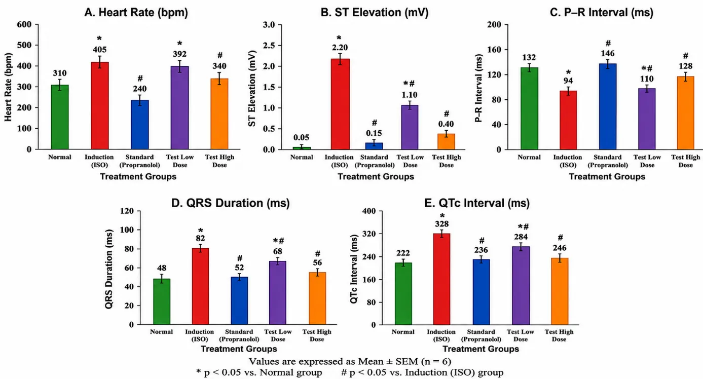
Fig 1. ECG Parameters
Effects of Rotula aquatica on heart weight in isoproterenol treated rats
Table no. 1 Effect on heart weight in isoproterenol-treated rats
|
Treatment |
Body weight (g) |
heart (g) |
|
Sham |
280.2 ± 5.2 |
0.61± 0.08 |
|
Control |
300.5 ± 6.0* |
0.85 ± 0.06* |
|
Low-dose |
172.8 ± 5.3 |
0.79 ± 0.05 |
|
High-dose |
184.6 ± 5.6# |
0.67 ± 0.04 |
|
Standard (Propranolol) |
193.1 ± 5.8# |
0.63 ± 0.06# |
Effects of Rotula aquatica on hemodynamic markers and cardiac function in isoproterenol-treated rats
Table no. 2 Effects of Rotula aquatica on hemodynamic markers and cardiac function in isoproterenol-treated rats
|
Hemodynamic marker |
sham |
control |
Low-dose |
High-dose |
Standard (propranolol) |
|
Heart Rate |
310 |
405 |
392 |
340 |
240 |
|
Systolic blood pressure |
110 |
89 |
85 |
83 |
81 |
|
Diastolic blood pressure |
69 |
79 |
78 |
74 |
63 |
|
Mean blood pressure |
90 |
81 |
78 |
69 |
61 |
Effect on Antioxidant Parameters
Table no. 3 effect on antioxidant parameters
|
Group SOD (% Activity) CAT (% Activity) LPO (nmol/mg protein) |
|||
|
Normal |
85 |
80 |
0.97 |
|
Induction |
40 |
35 |
3.77 |
|
Standard |
75 |
70 |
1.47 |
|
Low Dose |
55 |
50 |
1.93 |
|
High Dose |
65 |
60 |
1.69 |
Effect of rotula aquatica extract on antioxidant markers
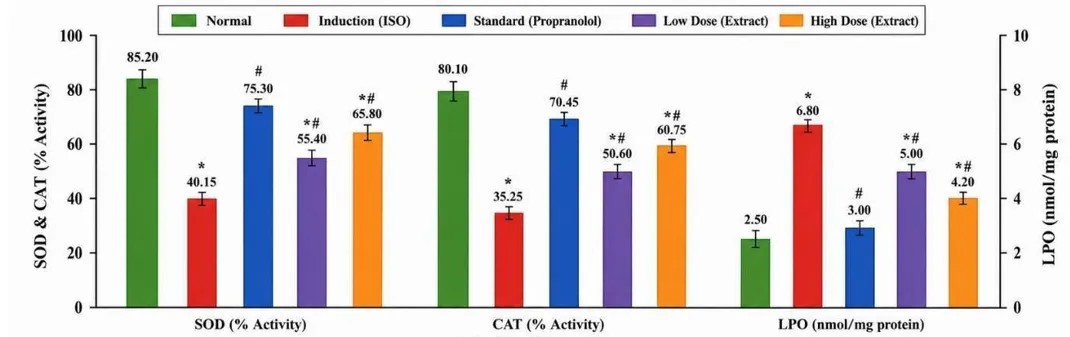
Fig.no.2 Effect on Oxidative marker
Effect on Cardiac Marker
Table no. 4 Effect on Cardiac Marker
|
Group |
CK-MB (U/L) |
LDH (U/L) |
SGOT (U/L) |
|
Normal |
95 |
170 |
30.80 |
|
Induction |
220 |
437 |
48.52 |
|
Standard |
110 |
210 |
32.40 |
|
Low Dose |
160 |
295 |
42.00 |
|
High Dose |
130 |
250 |
37.00 |
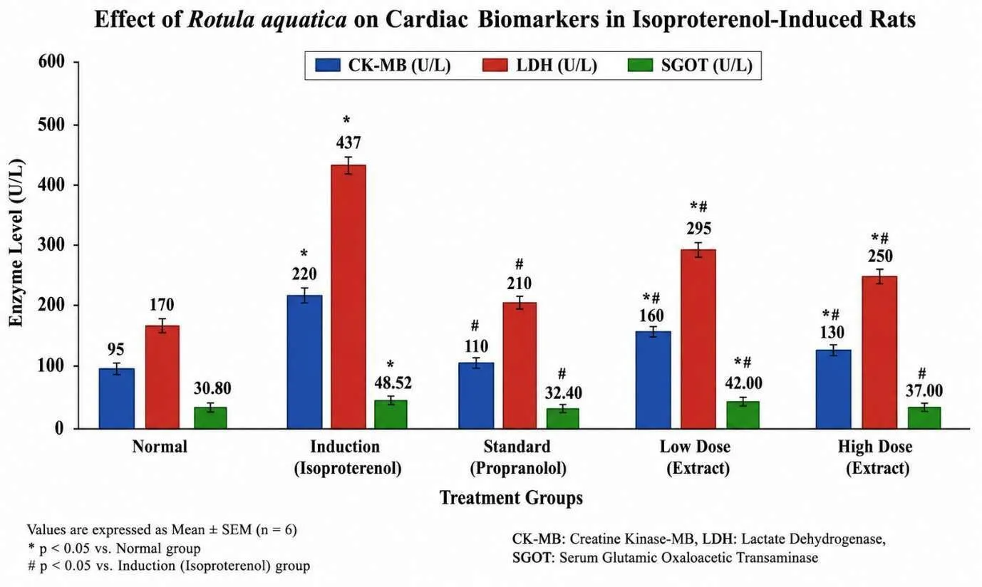
Figure no. 3 Effect on Cardiac Marker
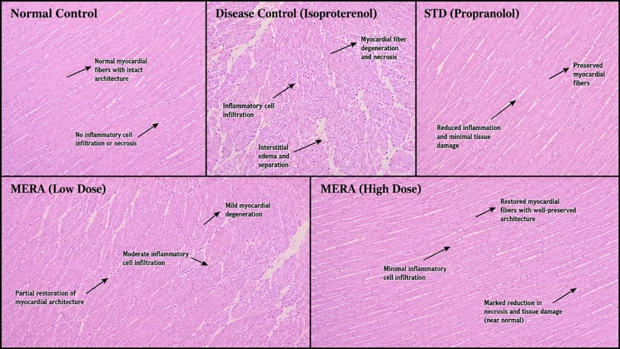
Histopathology
DISCUSSION
The present study demonstrated that the ethanolic extract of Rotula aquatica roots exerted significant cardioprotective activity against isoproterenol-induced myocardial injury in Wistar rats. Isoproterenol administration produced characteristic features of myocardial damage, including alterations in electrocardiographic parameters, elevation of serum cardiac biomarkers, increased oxidative stress, and histopathological abnormalities. Pretreatment with R. aquatica extract attenuated these changes in a dose-dependent manner, indicating its protective effect on the myocardium. The cardioprotective activity of R. aquatica is likely associated with its phytochemical constituents, particularly flavonoids, phenolic compounds, tannins, alkaloids, and saponins. These bioactive compounds are known to possess antioxidant and free radical scavenging properties that protect cardiac tissue against oxidative damage. The phytochemical screening performed in the present study confirmed the presence of these constituents, supporting their contribution to the observed pharmacological activity. Oxidative stress plays a central role in isoproterenol-induced cardiotoxicity by generating excessive reactive oxygen species, resulting in lipid peroxidation and myocardial cell injury. In the present study, treatment with R. aquatica significantly restored endogenous antioxidant defence by increasing superoxide dismutase and catalase activities while reducing lipid peroxidation. These findings suggest that the extract protects myocardial tissue by limiting oxidative stress and preserving cellular integrity. Electrocardiographic abnormalities such as ST-segment elevation and altered cardiac conduction observed in the isoproterenol-treated group were markedly improved following treatment with the extract. Furthermore, the reduction in serum CK-MB, LDH, and AST levels indicates stabilization of the cardiac cell membrane and decreased leakage of intracellular enzymes, reflecting reduced myocardial injury. Histopathological examination further supported the biochemical findings. Hearts from isoproterenol-treated animals exhibited myocardial degeneration, inflammatory cell infiltration, and disruption of muscle fibres, whereas extract-treated groups showed preservation of myocardial architecture with markedly reduced tissue damage. These observations confirm the protective effect of R. aquatica against structural cardiac injury. The overall cardioprotective effect of R. aquatica may therefore be attributed to the combined action of its phytoconstituents through antioxidant, membrane-stabilising, and cytoprotective mechanisms. Although the precise molecular pathways remain to be elucidated, the present findings suggest that the plant possesses promising therapeutic potential for preventing oxidative stress-mediated myocardial injury. In conclusion, the ethanolic extract of Rotula aquatica demonstrated significant cardioprotective activity against isoproterenol-induced cardiotoxicity by improving electrocardiographic changes, reducing cardiac biomarkers, enhancing antioxidant defence, and preserving myocardial histoarchitecture. Further studies involving isolation of active constituents and molecular investigations are warranted to clarify the exact mechanisms responsible for its cardioprotective effects.
CONCLUSION
The present study demonstrated that the ethanolic extract of Rotula aquatica roots exerts significant cardioprotective activity against isoproterenol-induced myocardial injury in Wistar rats. Pretreatment with the extract effectively attenuated electrocardiographic abnormalities, reduced serum cardiac biomarkers (CK-MB, LDH, and AST), restored antioxidant enzyme activities (SOD and CAT), decreased lipid peroxidation, and preserved normal myocardial histoarchitecture. These protective effects are likely attributable to the antioxidant potential of the phytochemical constituents present in the extract, particularly flavonoids, phenolic compounds, tannins, alkaloids, and saponins. Overall, the findings suggest that R. aquatica possesses promising therapeutic potential as a natural cardioprotective agent against oxidative stress-mediated myocardial injury. However, further studies are required to isolate and characterize the active phytoconstituents, elucidate their underlying molecular mechanisms, and establish their efficacy and safety through advanced preclinical and clinical investigations.
REFERENCES


Vaidehi Mhamane*, Sonal Vhankade, Vaibhav Salokhe, Feroz Momen, Godfrey Mathews, Sandeep Patil, Shankar Joshi, Cardioprotective Effect of Methanolic Extract of Rotula Aquatica Lour. Against Isoproterenol-Induced Myocardial Injury in Wistar Rats, Int. J. Med. Pharm. Sci., 2026, 2 (8), 93-101. https://doi.org/10.5281/zenodo.21771220
 10.5281/zenodo.21771220