

We use cookies to ensure our website works properly and to personalise your experience. Cookies policy
1Shri Ganpati Institute of Pharmaceutical Sciences and Research, Tembhurni
2Associate Professor, Shri Ganpati Institute of Pharmaceutical Sciences and Research Tembhurni
3Principal, Shri Ganpati Institute of Pharmaceutical Sciences and Research, Tembhurni
Banana (Musa acuminate) is a well-known plant valued not only as a nutritious food source but also for its traditional medicinal uses. For many years, different parts of the plant have been used in natural remedies to support digestion, promote wound healing, and improve overall health. These beneficial effects are largely linked to the presence of naturally occurring compounds such as flavonoids, phenolics, tannins, and other phytochemicals that help protect cells from oxidative damage. The present work focused on investigating the antioxidant potential of Musa acuminata leaves. Fresh leaves were collected, dried under shade, powdered, and extracted using petroleum ether, acetone, and ethanol through the maceration technique. The resulting extracts were examined for the presence of important phytochemical constituents and further evaluated using established in-vitro antioxidant models, including DPPH, nitric oxide, and hydroxyl radical scavenging assays. The study was undertaken to compare the efficiency of different extraction solvents and to assess the value of banana leaves as a natural source of antioxidant compounds. Growing concerns regarding the safety of synthetic antioxidants have increased interest in plant-derived alternatives. Therefore, Musa acuminata may represent a promising botanical resource for future development of herbal products and health-supporting formulations aimed at reducing oxidative stress.
Musa acuminata is a commonly cultivated banana plant grown extensively in warm climatic regions.Apart from being an important food crop, the plant is also recognized for its medicinal and nutritional importance. It is believed to have originated in Southeast Asia. Now extensively cultivate in countries like India, Indonesia, Malaysia, and Philippines. They are easily available have nutritional value, and health-promoting properties. banana has become an essential part of the daily diet as well as traditional healthcare practices. Musa acuminata is a large perennial herb characterized by broad green leaves, an underground rhizome, and a pseudostem formed by tightly packed leaf bases. The plant part like fruit, peel, leaves, stem, flowers, has traditionally been utilized for various medicinal purposes such as wound healing, digestion improvement, and treatment of infections. plant contains bioactive compounds including flavonoids, phenolic compounds, tannins, alkaloids, and vitamins, which contribute to its therapeutic potential. These phytochemicals are known to exhibit a variety of pharmacological activities. Nowadays considerable attention is given to antioxidant activity of Musa acuminata. Excess generation of reactive molecules present body can cause to oxidative damage which is associated with several chronic diseases. Natural antioxidants obtained from plants are considered safer and more beneficial compared to synthetic antioxidants. Previous studies have reported that extracts prepared from many parts of Musa acuminata, especially leaves, peels possess significant free radical scavenging activity in antioxidant assays such as DPPH and nitric oxide scavenging methods [1-8].
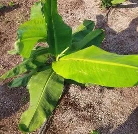
Figure: Musa acuminata
Kingdom: Plantae Division: Magnoliophyta Class: Liliopsida Order: Zingiberales Family: Musaceae Genus: Musa Species: Musa acuminata
2.1 Traditional Uses:
Musa acuminata has been used in traditional medicine because of nutritional and therapeutic properties. Different parts of the plant, including fruits, leaves, flowers, stem, and peel, are utilized for various medicinal applications. The plant is valued not only as a food source but also as an important medicinal plant in folk and herbal medicine. Ripe banana fruits are commonly consumed to improve digestion, provide energy, and reduce stomach irritation due to their soft texture and rich nutrient content. Unripe bananas are traditionally used to manage diarrhoea and intestinal disorders because of their high starch and tannin content. Banana flowers are also used to control blood sugar levels, reduce excessive menstrual bleeding, and treat ulcers. The leaves of Musa acuminata are traditionally applied to wounds, burns, and skin infections because of their cooling and soothing effects. Fresh leaf extracts and plant sap are also used to reduce inflammation and support wound healing. In addition, stem juice is consumed as a natural remedy for kidney stones and urinary disorders. Banana peel is traditionally used to relieve insect bites, skin irritation, and minor wounds [2-6, 9-11].
2.2 Reported Pharmacological Activities:
The plant exhibits antimicrobial activity to various bacteria and fungi, which may help in preventing infections and promoting wound healing. In addition, studies have reported anti-inflammatory effects, where plant extracts helped reduce inflammation and tissue damage. Research has further demonstrated the antidiabetic potential of banana flower and fruit extracts, which may help lower blood glucose levels and improve antioxidant defines in diabetic conditions. Musa acuminata has also shown antiulcer activity by protecting the stomach lining from ulcer formation. Other reported activities include wound-healing, anticancer, hepatoprotective, and antihypertensive effects [4-6, 10-12].
2.3 Chemical Constituents:
Musa acuminata contains a significant amount of natural chemical constituent responsible for its nutritional and medicinal properties. The fruits, peels, leaves, flowers, and stems, are rich in biological active compound like flavonoids, tannins, alkaloids, glycosides, saponin, vitamins, and minerals. Among these compounds, flavonoids and phenolic compounds are considered major natural antioxidants. In addition, Musa acuminata contains important minerals like as potassium, calcium, magnesium, phosphorus, and iron. Potassium is particularly important for maintaining normal blood pressure and proper muscle function. Studies have also reported the carotenoids, dopamine, serotonin, and other natural antioxidant compounds present in banana peels and leaves, which further enhance the medicinal importance of the plant [4-6,13,14].
2.4 Organoleptic Characteristics:
Musa acuminata is identified through organoleptic evaluation based on appearance, odor, taste, and texture. The leafs are large, elongated, green, and glossy upper surface and lighter lower surface showing visible veins. They are soft, flexible, and slightly fibrous. Fresh material has a mild herbal odor, while the leaves and pseudostem possess a slightly mucilaginous and mildly astringent taste due to tannins and other phytochemicals. The pseudostem is thick, succulent, pale green, and may contain brown or black spots. These features are important for identification, standardization, and quality evaluation.
Microscopic Characteristics
Microscopic examination of Musa acuminata helps in authentication and quality control. The leaf transverse section shows a single-layer upper and lower epidermis cover by a thin cuticle, with compact polygonal epidermal cells. Stomata are predominantly present on the lower epidermis and are generally paracytic in type. Mesophyll contains chlorophyll-rich parenchymatous cells, while vascular bundles with xylem and phloem are surrounded by sclerenchymatous fibres. In the pseudostem, scattered vascular bundles occur within mucilagerich soft ground tissue. Powder microscopy reveals epidermal fragments, fibres, spiral and annular vessels, starch grains, and chlorophyll-containing tissues [15-18].
2.5 Free Radicals:
Free radicals are unstable chemical species capable of reacting rapidly with body tissues. Because of their unstable nature, they readily react with other molecules in the body and may damage important cellular components like protein, DNA and lipid. Free radicals are naturally generate during normal metabolic activities like respiration and energy production. They may also be produced due to external factors such as pollution, cigarette smoke, radiation, stress, and unhealthy dietary habits. The most common free radicals include (ROS) and (RNS), like superoxide radical, hydroxyl radical, nitric oxide radical. Under normal conditions, the body possesses antioxidant defense systems that help neutralize these harmful compounds. However, excessive production of free radicals or reduced antioxidant protection can lead to oxidative stress, which has been associated with aging and several chronic diseases like cancer, cardiovascular disorders, diabetes [19-23].
2.6 Synthetic Antioxidants:
Synthetic antioxidants are man-made chemical compounds used to prevent oxidation and reduce damage caused by free radicals. They are commonly added to foods, medicines, cosmetics, and other products to improve stability and extend shelf life. Oxidation can cause spoilage of fats and oils, leading to unpleasant Odor, discoloration, bad taste, and loss of nutritional value. Synthetic antioxidants help slow these reactions by neutralizing free radicals and preventing oxidative chain reactions. Common synthetic antioxidants include (BHA) (BHT), (TBHQ), and (PG). However, prolonged or excessive use of synthetic antioxidants may produce adverse effects such as allergic reactions, liver toxicity, and possible carcinogenic risks. Due to these concerns, researchers are increasingly focusing on natural antioxidants obtained from plants, fruits, and vegetables as safer alternatives. Natural antioxidants contain bioactive compounds including flavonoids, tannins, and phenolic compounds that protect the body from oxidative damage. Musa acuminata has attracted significant attention because of its antioxidant and free radical scavenging properties associated with these phytochemicals [21,24-27].
2.7 Importance of Herbal Medicine:
Herbal Medicine therapies are widely preferred because they are economical and easily accessible and its comparatively lower incidence of side effects when used appropriately. Traditional systems of medicine such as Ayurveda, Siddha, and Unani have extensively utilized medicinal plants for maintaining health and treating chronic disorders. In recent years, interest in herbal medicine has increased considerably due to the side effects and drug resistance associated with many synthetic drugs. Consequently, researchers are exploring plant-based medicines as safer and more effective alternatives for future drug development. Musa acuminata is widely studied for its antioxidant and therapeutic potential because of the presence of valuable bioactive constituents [21,28-31].
2.8 Rationale for the Selection of Musa acuminata:
Musa acuminata was selected for the present study due to its well-known medicinal value and extensive use in traditional healthcare systems. Many plant part including the leafs, fruits, flowers, & pseudo stem, are traditionally use for treating wounds, ulcers, inflammation, diabetes, and digestive disorders. The plant contains important phytochemical like flavonoid, phenolic compounds, tannin, alkaloids, vitamins show the antioxidant potential and it show the ability to protect the body from oxidative stress. Scientific studies have also reported that Musa acuminata exhibits antioxidant, antimicrobial, anti-inflammatory, antidiabetic, and wound-heal activities, it is an important subject for herbal and pharmaceutical research. The plant is easily available, cost-effective, and nutritionally important. Due to concerns about side effects of a Synthetic antioxidants, there is increasing interest in natural sources, and Musa acuminata is considered a promising natural antioxidant source [15,16,21,32].
MATERIALS:
Plant Material:
The fresh leaves of Musa acuminata were collected from local area of Tembhurni, Solapur, India and washed with distilled water followed by air dried in the shade.
Chemicals and Reagents:
Acetic acid, Acetone, Ascorbic acid, Benedict’s reagent, Biuret reagent, Chloroform, concentrated hydrochloric acid, concentrated sulfuric acid [Produced by G S Laboratories Private Limited Pune, Maharashtra – 411052]
Copper sulfate, DPPH reagent, Dragendorff’s reagent, Ethanol, Ferric chloride solution, Ferrous sulfate, Hydrogen peroxide, Keller–Killiani reagent, Liebermann–Burchard reagent, Magnesium ribbon, Mayer’s reagent, Methanol, Molisch reagent, Naphthyl ethylene diamine dihydrochloride (NED), Ninhydrin reagent, [Produced by Aarati Scientific Company, Solapur Maharashtra, India 413002]
Nitric acid, Petroleum ether, Phosphate buffer solution, Potassium iodide, Salicylic acid, Salkowski reagent, Shinoda reagent, Sodium hydroxide, Sodium nitroprusside, Sulfanilamide [Produced by Research-Lab Fine Chem Industries, based in Mumbai 400 002 India.]
4. METHODOLOGY:
Procedure: (Maceration)
Healthy and fresh leaves of Musa acuminata was collected. Plant materials was authenticated by an expert to confirm its identity. Then collected leaves were washed properly with purified water for removal dirt, unwanted particles. Then cleaned leaves were dried under shade at room temp for 13-14 days to preserve their active constituents.
After complete drying, the leaves were converted into a coarse powder by a grinder. Take 25 gm of powder and then soaked in ethanol, acetone and petroleum ether for extraction by the maceration method.
The mixture is stand for 42–74 hours by occasional shaking for proper extraction of phytochemicals. The extract from different solvents obtained was filter by the Whatman filter paper for removel of solid particles.
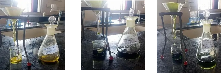
Fig: Pet. Ether extract Fig: Acetone extract Fig: Ethanol extract

The filtrate from the different solvents was concentrated by evaporating the solvent using a water bath. Finally, the dry extracts was collected and store into refrigerator for future phytochemical screening and antioxidant studies [41,44,46,54-56].
5.1 Percentage Yield of Extract:
The dried extract obtained from the petroleum ether, Acetone, Ethanol solvent was weighed carefully and the percentage yield is calculate
(%) Yield = (Weight of dried extract / Weight of crude Drug taken) × 100 [33,43-48].
Preliminary Phytochemical Screening
The extracts were screened for:
Using standard pharmacognostic procedures [36-42].
5.2. Antioxidant Assay:
5.2.1 DPPH Assay Principle:
The DPPH assay is one of the most commonly used methods for evaluating the antioxidant activity of plant extracts. DPPH is a stable free radical that exhibits a deep violet color in solution. Antioxidant compounds present in Musa acuminata extracts donate hydrogen atoms or electrons to DPPH, thereby neutralizing the free radicals. As a result, the violet color gradually changes to yellow. The reduction in color intensity indicates free radical scavenging ability of the extract and can be measured spectrophotometrically at 517 nm by using a UV-Visible spectrophotometer [7, 34].
Preparation of Stock Solution:
Take 20 mg of plant extract of different solvent of Musa acuminata and dissolve into the 20 ml of methanol, after mixing these solution 1000µg/mL concentrated solution was prepared.
Procedure:
Observation:
During the reaction, the deep violet color of DPPH solution gradually faded into yellow, indicate plant extract antioxidant potential [7, 34].
Calculation:
% Inhibition =Control Abs- Sample Abs/ Control Abs× 100 From percentage inhibition calculate the IC50 Value
5.2.2 Nitric Oxide Assay
Principle:
Nitric oxide assay is commonly used to evaluate the plant extracts antioxidant activity. In this method, sodium nitroprusside in phosphate buffer slowly releases nitric oxide radicals. These radicals react with oxygen and produce nitrite ions. The nitrite ions further react with sulfanilamide and NED reagent to form a pink-colored chromophore. If antioxidant compounds are present in the plant extract, they compete with oxygen and reduce the formation of nitrite ions. As a result, the intensity of the pink color decreases. The reduction in color intensity indicates nitric oxide activity which is measured spectrophotometrically at 546 nm [35,50, 49].
Preparation of Stock Solution:
Take 20 mg of plant extract of different solvent of Musa acuminata and dissolve into the 20 ml of distilled water,after mixing these solution 1000µg/mL concentrated solution was prepared.
Procedure: (used plant extract from Ethanol, Acetone, Petroleum ether)
Observation:
Tubes containing antioxidant-rich plant extracts show a lighter pink color due to inhibition of nitric oxide radicals [49, 50,35].
Calculation:
% Inhibition =Control Abs- Sample Abs/ Control Abs× 100 From percentage inhibition calculate the IC50 Value
5.2.3 Hydroxyl Radical Assay:
Principle:
Hydroxyl radicals are among the most harmful free radicals produced in the body. They damage important biological molecules like the DNA, lipids, protein that leads to oxidative stress and cellular injury. In this method, hydroxyl radicals are generated by the reaction between ferrous sulfate and hydrogen peroxide, commonly known as the Fenton reaction. The generated hydroxyl radicals react with salicylic acid and form-colored hydroxylated products. The intensity of the color formed is measured using a UV-visible spectrophotometer. When a plant extract containing antioxidant compounds is added to the reaction mixture, the antioxidants scavenge or neutralize the hydroxyl radicals. This reduces the formation of colored products, resulting in lower absorbance values. Therefore, a decrease in absorbance indicates stronger hydroxyl radical activity of that plant extract [51-53].
Preparation of Stock Solution:
Take 20 mg of plant extract of different solvent of Musa acuminata and dissolve into the 20 ml of distilled water after mixing these solution 1000µg/mL concentrated solution was prepared.
Procedure: (used plant extract from Ethanol, Acetone, Petroleum ether)
Observation:
Tubes containing antioxidant-rich plant extracts show lighter color intensity due to scavenging of hydroxyl radicals [51-53].
Calculation:
(%) Inhibition =Control Abs- Sample Abs/ Control Abs× 100 From percentage inhibition calculate the IC50 Value
RESULTS:
6.1. Percentage [%] Yield of Extracts:
A percentage yield of different solvent extracts of Musa acuminata was calculated after complete drying of the extracts. A known quantity of crude drug powder (25 gm) is extract separately from the solvents e.g-petroleum ether, acetone, ethanol. The petroleum ether extract showed a yield of 9.36% w/w, the acetone extract showed 9.76% w/w, and the ethanol extract showed the highest yield of 11.4% w/w.
Table 1: Percentage Yield of Different Extracts of Musa acuminata:
|
Sr. No |
Solvent Extract |
[%] Yield (% w/w) |
|
1 |
Petroleum Ether Extract |
9.36 % w/w |
|
2 |
Acetone Extract |
9.76 % w/w |
|
3 |
Ethanol Extract |
11.4 % w/w |
6.2. Phytochemical Screening of Different Extracts:
Table 2: Phytochemical Screening of Different Extracts of Musa acuminata:
|
Sr. No |
Phytochemical |
Petroleum Ether Extract |
Acetone Extract |
Ethanol Extract |
Inference |
|
1 |
Alkaloids |
Absent |
Present |
Present |
Alkaloids are present mainly in polar extracts. |
|
2 |
Carbohydrates |
Absent |
Present |
Present |
Carbohydrates are present in acetone and ethanol extract |
|
3 |
Glycoside |
Absent |
Present |
Present |
Glycosides are present in polar solvent extracts. |
|
4 |
Proteins & Amino Acid |
Absent |
Absent |
Present |
Proteins and amino acids are mainly present in ethanol extract. |
|
5 |
Phenols & Tannin |
Absent |
Present |
Present |
Phenolic compounds are present in acetone and ethanol extracts. |
|
6 |
Flavonoid |
Absent |
Present |
Present |
Flavonoids are present in moderately polar and polar extracts. |
|
7 |
Saponins |
Absent |
Absent |
Present |
Saponins are mainly present in ethanol extract. |
|
8 |
Phytosterols |
Present |
Present |
Present |
Phytosterols are present in all extracts. |
6.3 Antioxidant Activity:
1. Nitric oxide Radicals scavenging assay:
Table 3: Nitric oxide Radicals scavenging assay of different extract
|
Sr. No |
Test Extracts/STD Drug Conc. µg/mL) |
|
% Inhibition |
|
|
|
Petroleum ether extract |
Acetone extract |
Ethanol extract |
STD Drug Ascorbic acid |
||
|
1 |
100 |
23.5±5.0 |
26±5.5 |
31.1±4.0 |
- |
|
2 |
200 |
31.4±3.3 |
34±4.3 |
38.3±3.1 |
- |
|
3 |
400 |
35.9±4.7 |
46.2±4.3 |
44.5±4.6 |
- |
|
4 |
600 |
45.0±4.1 |
58.7±5.9 |
58.1±3.1 |
- |
|
5 |
800 |
52.6±6.1 |
66.5±4.4 |
70.5±4.7 |
- |
|
6 |
1000 |
63.0±6.0 |
75.2±4.5 |
77.3±4.3 |
- |
|
7 |
10 |
- |
- |
- |
32.88±2.12 |
|
8 |
20 |
- |
- |
- |
45.07±1.33 |
|
9 |
40 |
- |
- |
- |
49.15±1.77 |
|
10 |
IC50 Value (µg/mL) |
710±122.4 |
495±84.9 |
453±55.8 |
45±9.1 |
2. Hydroxyl radicals scavenging assay:
Table 4: Hydroxyl radicals scavenging assay of different extract:
|
|
|
%Inhibition |
|
||
|
Sr. No |
Test Extracts/STD Drug Conc. µg/mL) |
Petroleum ether extract |
Acetone extract |
Ethanol extract |
STD Drug Ascorbic acid |
|
1 |
100 |
32.2±3.9 |
30.4±3.9 |
36.6±3.4 |
- |
|
2 |
200 |
41.7±3.5 |
40.±3.3 |
43.7±3 |
- |
|
3 |
400 |
45.4±4.2 |
47±3.6 |
53.6±3.8 |
- |
|
4 |
600 |
51.6±4.1 |
55±3.6 |
58.9±3.1 |
- |
|
5 |
800 |
58.3±4.0 |
63.4±3.8 |
69.5±3.4 |
- |
|
6 |
1000 |
69.7±5.3 |
70.8±3.3 |
77.8±3.3 |
- |
|
7 |
10 |
- |
- |
- |
32.5±3.5 |
|
8 |
20 |
- |
- |
- |
43.1±3.1 |
|
9 |
40 |
- |
- |
- |
45.6±3.7 |
|
10 |
IC50 Value (µg/mL) |
523.1±112.1 |
520±83.6 |
490±77.5 |
47.8±3.43 |
3. DPPH Radicals scavenging assay:
Table 5: DPPH Radicals scavenging assay:
|
|
|
% Inhibition |
|
||
|
Sr. No |
Test Extracts/STD Drug Conc. µg/mL) |
Petroleum ether extract |
Acetone extract |
Ethanol extract |
STD drug Ascorbic acid |
|
1 |
100 |
2.20±1.1 |
15.38±1.8 |
28.10±9.3 |
- |
|
2 |
200 |
6.44±2.5 |
27.61±1.6 |
37.77±6.1 |
- |
|
3 |
400 |
18.74±2.4 |
35.54±2.9 |
47.63±7.5 |
- |
|
4 |
600 |
29.45±3.4 |
47.85±2.0 |
58.89±6.8 |
- |
|
5 |
800 |
39.71±4.7 |
54.84±1.9 |
67.81±4.4 |
- |
|
6 |
1000 |
47.78±2.1 |
60.12±2.4 |
72.85±3.2 |
- |
|
7 |
10 |
- |
- |
- |
20.49±1.8 |
|
8 |
20 |
- |
- |
- |
33.19±1.4 |
|
9 |
40 |
- |
- |
- |
45.64±0.1 |
|
10 |
IC50 Value (µg/mL) |
955±42.5 |
721±30.5 |
463±131.7 |
44.27±0.5 |
DISCUSSION:
The antioxidant activity of different leaf extracts of Musa acuminata was assessed using DPPH, nitric oxide, and hydroxyl radical scavenging assays. All extracts exhibited concentration-dependent antioxidant effects, indicating the presence of bioactive compounds capable of neutralizing free radicals and reducing oxidative stress [7,19–20]. Among the tested extracts, the ethanol extract showed the highest antioxidant activity in all assays, followed by the acetone extract, while the petroleum ether extract displayed the lowest activity. The superior performance of the ethanol extract may be attributed to its higher content of phenolics, flavonoids, tannins, glycosides, and saponins, which are known for their free radical scavenging properties [5,6,14,34–35,49–53]. The findings are consistent with previous reports highlighting the medicinal and antioxidant potential of Musa acuminata. The greater effectiveness of ethanol as an extraction solvent can be explained by its polarity, which facilitates the recovery of antioxidant-rich phytochemicals [41,43,45,47]. Although the extracts were less potent than ascorbic acid, the results confirm that Musa acuminata leaves possess significant natural antioxidant potential. These findings support their traditional use and suggest their possible application in herbal medicines and nutraceuticals for managing oxidative stress-related disorders. Further studies are required to isolate active compounds and evaluate their safety and therapeutic efficacy [21,23–31].
CONCLUSION:
The study confirmed that the leaves of Musa acuminata possess significant antioxidant activity due to the presence of important phytochemicals such as flavonoids, phenols, and tannins. Different extracts prepared using petroleum ether, acetone, and ethanol were evaluated by nitric oxide, hydroxyl radical, DPPH, and scavenging assays. Among all the extracts, the ethanol extract showed the highest antioxidant activity with lower IC₅₀ values, while the petroleum ether extract showed the least activity. The study suggests that Musa acuminata leaves can serve as a promising natural source of antioxidants and may have potential applications in herbal medicine and pharmaceutical formulations for managing oxidative stress-related disorders.
REFERENCES




Vikas Nale*, Vaibhav Karande, Rohan Kolhe, Dhanshri Karande, R. R. Bendgude, Comparative Analysis of Different Extracts of Musa Acuminata for In-Vitro Antioxidant Activity, Int. J. Med. Pharm. Sci., 2026, 2 (7), 87-97. https://doi.org/10.5281/zenodo.21110005
 10.5281/zenodo.21110005