

We use cookies to ensure our website works properly and to personalise your experience. Cookies policy
Faculty of Pharmacy, Mansarovar Global University, Sehore, M.P. - India 466111
Background: Fenofibrate is a highly lipophilic Biopharmaceutics Classification System (BCS) Class II drug whose oral performance is limited by poor aqueous solubility and dissolution. This study developed fenofibrate-loaded liposomes to improve dissolution while providing controlled drug release. Methods: Eight formulations (F1–F8) containing a constant amount of fenofibrate and varying soya lecithin/cholesterol concentrations were prepared by thin-film hydration followed by probe sonication. Formulations were characterized for particle size, polydispersity index (PDI), zeta potential, entrapment efficiency, drug content, morphology, in vitro release, release kinetics and accelerated stability. Results: F5, containing 100 mg fenofibrate, 300 mg soya lecithin and 30 mg cholesterol, was optimal, with particle size 168.9 ± 2.1 nm, PDI 0.196 ± 0.01, zeta potential −36.5 ± 0.9 mV, entrapment efficiency 91.82 ± 0.82% and drug content 99.14 ± 0.41%. The optimized formulation released 98.84 ± 1.21% drug at 24 h compared with 72.18 ± 1.48% from pure fenofibrate. Release fitted the Higuchi model best (R² = 0.9956), while a Korsmeyer–Peppas exponent of 0.61 indicated anomalous transport. After three months at 40 ± 2°C/75 ± 5% RH, changes in major quality attributes were reported as statistically nonsignificant (p > 0.05). Conclusion: Thin-film-hydrated fenofibrate liposomes, particularly F5, produced nanosized, homogeneous and physically stable vesicles with high drug entrapment and substantially improved in vitro dissolution. These findings support further in vivo evaluation of the system as an oral delivery approach for fenofibrate.
Poor aqueous solubility remains a major formulation barrier for oral drug products because dissolution in gastrointestinal fluids is a prerequisite for absorption. The Biopharmaceutics Classification System (BCS) identifies Class II compounds as drugs with low solubility and high permeability; for these compounds, dissolution is commonly the rate-limiting step in oral absorption [1,2]. Numerous formulation strategies have therefore been investigated to increase apparent solubility, maintain drug in a solubilized state and improve the reproducibility of gastrointestinal absorption [3,4]. Lipid-based drug delivery systems are particularly useful for lipophilic molecules. Following oral administration, lipid excipients can undergo digestion and interact with bile salts and endogenous phospholipids to generate colloidal structures capable of maintaining poorly soluble drugs in a solubilized or dispersed state [3,5]. These systems may improve wetting and dissolution, reduce precipitation and, depending on the drug and formulation, facilitate intestinal and lymphatic transport [5,6]. Liposomes are spherical vesicles composed of one or more phospholipid bilayers enclosing an aqueous compartment. Since their early description as phospholipid vesicles, they have become important pharmaceutical carriers because hydrophilic molecules can be located in the aqueous core whereas lipophilic compounds can partition into the lipid bilayer [7,8]. Their biocompatibility, compositional flexibility and ability to modify drug dissolution and release have stimulated considerable interest in oral delivery, although gastrointestinal stability and scale-up remain important translational considerations [8,9]. Fenofibrate is a lipophilic antihyperlipidemic drug and a representative BCS Class II compound. Its very poor aqueous solubility results in dissolution-limited absorption and has motivated the development of micronized, nanoparticulate, self-emulsifying and other lipid-based formulations [10–12]. Previous studies have demonstrated that lipid nanocarriers can improve the dissolution and oral performance of fenofibrate [12–15]. Liposomal encapsulation is especially attractive because fenofibrate can be incorporated into the hydrophobic region of the phospholipid bilayer while cholesterol can modulate membrane packing and stability [14,16]. Accordingly, the present study was designed to formulate fenofibrate-loaded liposomes by the thin-film hydration technique, optimize the phospholipid-to-cholesterol composition, and characterize the resulting vesicles for particle size, PDI, zeta potential, entrapment efficiency, drug content and morphology. The optimized formulation was further compared with pure fenofibrate for in vitro drug release, subjected to kinetic modelling, and evaluated under accelerated stability conditions. The study was intended to determine whether liposomal incorporation could overcome the dissolution limitation of fenofibrate and provide a stable sustained-release lipid-based system.
2. Materials and Methodology
2.1 Materials and preformulation studies
Fenofibrate was used as the model poorly water-soluble drug, while soya lecithin and cholesterol served as the principal liposomal components. Chloroform and methanol were used during thin-film formation and phosphate buffers were used during hydration and release testing. Before formulation, the drug was evaluated for organoleptic characteristics, melting point, qualitative solubility and UV spectrophotometric behaviour. Fenofibrate showed a mean melting point of 80.07 ± 0.25°C and was practically insoluble in distilled water but freely soluble in methanol and ethanol and very freely soluble in chloroform. A UV absorption maximum of 290 nm was selected for quantitative analysis. Calibration over 2–20 μg/mL in methanol showed linearity with R² = 0.9994. Drug–excipient compatibility was evaluated by FTIR spectroscopy as described in the thesis formulation protocol.
2.2 Formulation design
Eight trial formulations (F1–F8) were prepared by varying the quantities of soya lecithin and cholesterol while maintaining fenofibrate at 100 mg. The organic solvent volume and hydration medium were kept constant. The formulation compositions are summarized in Table 1.
Table 1. Composition of fenofibrate-loaded liposomal formulations F1–F8. Chloroform:methanol (2:1 v/v), 10 mL, was used for each formulation.
|
Code |
Fenofibrate (mg) |
Soya lecithin (mg) |
Cholesterol (mg) |
Hydration medium |
|
F1 |
100 |
200 |
20 |
Phosphate buffer pH 7.4 |
|
F2 |
100 |
200 |
40 |
Phosphate buffer pH 7.4 |
|
F3 |
100 |
250 |
25 |
Phosphate buffer pH 7.4 |
|
F4 |
100 |
250 |
50 |
Phosphate buffer pH 7.4 |
|
F5 |
100 |
300 |
30 |
Phosphate buffer pH 7.4 |
|
F6 |
100 |
300 |
60 |
Phosphate buffer pH 7.4 |
|
F7 |
100 |
350 |
35 |
Phosphate buffer pH 7.4 |
|
F8 |
100 |
350 |
70 |
Phosphate buffer pH 7.4 |
2.3 Preparation of fenofibrate-loaded liposomes
Fenofibrate, soya lecithin and cholesterol were dissolved in approximately 10 mL chloroform:methanol (2:1 v/v) in a 250-mL round-bottom flask. Organic solvent was removed using a rotary vacuum evaporator at 45 ± 2°C, 100 rpm and 600–650 mmHg for 30–40 min to produce a thin lipid film. The flask was maintained under vacuum for an additional 30 min to remove residual solvent. The film was hydrated with 20 mL phosphate buffer (pH 7.4) preheated to 45°C while rotating at 60 rpm for 45 min, and the dispersion was then allowed to stand for 2 h. The multilamellar dispersion was probe-sonicated at 40% amplitude using 5-s ON/5-s OFF pulses for a total of 10 min while maintaining the temperature below 30°C in an ice bath. Preparations were stored in amber glass containers at 4 ± 2°C. All formulations were prepared in triplicate [8,14].
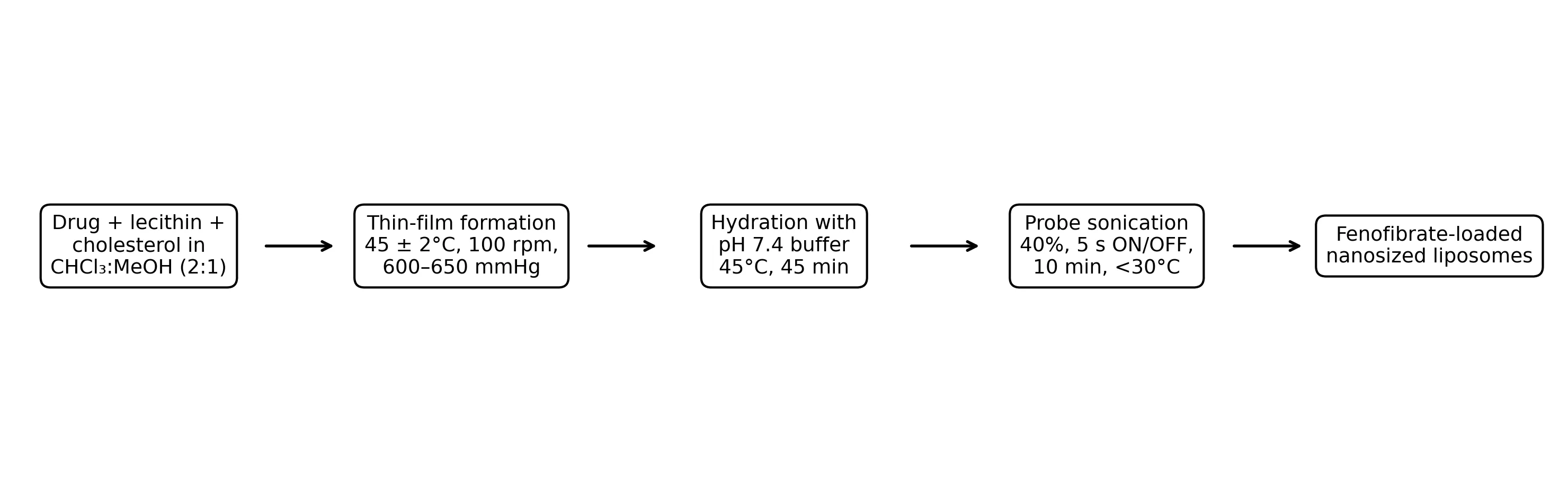
Figure 1. Schematic representation of the thin-film hydration and sonication procedure used to prepare fenofibrate-loaded liposomes.
2.4 Physicochemical characterization
Fresh dispersions were visually examined for colour, homogeneity, sedimentation, phase separation and aggregation. Particle size and PDI were determined by dynamic light scattering using a Malvern Zetasizer Nano ZS90 after dilution of approximately 100 μL dispersion with 10 mL filtered distilled water. Measurements were performed at 25 ± 1°C in triplicate. Zeta potential was determined in a folded capillary cell at 25°C using appropriately diluted samples, with three independent readings. Entrapment efficiency was determined by centrifuging 5 mL of liposomal dispersion at 15,000 rpm for 45 min at 4°C. Free fenofibrate in the supernatant was diluted with methanol and quantified at 290 nm. Entrapped drug was calculated by difference from the total drug added. Drug content was measured by disrupting an amount of dispersion equivalent to 10 mg fenofibrate with methanol, sonicating for 15 min, making the volume to 100 mL, filtering and measuring the appropriate dilution at 290 nm. Entrapment efficiency was expressed as: EE (%) = [(total drug − free drug)/total drug] × 100.
2.5 Surface morphology
The optimized formulation was examined by scanning electron microscopy (SEM). A small quantity of dispersion was placed on an aluminium stub, dried under vacuum, sputter-coated with gold and examined at suitable magnification to assess vesicle shape, surface characteristics and aggregation [17].
2.6 In vitro drug release
Drug release was evaluated by the dialysis-bag diffusion technique. Liposomal dispersion equivalent to 100 mg fenofibrate was placed in a pre-soaked dialysis membrane and suspended in 900 mL phosphate buffer (pH 6.8) containing 0.5% Tween 80 at 37 ± 0.5°C. The paddle speed was 50 rpm. At 0.5, 1, 2, 3, 4, 6, 8, 10, 12 and 24 h, 5-mL samples were withdrawn and replaced with equal volumes of fresh medium. Samples were filtered through a 0.45-μm membrane, diluted and analyzed at 290 nm. The optimized formulation was compared with pure fenofibrate under identical conditions.
2.7 Drug-release kinetic analysis
Release data for the optimized formulation were fitted to zero-order, first-order, Higuchi, Korsmeyer–Peppas and Hixson–Crowell models. Model fit was compared using the regression coefficient (R²), and the Korsmeyer–Peppas release exponent (n) was used to support interpretation of the release mechanism [18,19].
2.8 Accelerated stability study and statistical analysis
The optimized formulation was packed in airtight amber-coloured glass containers and stored at 40 ± 2°C/75 ± 5% relative humidity for three months in accordance with the stability protocol reported in the thesis. Samples were evaluated initially and after 1, 2 and 3 months for appearance, particle size, PDI, zeta potential, entrapment efficiency, drug content and 24-h release. The thesis reports one-way ANOVA for stability data, with p > 0.05 interpreted as a nonsignificant change.
3. RESULTS AND DISCUSSION
3.1 Preformulation findings
Fenofibrate was obtained as a white to off-white, odourless crystalline powder. Its mean melting point (80.07 ± 0.25°C) was within the reported range of 79–82°C, supporting acceptable identity and purity. The drug was practically insoluble in water and showed markedly greater solubility in organic solvents, consistent with its BCS Class II behaviour [1,10]. The strong solubility in chloroform and methanol supported use of the 2:1 organic solvent system during lipid-film formation. UV analysis showed λmax at 290 nm and excellent linearity from 2 to 20 μg/mL (R² = 0.9994), providing the analytical basis for drug-content, entrapment and release measurements.
3.2 Effect of lipid composition and selection of the optimized formulation
All formulations formed milky-white liposomal dispersions, but physical quality varied with lipid composition. F5 produced a smooth homogeneous dispersion without visible aggregation or sedimentation, whereas the highest-lipid formulation F8 showed slight aggregation. This pattern indicates that increasing lipid concentration was beneficial only to an optimum level; excessive phospholipid/cholesterol likely increased dispersion viscosity and favoured vesicle interaction or fusion.
Particle size decreased from 268.5 ± 4.2 nm for F1 to a minimum of 168.9 ± 2.1 nm for F5, then increased to 236.5 ± 3.9 nm for F8. The PDI showed a corresponding improvement, reaching 0.196 ± 0.01 for F5. A low PDI indicates a narrow and reproducible vesicle-size distribution, an important attribute for nanocarrier performance [17,20]. The negative zeta potentials ranged from −20.6 ± 1.1 to −36.5 ± 0.9 mV. F5 had the greatest magnitude (−36.5 ± 0.9 mV), providing electrostatic repulsion that is consistent with improved colloidal stability.
Entrapment efficiency increased from 68.42 ± 1.26% in F1 to 91.82 ± 0.82% in F5, then decreased with further increases in lipid/cholesterol. The initial improvement is consistent with greater phospholipid bilayer volume available for partitioning of lipophilic fenofibrate, whereas excessive cholesterol may reduce the space available for drug accommodation or increase bilayer rigidity. F5 also provided the highest drug content (99.14 ± 0.41%). Similar dependence of liposomal characteristics on lipid composition and processing variables has been reported for fenofibrate and other poorly soluble compounds [8,14,16].
Table 2. Physicochemical characteristics of fenofibrate-loaded liposomes (mean ± SD, n = 3 as reported in the thesis).
|
Formulation |
Particle size (nm) |
PDI |
Zeta potential (mV) |
EE (%) |
Drug content (%) |
|
F1 |
268.5 ± 4.2 |
0.382 ± 0.02 |
-20.6 ± 1.1 |
68.42 ± 1.26 |
91.54 ± 0.84 |
|
F2 |
254.8 ± 3.8 |
0.341 ± 0.03 |
-24.8 ± 1.3 |
73.68 ± 1.14 |
93.21 ± 0.72 |
|
F3 |
221.7 ± 2.6 |
0.284 ± 0.02 |
-28.4 ± 1.2 |
81.56 ± 1.03 |
95.83 ± 0.64 |
|
F4 |
196.4 ± 2.3 |
0.241 ± 0.01 |
-31.2 ± 1.0 |
86.48 ± 0.95 |
97.26 ± 0.56 |
|
F5 |
168.9 ± 2.1 |
0.196 ± 0.01 |
-36.5 ± 0.9 |
91.82 ± 0.82 |
99.14 ± 0.41 |
|
F6 |
181.3 ± 2.8 |
0.214 ± 0.02 |
-34.8 ± 1.2 |
89.16 ± 0.93 |
98.42 ± 0.52 |
|
F7 |
209.4 ± 3.2 |
0.268 ± 0.02 |
-30.6 ± 1.3 |
84.74 ± 1.16 |
96.83 ± 0.67 |
|
F8 |
236.5 ± 3.9 |
0.316 ± 0.03 |
-27.2 ± 1.5 |
80.68 ± 1.34 |
95.12 ± 0.73 |
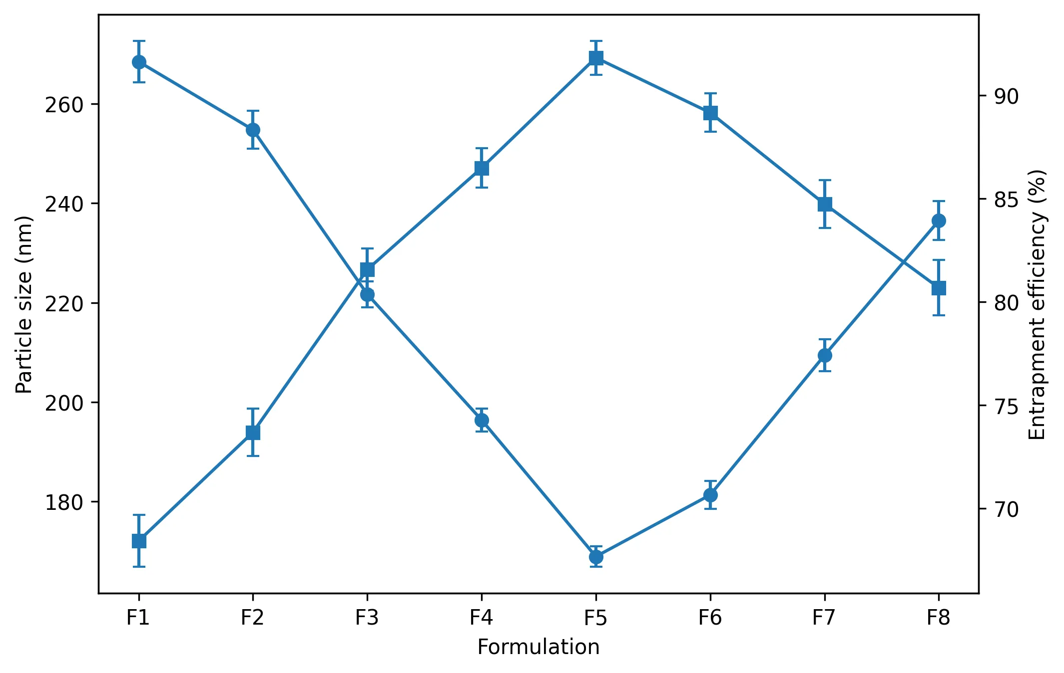
Figure 2. Effect of formulation composition on particle size and entrapment efficiency. F5 showed the minimum particle size and maximum entrapment efficiency.
3.3 Morphological characteristics
SEM examination of F5 confirmed formation of discrete, predominantly spherical vesicular structures with smooth surfaces and no obvious large aggregates. The morphological observations were consistent with the low PDI and relatively high magnitude of zeta potential. The DLS diameter of 168.9 nm represents the hydrodynamic diameter of hydrated particles, whereas SEM evaluates dried structures; therefore, exact equivalence between the two measurements is not expected.
3.4 In vitro drug-release performance
All eight formulations showed progressive drug release over 24 h, with the extent of release strongly dependent on lipid composition. At 24 h, release ranged from 82.64 ± 1.54% for F1 to 98.84 ± 1.21% for F5. Formulations F6–F8 released slightly less drug despite their higher lipid content, consistent with formation of a more rigid or diffusion-resistant bilayer and their larger vesicle sizes. Thus, the same composition that optimized particle size and entrapment also produced the most favourable dissolution profile. The superiority of liposomal incorporation was particularly evident when F5 was compared directly with pure fenofibrate. At 0.5 h, pure drug and F5 released 4.26 ± 0.22% and 12.85 ± 0.31%, respectively. The difference persisted throughout the study: at 12 h the values were 64.42 ± 1.26% and 95.74 ± 1.14%, and at 24 h they were 72.18 ± 1.48% and 98.84 ± 1.21%. The absolute improvement at 24 h was 26.66 percentage points. This enhanced dissolution can reasonably be related to the nanoscale vesicle size, increased effective interfacial area, improved drug dispersion and wetting by phospholipids. These observations are consistent with previous reports in which lipid-based nanoformulations improved fenofibrate dissolution and oral exposure [11–15,21].
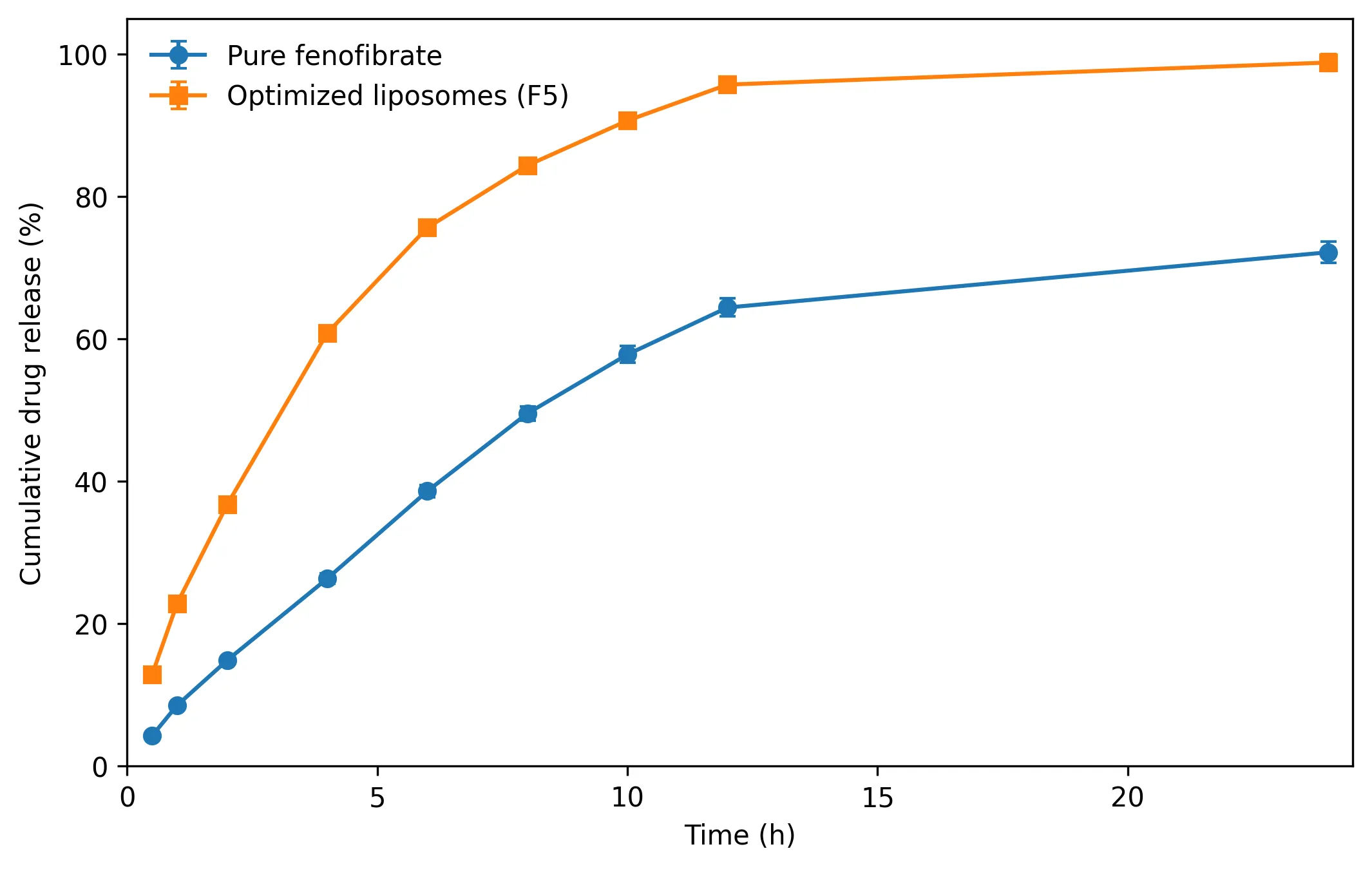
Figure 3. Comparative in vitro release of pure fenofibrate and optimized liposomal formulation F5 (mean ± SD).
3.5 Drug-release kinetics
Kinetic modelling provided further insight into release from F5. The Higuchi model produced the highest regression coefficient (R² = 0.9956), followed by the Korsmeyer–Peppas model (R² = 0.9918), first-order model (R² = 0.9841), Hixson–Crowell model (R² = 0.9738) and zero-order model (R² = 0.9624). The predominance of Higuchi behaviour supports diffusion through the phospholipid bilayer as the major rate-controlling process. The Korsmeyer–Peppas exponent of n = 0.61 was interpreted in the thesis as anomalous/non-Fickian transport, suggesting a contribution from both diffusion and structural relaxation or reorganization of the lipid membrane [18,19].
Table 3. Release-kinetic modelling of optimized formulation F5.
|
Model |
Regression equation / parameter |
R² |
Interpretation |
|
Zero order |
Y = 4.18X + 32.47 |
0.9624 |
Good fit; not dominant |
|
First order |
Y = −0.084X + 1.985 |
0.9841 |
Concentration-dependent component |
|
Higuchi |
Y = 23.84X + 3.72 |
0.9956 |
Best fit; diffusion-dominated |
|
Korsmeyer–Peppas |
n = 0.61 |
0.9918 |
Anomalous/non-Fickian transport |
|
Hixson–Crowell |
— |
0.9738 |
Secondary contribution |
3.6 Accelerated stability
F5 retained its principal quality attributes during three months of accelerated storage. Particle size increased modestly from 168.9 ± 2.1 to 178.2 ± 2.8 nm and PDI from 0.196 ± 0.01 to 0.218 ± 0.02. Zeta potential changed from −36.5 ± 0.9 to −34.6 ± 1.2 mV, remaining sufficiently negative to support electrostatic stabilization. Entrapment efficiency decreased from 91.82 ± 0.82% to 88.96 ± 0.88%, while drug content remained high at 97.86 ± 0.53% after three months. Twenty-four-hour release decreased only from 98.84 ± 1.21% to 96.94 ± 1.28%. The thesis reports that none of these changes was statistically significant by one-way ANOVA (p > 0.05). The modest increase in vesicle size and small reductions in surface charge and entrapment are compatible with limited vesicle interaction and gradual drug leakage during storage. Importantly, no phase separation or sedimentation was reported, and the formulation retained >96% 24-h release. These data indicate acceptable short-term physicochemical stability under the accelerated conditions examined, although longer-term stability and additional chemical degradation assays would be required before making definitive shelf-life claims.
Table 4. Accelerated stability summary for F5 after storage at 40 ± 2°C/75 ± 5% RH.
|
Parameter |
Initial |
3 months |
% change |
Thesis statistical result |
|
Particle size (nm) |
168.9 |
178.2 |
5.51 |
p > 0.05 |
|
PDI |
0.196 |
0.218 |
11.22 |
p > 0.05 |
|
Zeta potential (mV) |
−36.5 |
−34.6 |
5.20 |
p > 0.05 |
|
Entrapment efficiency (%) |
91.82 |
88.96 |
3.11 |
p > 0.05 |
|
Drug content (%) |
99.14 |
97.86 |
1.29 |
p > 0.05 |
|
24-h drug release (%) |
98.84 |
96.94 |
1.92 |
p > 0.05 |
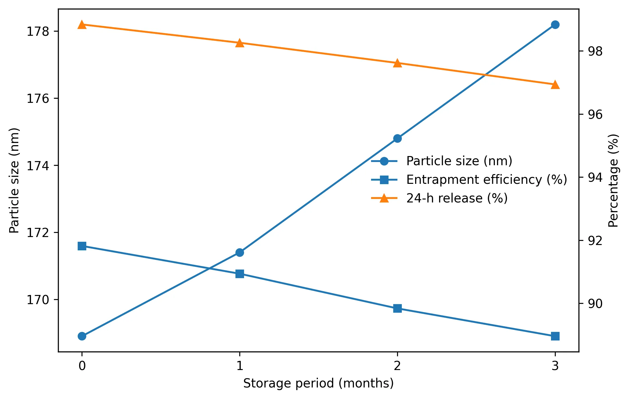
Figure 4. Changes in particle size, entrapment efficiency and 24-h release during three months of accelerated storage.
3.7 Overall interpretation and study limitations
Taken together, the formulation-screening data show a clear optimum at F5 rather than a simple linear benefit from increasing lipid content. The 300:30 mg soya lecithin-to-cholesterol composition generated the smallest vesicles, narrowest size distribution, greatest magnitude of zeta potential, highest drug entrapment and drug content, and the greatest cumulative release. This convergence of independent quality attributes strengthens the selection of F5 as the optimized formulation. The present work demonstrates enhanced in vitro dissolution and controlled release, but it does not itself establish improved oral bioavailability. Although previous studies have shown that lipid-based fenofibrate formulations can improve systemic exposure [12,13,21], confirmation for the present formulation requires pharmacokinetic and, where relevant, pharmacodynamic studies. Additional work should also address long-term stability, residual organic solvents, phospholipid oxidation, gastrointestinal robustness and scale-up before translational conclusions are drawn.
CONCLUSION
Fenofibrate-loaded liposomes were successfully developed by thin-film hydration followed by probe sonication. Systematic variation of soya lecithin and cholesterol demonstrated that lipid composition strongly influenced vesicle size, distribution, surface charge, drug entrapment and release. F5 (100 mg fenofibrate, 300 mg soya lecithin and 30 mg cholesterol) was identified as the optimized formulation, exhibiting a particle size of 168.9 ± 2.1 nm, PDI of 0.196 ± 0.01, zeta potential of −36.5 ± 0.9 mV, entrapment efficiency of 91.82 ± 0.82% and drug content of 99.14 ± 0.41%. F5 achieved 98.84 ± 1.21% cumulative release at 24 h compared with 72.18 ± 1.48% from pure fenofibrate. Release was best described by the Higuchi model (R² = 0.9956), with a Korsmeyer–Peppas exponent of 0.61 supporting anomalous transport. The formulation also retained acceptable physicochemical characteristics during three months of accelerated storage. Overall, liposomal incorporation substantially improved the in vitro dissolution behaviour of fenofibrate and produced a stable sustained-release vesicular system. Further in vivo pharmacokinetic evaluation is required to determine whether these in vitro advantages translate into improved oral bioavailability.
REFERENCES

Ekta Kushwaha*, Shivang Tripathi, Satish Kumar Sarankar, Development and Characterization of Fenofibrate-Loaded Liposomes for Enhanced Dissolution and Sustained Oral Drug Release, Int. J. Med. Pharm. Sci., 2026, 2 (9), 577-585. https://doi.org/10.5281/zenodo.22999510
 10.5281/zenodo.22999510