
We use cookies to ensure our website works properly and to personalise your experience. Cookies policy
Research Scholar, Desh Bhagat University, School of Pharmacy, Mandi Gobindgarh, Punjab
Liposomes are among the most extensively investigated nanocarriers for targeted drug delivery owing to their biocompatibility, biodegradability, ability to encapsulate both hydrophilic and lipophilic drugs, and potential for surface modification. Since the approval of liposomal formulations such as Doxil®, AmBisome®, and DaunoXome®, liposome-based drug delivery has transformed the therapeutic landscape of cancer, infectious diseases, and inflammatory disorders. Advances in nanotechnology have enabled the development of stealth liposomes, ligand-targeted liposomes, stimuli-responsive liposomes, and multifunctional theranostic liposomes. Despite significant progress, challenges including rapid clearance, limited drug loading, manufacturing complexity, scale-up issues, and regulatory concerns continue to restrict widespread clinical translation. This review discusses the structure and classification of liposomes, mechanisms of targeted delivery, formulation strategies, therapeutic applications, clinically approved products, current challenges, and future prospects of liposome-mediated drug delivery.
The successful treatment of many diseases depends not only on the therapeutic activity of a drug but also on its ability to reach the desired site of action at an effective concentration. Conventional drug delivery approaches, including oral and parenteral dosage forms, often face significant limitations such as poor bioavailability, rapid degradation, non-specific distribution, and frequent dosing requirements. These challenges can result in reduced therapeutic efficacy and increased systemic toxicity, particularly in the treatment of chronic diseases and cancer. Consequently, there has been growing interest in developing advanced drug delivery systems capable of improving drug stability, enhancing bioavailability, and delivering therapeutic agents more selectively to diseased tissues while minimizing adverse effects on healthy organs (Liu et al., 2022; Rommasi & Esfandiari, 2021). The emergence of nanotechnology has significantly transformed the field of pharmaceutical sciences by enabling the design of nanoscale carriers that can improve the pharmacokinetic and pharmacodynamic properties of drugs. Various nanocarrier systems, including polymeric nanoparticles, dendrimers, micelles, solid lipid nanoparticles, and liposomes, have been investigated for targeted and controlled drug delivery. Among these platforms, liposomes have attracted particular attention because of their biocompatibility, biodegradability, low toxicity, and ability to encapsulate both hydrophilic and lipophilic therapeutic agents. These characteristics make liposomes highly versatile carriers for a broad range of pharmaceutical and biomedical applications, including drug delivery, gene therapy, vaccine development, and diagnostic imaging (Wang et al., 2023; Gatto et al., 2024). Liposomes were first described by Alec D. Bangham and colleagues in 1965 during studies involving phospholipid behavior in aqueous environments. They observed that phospholipids spontaneously formed closed bilayer vesicles that closely resembled biological cell membranes, leading to the development of the liposome concept (Bangham et al., 1965). Since then, liposomes have evolved from simple membrane models into sophisticated nanocarriers with significant clinical relevance. Advances in formulation technologies have enabled the development of long-circulating liposomes, ligand-targeted liposomes, and stimuli-responsive systems designed to improve drug accumulation at specific disease sites. Such innovations have enhanced the therapeutic potential of liposomal formulations by improving drug retention, prolonging circulation time, and facilitating site-specific delivery (Samad et al., 2007; Fathi & Oyelere, 2016). Over the past three decades, liposomal drug delivery systems have achieved considerable success in clinical practice, particularly in oncology, infectious diseases, and inflammatory disorders. Several liposomal products, including Doxil®, AmBisome®, and Vyxeos®, have received regulatory approval and demonstrated improved therapeutic outcomes compared with conventional formulations. More recently, the growing demand for precision medicine has further accelerated research into multifunctional liposomes capable of combining targeting, imaging, and therapeutic functions within a single platform. Despite challenges related to stability, large-scale manufacturing, and regulatory approval, liposomes remain among the most extensively studied and clinically successful nanocarriers, continuing to play a central role in the advancement of modern nanomedicine and targeted drug delivery strategies (Liu et al., 2022; Gatto et al., 2024; Izadiyan et al., 2025).
2. Structure and Composition Of Liposomes
The remarkable success of liposomes as drug delivery vehicles is largely attributed to their unique structural organization and versatile composition. Liposomes are spherical vesicular systems composed primarily of phospholipids arranged in one or more concentric bilayers surrounding an aqueous interior. Their architecture closely resembles that of biological cell membranes, which contributes to their excellent biocompatibility and low toxicity. This biomimetic nature allows liposomes to interact effectively with biological systems while protecting encapsulated therapeutic agents from premature degradation. Furthermore, the structural flexibility of liposomes enables the incorporation of a wide variety of molecules, ranging from small-molecule drugs and proteins to nucleic acids and imaging agents, making them highly adaptable platforms for targeted drug delivery applications (Liu et al., 2022; Kim, 2016). The physicochemical properties of liposomes are strongly influenced by their lipid composition, size, surface charge, and membrane fluidity. These characteristics determine critical parameters such as drug loading capacity, circulation time, biodistribution, cellular uptake, and release behavior. By carefully modifying liposomal components, researchers can tailor formulations to meet specific therapeutic requirements, thereby enhancing drug efficacy and reducing unwanted side effects. Modern liposomal systems often incorporate cholesterol, polyethylene glycol (PEG), targeting ligands, and other functional molecules to improve stability, prolong systemic circulation, and facilitate selective delivery to diseased tissues (Gatto et al., 2024; Wang et al., 2023).
2.1 Basic Structure of Liposomes
Liposomes are composed of four major structural elements: a phospholipid bilayer, an aqueous core, cholesterol molecules, and surface-modifying agents. Each of these components plays a distinct role in determining the performance and therapeutic potential of the liposomal formulation.
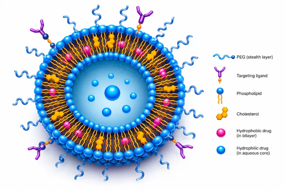
Figure 1: Generalized structure of a targeted PEGylated liposome showing the phospholipid bilayer, aqueous core, cholesterol molecules, encapsulated therapeutic agents, PEG coating, and targeting ligands designed to enhance circulation time and site-specific drug delivery.
Phospholipid Bilayer
The phospholipid bilayer forms the fundamental structural framework of liposomes. Phospholipids are amphiphilic molecules consisting of a hydrophilic (water-attracting) polar head and one or more hydrophobic (water-repelling) fatty acid tails. When exposed to an aqueous environment, these molecules spontaneously arrange themselves into bilayer structures with the hydrophobic tails facing inward and the hydrophilic heads facing outward toward the surrounding water. This self-assembly process results in the formation of a stable vesicular structure capable of encapsulating therapeutic agents (Samad et al., 2007). The lipid bilayer serves multiple functions. It acts as a protective barrier that shields encapsulated drugs from enzymatic degradation and environmental factors, while also controlling the rate of drug release. Lipophilic drugs are typically incorporated within the hydrophobic region of the bilayer, whereas hydrophilic drugs are entrapped in the aqueous core. The composition and fluidity of the bilayer significantly influence liposome stability, permeability, and interactions with biological membranes (Kim, 2016).
Aqueous Core
At the center of the liposome lies an aqueous compartment capable of entrapping water-soluble therapeutic molecules. The aqueous core serves as a reservoir for hydrophilic drugs, peptides, proteins, nucleic acids, and other biomacromolecules. Encapsulation within this compartment protects sensitive therapeutic agents from degradation and facilitates controlled release following administration. The size of the aqueous core varies depending on liposome type and preparation method, directly affecting drug-loading efficiency and therapeutic performance (Rommasi & Esfandiari, 2021). The ability to simultaneously accommodate hydrophilic compounds in the aqueous core and lipophilic compounds within the lipid bilayer represents one of the most significant advantages of liposomal drug delivery systems. This dual-loading capability distinguishes liposomes from many other nanocarrier platforms and broadens their applicability across diverse therapeutic areas.
Cholesterol
Cholesterol is frequently incorporated into liposomal formulations to enhance membrane stability and optimize physicochemical properties. Positioned between phospholipid molecules within the bilayer, cholesterol regulates membrane fluidity and reduces permeability. Its presence strengthens the lipid membrane, minimizing drug leakage and preventing premature release during storage and circulation (Liu et al., 2022). In addition to improving structural integrity, cholesterol increases resistance to mechanical stress and serum-induced destabilization. Appropriate cholesterol concentrations can significantly extend the shelf life of liposomal formulations and improve their in vivo performance. However, excessive cholesterol content may reduce membrane flexibility and negatively affect drug encapsulation efficiency; therefore, optimization of cholesterol concentration is a critical aspect of liposome formulation development (Gatto et al., 2024).
Surface Modifiers
Surface modification has emerged as a powerful strategy for enhancing the therapeutic performance of liposomal drug delivery systems. Various polymers, ligands, antibodies, peptides, and other functional molecules can be attached to the outer surface of liposomes to alter their biological behavior. These modifications are designed to improve circulation time, evade immune recognition, enhance cellular uptake, and facilitate targeted drug delivery (Fathi & Oyelere, 2016). Among surface modifiers, polyethylene glycol (PEG) is one of the most widely used. PEG forms a hydrophilic protective layer around the liposome, reducing protein adsorption and recognition by the reticuloendothelial system (RES). This modification produces so-called "stealth liposomes," which exhibit prolonged circulation times and improved accumulation at target sites. Additionally, targeting ligands such as antibodies, folic acid, transferrin, aptamers, and peptides can be attached to the liposome surface to promote receptor-mediated uptake by specific cells, thereby enhancing therapeutic selectivity and efficacy (Wang et al., 2023).
2.2 Major Components of Liposomal Formulations
The performance of liposomal drug delivery systems depends largely on the selection and optimization of their constituent components. Each ingredient contributes specific functional characteristics that influence stability, targeting ability, drug-loading efficiency, and pharmacokinetic behavior.
Table 1: Major Components of Liposomes and their Function
|
Component |
Primary Function |
|
Phosphatidylcholine |
Formation of phospholipid bilayer and vesicle structure |
|
Cholesterol |
Enhances membrane rigidity, stability, and drug retention |
|
Polyethylene Glycol (PEG) |
Prolongs circulation time by reducing RES clearance |
|
Targeting Ligands |
Enables receptor-mediated active targeting of specific tissues or cells |
|
Surfactants |
Improves membrane permeability and formulation stability |
|
Charged Lipids |
Modifies surface charge and cellular interactions |
|
Antioxidants |
Prevents lipid oxidation and enhances storage stability |
Phosphatidylcholine
Phosphatidylcholine is the most commonly used phospholipid in liposomal formulations due to its biocompatibility and structural similarity to natural cellular membranes. It provides the primary framework for bilayer formation and contributes significantly to liposome stability and drug encapsulation efficiency (Samad et al., 2007).
Polyethylene Glycol (PEG)
PEGylation has become a standard approach in advanced liposomal formulations. The hydrophilic PEG chains create a steric barrier around the liposome surface, reducing plasma protein adsorption and delaying immune system recognition. This modification prolongs systemic circulation and enhances passive accumulation in tumors through the enhanced permeability and retention (EPR) effect (Liu et al., 2022).
Targeting Ligands
Targeting ligands are incorporated to improve the specificity of drug delivery. These molecules recognize and bind to receptors that are overexpressed on diseased cells, facilitating receptor-mediated endocytosis and increased intracellular drug delivery. Common ligands include folic acid, transferrin, antibodies, peptides, and aptamers. Such targeted systems have shown considerable promise in cancer therapy and precision medicine applications (Fathi & Oyelere, 2016; Gatto et al., 2024).
Surfactants
Surfactants are occasionally incorporated into liposomal formulations to improve membrane flexibility, enhance drug permeation, and increase formulation stability. Depending on their concentration and chemical nature, surfactants can influence vesicle size, drug release profiles, and interactions with biological membranes. Their incorporation is particularly beneficial in specialized liposomal systems designed for transdermal, pulmonary, or mucosal drug delivery (Kim, 2016).
3. Classification of Liposomes
Liposomes are highly versatile vesicular carriers that can be tailored to meet specific therapeutic requirements through modifications in size, membrane structure, and surface composition. Their physicochemical characteristics significantly influence drug loading capacity, circulation behavior, biodistribution, cellular uptake, and therapeutic efficacy. Consequently, liposomes are commonly classified according to their size, lamellarity (number of lipid bilayers), and functional properties. Understanding these classifications is essential for selecting appropriate liposomal systems for targeted drug delivery applications and optimizing their performance in clinical settings (Liu et al., 2022; Kim, 2016).
3.1 Classification Based on Size
The size of liposomes plays a critical role in determining their biological behavior, including circulation time, tissue penetration, cellular uptake, and drug release kinetics. Depending on their diameter, liposomes are generally categorized into small unilamellar vesicles (SUVs), large unilamellar vesicles (LUVs), and giant unilamellar vesicles (GUVs).
Small Unilamellar Vesicles (SUVs)
Small unilamellar vesicles are single-bilayer liposomes with diameters typically ranging from 20 to 100 nm. Due to their small size, SUVs possess a high surface-area-to-volume ratio, which facilitates efficient interaction with biological membranes and enhances cellular uptake. These liposomes often exhibit prolonged circulation times and improved penetration into tissues, making them particularly useful for targeted drug delivery and anticancer applications. However, their relatively small aqueous core may limit the encapsulation efficiency of hydrophilic drugs (Samad et al., 2007; Wang et al., 2023).
Large Unilamellar Vesicles (LUVs)
Large unilamellar vesicles generally range from 100 to 1000 nm in diameter and consist of a single phospholipid bilayer surrounding a comparatively larger aqueous compartment. The increased internal volume allows greater encapsulation of water-soluble drugs, proteins, and nucleic acids. LUVs are frequently employed in controlled-release formulations and gene delivery applications due to their higher drug-loading capacity. Their size can also be optimized to balance circulation time and therapeutic payload delivery (Rommasi & Esfandiari, 2021).
Giant Unilamellar Vesicles (GUVs)
Giant unilamellar vesicles are typically larger than 1 µm in diameter and are mainly utilized in membrane biology studies and experimental research. Because their dimensions resemble those of living cells, GUVs serve as valuable model systems for investigating membrane dynamics, drug transport mechanisms, and lipid-protein interactions. Although they are less commonly used for systemic drug delivery, they provide important insights into liposome behavior and cellular interactions (Kim, 2016).
3.2 Classification Based on Lamellarity
Lamellarity refers to the number of phospholipid bilayers present within a liposomal vesicle. This structural characteristic influence drug encapsulation efficiency, membrane stability, release behavior, and biological performance.
Unilamellar Liposomes
Unilamellar liposomes contain a single phospholipid bilayer enclosing an aqueous core. Depending on their size, they may be classified as small, large, or giant unilamellar vesicles. These liposomes are widely used in pharmaceutical formulations because they exhibit predictable drug release patterns and efficient cellular uptake. Their relatively simple structure also facilitates formulation reproducibility and quality control (Liu et al., 2022).
Oligolamellar Liposomes
Oligolamellar liposomes consist of a few concentric phospholipid bilayers, usually ranging from two to five layers. They exhibit intermediate properties between unilamellar and multilamellar vesicles, offering enhanced stability while maintaining reasonable drug-loading capacity. Oligolamellar systems are often investigated for sustained drug release applications and specialized therapeutic formulations (Gatto et al., 2024).
Multilamellar Liposomes
Multilamellar vesicles (MLVs) are composed of multiple phospholipid bilayers arranged in a concentric "onion-like" structure. These liposomes generally possess larger diameters and greater membrane content than unilamellar systems. The multiple bilayers provide increased structural stability and enable the incorporation of substantial amounts of lipophilic drugs within the membrane layers. However, their larger size may reduce tissue penetration and cellular internalization compared with smaller liposomal systems (Samad et al., 2007).
3.3 Classification Based on Functionality
Advances in lipid engineering and surface modification technologies have led to the development of specialized liposomal systems designed to address specific therapeutic challenges. Functionalized liposomes exhibit enhanced targeting capabilities, improved pharmacokinetics, and controlled drug release characteristics.
Conventional Liposomes
Conventional liposomes are the earliest generation of liposomal drug delivery systems and are primarily composed of phospholipids and cholesterol. These liposomes effectively encapsulate therapeutic agents and improve drug solubility; however, they are rapidly recognized and removed from circulation by the reticuloendothelial system (RES). Consequently, their clinical utility may be limited by short circulation times and reduced accumulation at target sites (Kim, 2016).
Stealth Liposomes
Stealth liposomes, also known as PEGylated liposomes, are designed to evade immune recognition and prolong systemic circulation. Surface modification with polyethylene glycol (PEG) creates a hydrophilic protective layer around the liposome, reducing protein adsorption and uptake by macrophages. This "stealth" behavior enhances blood circulation time and increases the likelihood of passive accumulation at disease sites through the enhanced permeability and retention (EPR) effect. The success of formulations such as Doxil® has demonstrated the clinical value of PEGylated liposomes in cancer therapy (Liu et al., 2022; Wang et al., 2023).
Immunoliposomes
Immunoliposomes are antibody-conjugated liposomal systems developed for active targeting of specific cells or tissues. Antibodies or antibody fragments attached to the liposome surface recognize and bind selectively to target receptors expressed on diseased cells. This receptor-mediated interaction promotes enhanced cellular uptake and improved therapeutic selectivity. Immunoliposomes have shown considerable promise in cancer treatment, where they can deliver cytotoxic drugs directly to tumor cells while minimizing damage to healthy tissues (Fathi & Oyelere, 2016; Gatto et al., 2024).
Cationic Liposomes
Cationic liposomes contain positively charged lipids that facilitate electrostatic interactions with negatively charged nucleic acids such as DNA, siRNA, and mRNA. These systems have emerged as important non-viral vectors for gene delivery and genetic therapies. Their ability to condense nucleic acids and promote cellular internalization makes them attractive platforms for gene editing, RNA interference, and vaccine development. Nevertheless, challenges such as cytotoxicity and serum instability require careful formulation optimization (Rommasi & Esfandiari, 2021).
Stimuli-Responsive Liposomes
Stimuli-responsive liposomes represent an advanced generation of smart drug delivery systems capable of releasing their payload in response to specific internal or external triggers. These systems are designed to improve therapeutic precision by ensuring that drug release occurs preferentially at the target site.
pH-Sensitive Liposomes
These liposomes respond to acidic environments commonly found in tumors, inflamed tissues, and intracellular endosomes. Exposure to lower pH values destabilizes the lipid membrane, triggering drug release at the desired location (Wang et al., 2023).
Temperature-Sensitive Liposomes
Temperature-sensitive liposomes release their contents when exposed to elevated temperatures, typically between 40–42°C. They are often used in combination with hyperthermia-based cancer therapies to enhance localized drug delivery and treatment efficacy (Gatto et al., 2024).
Ultrasound-Sensitive Liposomes
Ultrasound-responsive liposomes utilize externally applied ultrasonic waves to induce membrane disruption and controlled drug release. This non-invasive approach enables spatial and temporal control of therapeutic delivery, improving treatment precision and reducing systemic exposure (Liu et al., 2022).
Redox-Sensitive Liposomes
Redox-sensitive liposomes exploit differences in oxidative and reductive conditions between healthy and diseased tissues. Elevated intracellular glutathione levels found in many cancer cells can trigger the breakdown of redox-sensitive linkages, resulting in selective drug release within target cells (Fathi & Oyelere, 2016).
Table 3. Classification of Liposomes
|
Classification Basis |
Type |
Characteristics |
|
Size |
SUVs |
20–100 nm, high cellular uptake |
|
LUVs |
100–1000 nm, larger drug-loading capacity |
|
|
GUVs |
>1 µm, membrane model systems |
|
|
Lamellarity |
Unilamellar |
Single lipid bilayer |
|
Oligolamellar |
Few concentric bilayers |
|
|
Multilamellar |
Multiple bilayers, enhanced stability |
|
|
Functionality |
Conventional |
Basic phospholipid vesicles |
|
Stealth |
PEGylated, prolonged circulation |
|
|
Immunoliposomes |
Antibody-mediated targeting |
|
|
Cationic |
Gene and nucleic acid delivery |
|
|
Stimuli-responsive |
Triggered drug release |
4. Methods of Liposome Preparation
The therapeutic performance of liposomal drug delivery systems is strongly influenced by the method used for their preparation. Parameters such as vesicle size, lamellarity, encapsulation efficiency, drug loading capacity, membrane stability, and release characteristics are largely determined during the manufacturing process. Over the years, numerous preparation techniques have been developed to produce liposomes with specific physicochemical properties tailored to different pharmaceutical applications. These methods can generally be categorized into conventional techniques and advanced manufacturing approaches. While conventional methods remain widely employed in laboratory-scale research due to their simplicity and cost-effectiveness, modern techniques have emerged to address challenges related to scalability, reproducibility, and industrial production (Liu et al., 2022; Kim, 2016).
4.1 Conventional Methods of Liposome Preparation
Conventional liposome preparation methods are among the most extensively used techniques in pharmaceutical research and development. These approaches are relatively simple, require minimal specialized equipment, and provide flexibility for encapsulating a wide range of therapeutic agents.
Thin-Film Hydration Method
The thin-film hydration method, also known as the Bangham method, is the most widely used and historically significant technique for liposome preparation. Developed from the pioneering work of Bangham and colleagues, this method involves dissolving phospholipids and cholesterol in an organic solvent such as chloroform or methanol. The solvent is subsequently removed under reduced pressure using a rotary evaporator, forming a thin lipid film on the walls of a round-bottom flask. Hydration of this dry lipid film with an aqueous solution leads to spontaneous swelling and the formation of multilamellar liposomes (Bangham et al., 1965; Samad et al., 2007). The method is highly versatile and can be applied to a broad range of lipid compositions and therapeutic agents. Following hydration, the liposomes may undergo sonication, extrusion, or homogenization to reduce particle size and improve uniformity.
Advantages
Limitations
Despite these limitations, the thin-film hydration method remains one of the most commonly employed techniques in liposomal formulation research because of its simplicity and reproducibility (Kim, 2016).
Reverse-Phase Evaporation Method
The reverse-phase evaporation method was developed to improve the encapsulation efficiency of hydrophilic compounds. In this technique, phospholipids are dissolved in an organic solvent and mixed with an aqueous phase containing the drug. Emulsification produces a water-in-oil emulsion, which is subsequently subjected to solvent evaporation under reduced pressure. As the organic solvent is removed, phospholipid molecules reorganize to form liposomes with large aqueous compartments (Liu et al., 2022). This technique is particularly useful for encapsulating proteins, peptides, nucleic acids, and other water-soluble therapeutic agents because it provides higher internal aqueous volume compared with conventional thin-film hydration methods.
Advantages
Limitations
Because of its superior loading capacity, reverse-phase evaporation remains an important technique for preparing liposomes intended for biological and biopharmaceutical applications (Rommasi & Esfandiari, 2021).
Solvent Injection Methods
Solvent injection methods involve the rapid mixing of lipid solutions with an aqueous phase, leading to spontaneous liposome formation. These methods are considered relatively simple and scalable compared with other conventional techniques.
Ethanol Injection Method
In the ethanol injection technique, phospholipids are dissolved in ethanol and rapidly injected into an aqueous medium under continuous stirring. Upon contact with water, the ethanol becomes diluted, causing the phospholipids to self-assemble into liposomal vesicles. The remaining ethanol is subsequently removed through evaporation or dialysis (Samad et al., 2007).
Advantages
Limitations
Ether Injection Method
In this method, lipids are dissolved in diethyl ether and slowly injected into a heated aqueous phase. As the ether evaporates, phospholipids organize into liposomal vesicles. The evaporation process promotes the formation of relatively uniform liposomes with controlled particle sizes (Kim, 2016).
Advantages
Limitations
4.2 Advanced Liposome Preparation Techniques
Although conventional methods are valuable for laboratory research, they often face challenges related to scalability, reproducibility, and batch-to-batch variability. To overcome these limitations, advanced manufacturing technologies have been developed to improve process control and facilitate industrial production.
Microfluidics-Based Preparation
Microfluidic technology has emerged as one of the most promising approaches for the preparation of liposomal nanocarriers. This method utilizes microscale channels that allow precise control over fluid mixing, enabling highly reproducible liposome formation. Lipid and aqueous phases are introduced into microchannels where rapid and controlled mixing results in the spontaneous assembly of liposomes with narrow size distributions (Gatto et al., 2024). Compared with conventional methods, microfluidics provides superior control over particle size, encapsulation efficiency, and formulation reproducibility. It is particularly advantageous for the development of liposomes intended for personalized medicine and nucleic acid delivery systems.
Advantages
Limitations
Supercritical Fluid Technology
Supercritical fluid technology utilizes supercritical carbon dioxide (SC-CO₂) as a solvent or anti-solvent for liposome production. Under supercritical conditions, carbon dioxide exhibits properties of both liquids and gases, enabling efficient lipid processing while reducing the use of toxic organic solvents (Liu et al., 2022). This environmentally friendly approach offers excellent control over particle formation and has attracted increasing attention for large-scale pharmaceutical manufacturing.
Advantages
Limitations
Continuous Manufacturing Platforms
Traditional batch manufacturing processes often suffer from variability and limited scalability. Continuous manufacturing platforms have emerged as a modern alternative that allows uninterrupted production of liposomal formulations under tightly controlled conditions. Continuous systems integrate lipid mixing, vesicle formation, size reduction, purification, and quality monitoring into a single automated process (Ilić-Stojanović et al., 2024). The pharmaceutical industry increasingly recognizes continuous manufacturing as a key strategy for improving efficiency, reducing production costs, and ensuring consistent product quality.
Advantages
Limitations
Quality-by-Design (QbD) Approaches
Quality-by-Design (QbD) is not a specific preparation method but a systematic development strategy increasingly applied to liposomal manufacturing. QbD focuses on understanding the relationship between formulation variables, process parameters, and critical quality attributes to ensure consistent product performance (Liu et al., 2022). Using statistical design of experiments (DoE), risk assessment, and process analytical technologies, QbD enables manufacturers to identify optimal operating conditions and achieve robust, reproducible liposomal formulations.
Advantages
Limitations
Table 4. Comparison of Major Liposome Preparation Methods
|
Method |
Principle |
Advantages |
Limitations |
|
Thin-Film Hydration |
Lipid film hydration |
Simple, widely used |
Broad size distribution |
|
Reverse-Phase Evaporation |
Emulsion solvent removal |
High encapsulation efficiency |
Organic solvent exposure |
|
Ethanol Injection |
Solvent dilution |
Rapid and simple |
Residual solvent concerns |
|
Ether Injection |
Solvent evaporation |
Uniform vesicles |
Use of volatile solvents |
|
Microfluidics |
Controlled micro-mixing |
Precise size control |
Specialized equipment |
|
Supercritical Fluid |
SC-CO₂ processing |
Solvent-free production |
High equipment cost |
|
Continuous Manufacturing |
Automated continuous production |
Scalable and reproducible |
Complex implementation |
|
Quality-by-Design |
Systematic process optimization |
Enhanced quality assurance |
Data-intensive approach |
5. Drug Loading Strategies
The therapeutic success of liposomal drug delivery systems depends not only on the composition and structural characteristics of liposomes but also on the efficiency with which therapeutic agents are incorporated into the vesicles. Drug loading is a critical step in liposome formulation because it directly influences encapsulation efficiency, drug retention, release kinetics, stability, and overall therapeutic performance. An ideal loading strategy should maximize the amount of drug incorporated into the liposome while minimizing drug leakage during storage and circulation. Depending on the physicochemical properties of the drug and the intended application, therapeutic agents can be incorporated into liposomes using either passive loading or active (remote) loading approaches (Liu et al., 2022; Gatto et al., 2024). Over the past several decades, significant advances in drug-loading technologies have improved the clinical effectiveness of liposomal formulations. While passive loading remains a simple and widely used method for many compounds, active loading techniques have revolutionized liposomal drug delivery by achieving exceptionally high encapsulation efficiencies and enhanced drug retention. Several clinically approved liposomal products currently employ active loading strategies to optimize therapeutic outcomes and improve formulation stability (Rommasi & Esfandiari, 2021).
5.1 Passive Loading
Passive loading is the simplest and most traditional method of incorporating drugs into liposomes. In this approach, the therapeutic agent is introduced during the liposome preparation process and becomes entrapped as the vesicles spontaneously form. Drug encapsulation occurs simultaneously with lipid self-assembly, making passive loading a straightforward and cost-effective technique for laboratory-scale and commercial applications (Samad et al., 2007). During liposome formation, hydrophilic drugs dissolve in the aqueous phase and become encapsulated within the internal aqueous core, whereas lipophilic drugs partition into the phospholipid bilayer because of their affinity for the hydrophobic membrane environment. The amount of drug encapsulated depends largely on the drug's solubility, liposome size, internal aqueous volume, lipid composition, and preparation method (Kim, 2016). Passive loading is particularly suitable for hydrophilic molecules such as small drug compounds, peptides, proteins, and certain biological agents. However, the efficiency of drug encapsulation is often limited because only a fraction of the available aqueous phase becomes enclosed within the liposome during vesicle formation. Consequently, many passively loaded formulations exhibit relatively low encapsulation efficiencies and may experience premature drug leakage during storage or circulation (Liu et al., 2022).
Advantages of Passive Loading
Limitations of Passive Loading
Despite these limitations, passive loading remains widely used in pharmaceutical research because of its simplicity and versatility, particularly during formulation screening and early-stage development (Wang et al., 2023).
5.2 Active (Remote) Loading
Active loading, also known as remote loading, is a more advanced strategy that allows drugs to be incorporated into preformed liposomes after vesicle formation. Unlike passive loading, where drug entrapment occurs during liposome assembly, active loading utilizes transmembrane chemical gradients to drive therapeutic molecules into the liposomal interior. This approach has significantly improved the efficiency and stability of liposomal drug formulations and is widely regarded as one of the most important innovations in liposome technology (Liu et al., 2022). The fundamental principle of remote loading involves creating a concentration gradient across the liposomal membrane. Drug molecules diffuse across the lipid bilayer in an uncharged form and subsequently become trapped within the liposome through ionization, precipitation, or complex formation. This mechanism enables the accumulation of large quantities of drug inside the vesicle while minimizing drug leakage. Active loading is particularly effective for weakly basic drugs such as doxorubicin, daunorubicin, vincristine, and irinotecan, which can readily cross lipid membranes in their neutral form before becoming trapped within the liposome interior (Barenholz, 2012).
pH Gradient Loading
One of the most widely employed active loading techniques utilizes a transmembrane pH gradient. In this method, liposomes are prepared with an acidic internal environment and a neutral external medium. Weakly basic drugs diffuse across the lipid membrane into the acidic interior, where they become protonated and unable to diffuse back across the membrane. As a result, drug molecules accumulate within the liposome at concentrations substantially higher than those achievable through passive loading (Fathi & Oyelere, 2016). This strategy offers several advantages, including enhanced drug retention, controlled release behavior, and improved formulation stability. pH-gradient loading has been extensively applied in the development of anticancer liposomal formulations because many chemotherapeutic agents possess weakly basic properties suitable for this mechanism.
Ammonium Sulfate Gradient Loading
The ammonium sulfate gradient method represents one of the most successful active loading techniques and is widely used in commercially approved liposomal products. In this approach, liposomes are initially prepared with a high internal concentration of ammonium sulfate, while the external solution contains a lower concentration. Following establishment of the gradient, weakly basic drug molecules diffuse into the liposome and form insoluble complexes or precipitates within the aqueous core. The resulting drug precipitation creates a powerful trapping mechanism that enables exceptionally high encapsulation efficiencies and prolonged drug retention. Because the drug remains stably retained within the liposome until release at the target site, this approach significantly enhances therapeutic performance and reduces premature drug leakage (Barenholz, 2012; Liu et al., 2022).
Advantages of Active (Remote) Loading
Compared with passive loading, active loading provides several important advantages:
High Encapsulation Efficiency
Remote loading can achieve encapsulation efficiencies exceeding 90% for many therapeutic agents. This significantly improves drug utilization and reduces manufacturing waste.
Improved Drug Retention
Drugs loaded through transmembrane gradient techniques are more effectively retained within liposomes, reducing premature leakage during storage and systemic circulation.
Enhanced Stability
Active loading often produces highly stable drug-liposome complexes that maintain therapeutic payloads for extended periods.
Higher Drug-to-Lipid Ratios
Greater quantities of drug can be incorporated into liposomes without compromising vesicle integrity.
Improved Clinical Performance
Enhanced retention and controlled release contribute to superior pharmacokinetic profiles and therapeutic outcomes.
Clinical Example: Doxil®
One of the most successful examples of active loading is Doxil®, the first FDA-approved nanomedicine and pegylated liposomal formulation of doxorubicin. Doxil® utilizes an ammonium sulfate gradient to achieve highly efficient loading of doxorubicin into PEGylated liposomes. The drug forms stable complexes within the liposomal core, resulting in exceptional encapsulation efficiency and prolonged retention during circulation (Barenholz, 2012). The incorporation of PEG on the liposome surface further extends circulation time by reducing recognition and clearance by the reticuloendothelial system. Compared with conventional doxorubicin formulations, Doxil® demonstrates reduced cardiotoxicity, improved tumor accumulation, and enhanced therapeutic efficacy. Its clinical success has established active loading as a benchmark strategy for the development of modern liposomal drug delivery systems (Liu et al., 2022; Wang et al., 2023).
Table 5. Comparison of Drug Loading Strategies
|
Parameter |
Passive Loading |
Active (Remote) Loading |
|
Loading Stage |
During liposome formation |
After liposome formation |
|
Driving Force |
Physical entrapment |
Chemical gradient |
|
Encapsulation Efficiency |
Low to moderate |
High to very high |
|
Drug Retention |
Moderate |
Excellent |
|
Formulation Complexity |
Simple |
More complex |
|
Suitable Drugs |
Hydrophilic and lipophilic compounds |
Weak acids and weak bases |
|
Commercial Use |
Limited |
Widely used in approved products |
|
Example |
Basic liposomal formulations |
Doxil®, Myocet®, Marqibo® |
6. Mechanisms Of Targeted Drug Delivery
The ability of liposomes to selectively deliver therapeutic agents to diseased tissues is one of the primary reasons for their success as nanocarriers. Targeted drug delivery aims to increase drug accumulation at the desired site while minimizing exposure to healthy tissues, thereby improving therapeutic efficacy and reducing adverse effects. In conventional drug administration, a large proportion of the administered dose is distributed throughout the body, often leading to systemic toxicity and suboptimal treatment outcomes. Liposomal formulations address this challenge by exploiting both biological and molecular targeting mechanisms to enhance site-specific drug delivery. Generally, liposome-mediated targeting can be classified into two major approaches: passive targeting and active targeting. While passive targeting relies on the unique physiological characteristics of diseased tissues, active targeting utilizes specific molecular interactions between ligands on the liposome surface and receptors expressed on target cells (Gatto et al., 2024; Liu et al., 2022). The effectiveness of targeted drug delivery depends on several factors, including liposome size, surface charge, circulation time, vascular permeability, receptor expression levels, and intracellular trafficking pathways. Advances in nanotechnology and surface engineering have enabled the development of sophisticated liposomal systems capable of achieving improved tissue selectivity and controlled intracellular drug release. These innovations have significantly expanded the application of liposomes in cancer therapy, inflammatory diseases, gene delivery, and precision medicine (Wang et al., 2023).
6.1 Passive Targeting
Passive targeting is the simplest and most widely utilized mechanism of liposome-mediated drug delivery. Unlike active targeting strategies, passive targeting does not require the attachment of specific ligands or recognition molecules to the liposome surface. Instead, it exploits the abnormal physiological characteristics of diseased tissues, particularly tumors and sites of inflammation, to facilitate the preferential accumulation of liposomal carriers (Rommasi & Esfandiari, 2021). The success of passive targeting largely depends on the prolonged circulation of liposomes in the bloodstream and their ability to extravasate through abnormal vascular structures present in pathological tissues. Long-circulating liposomes, especially PEGylated formulations, remain in systemic circulation for extended periods, increasing the probability of reaching target sites and accumulating within diseased tissues (Liu et al., 2022).
Enhanced Permeability and Retention (EPR) Effect
The most important mechanism underlying passive targeting is the Enhanced Permeability and Retention (EPR) effect, first described by Matsumura and Maeda in 1986. Rapidly growing tumors require extensive vascularization to sustain their metabolic demands. However, the newly formed blood vessels are often structurally abnormal, characterized by irregular architecture, defective endothelial cell junctions, and increased permeability. These abnormalities create gaps within the vascular walls that allow nanosized carriers, including liposomes, to penetrate and accumulate within tumor tissues more readily than in normal tissues (Matsumura & Maeda, 1986). In addition to increased vascular permeability, tumors typically exhibit impaired lymphatic drainage. As a result, liposomes that enter the tumor microenvironment tend to remain trapped for prolonged periods, leading to enhanced retention of the therapeutic agent. This combination of increased permeability and reduced clearance forms the basis of the EPR effect and contributes significantly to the accumulation of liposomal formulations in solid tumors (Wang et al., 2023).
Role of Tumor Vasculature
Tumor blood vessels differ markedly from healthy vasculature. They are often tortuous, dilated, and poorly organized, resulting in increased vascular leakiness. The pore sizes within tumor vessels may range from 100 to 800 nm, depending on tumor type and stage. Such pores allow appropriately sized liposomes to extravasate from the bloodstream into the tumor interstitium, where they can gradually release their therapeutic payload (Gatto et al., 2024).
Leaky Blood Vessels and Liposome Accumulation
The leaky nature of tumor vasculature creates an opportunity for nanocarriers to selectively accumulate within cancerous tissues. Liposomes with diameters ranging from 50 to 200 nm are generally considered optimal for exploiting the EPR effect because they are small enough to penetrate abnormal vasculature while remaining large enough to avoid rapid renal clearance. PEGylated liposomes are particularly effective in this regard because their prolonged circulation increases the likelihood of reaching and accumulating in tumor tissues (Fathi & Oyelere, 2016).
Advantages of Passive Targeting
Passive targeting offers several practical advantages:
The clinical success of formulations such as Doxil® demonstrates the potential of passive targeting to improve therapeutic outcomes while reducing systemic toxicity (Barenholz, 2012).
Limitations of Passive Targeting
Despite its advantages, passive targeting has important limitations.
Because of these limitations, researchers increasingly combine passive and active targeting strategies to improve treatment efficacy (Danhier, 2016).
6.2 Active Targeting
Active targeting represents a more sophisticated approach to liposome-mediated drug delivery. In this strategy, specific recognition molecules, known as ligands, are attached to the liposome surface to facilitate selective binding to target cells. These ligands recognize receptors that are overexpressed on diseased tissues but minimally expressed on normal cells, thereby enhancing therapeutic specificity and cellular uptake (Fathi & Oyelere, 2016).
Unlike passive targeting, which primarily improves tissue-level accumulation, active targeting promotes direct interaction between liposomes and target cells. Following receptor recognition, the liposome is internalized through receptor-mediated processes, allowing intracellular delivery of therapeutic agents and potentially improving treatment outcomes (Gatto et al., 2024).
Ligands Used in Active Targeting
A variety of ligands have been investigated for active targeting applications. The choice of ligand depends on the target receptor, disease type, and therapeutic objective.
Table 6: Ligands used in Active Targeting
|
Ligand |
Target Receptor/Marker |
Major Applications |
|
Folate |
Folate receptor |
Ovarian, breast, lung, and colorectal cancers |
|
Transferrin |
Transferrin receptor |
Brain tumors and rapidly proliferating cancers |
|
Antibodies |
HER2, EGFR, CD44 |
Targeted cancer therapy |
|
Aptamers |
Cancer-specific biomarkers |
Precision oncology |
|
Peptides |
Integrins (αvβ3, αvβ5) |
Tumor angiogenesis and metastasis |
Folate-Based Targeting
Folate receptors are overexpressed in many types of cancer, including ovarian, breast, lung, and colorectal tumors. Liposomes conjugated with folic acid can selectively bind to these receptors, enhancing drug uptake by malignant cells while minimizing effects on healthy tissues. Because folic acid is inexpensive, stable, and non-immunogenic, it remains one of the most extensively studied targeting ligands (Wang et al., 2023).
Transferrin-Based Targeting
Transferrin receptors are frequently overexpressed in rapidly dividing cells due to their increased iron requirements. Transferrin-conjugated liposomes exploit this characteristic to enhance drug delivery to tumor tissues and, in some cases, facilitate transport across biological barriers such as the blood-brain barrier (Gatto et al., 2024).
Antibody-Mediated Targeting
Monoclonal antibodies provide highly specific recognition of disease-associated receptors such as HER2 and epidermal growth factor receptor (EGFR). Liposomes modified with antibodies, commonly referred to as immunoliposomes, can selectively deliver therapeutic agents to receptor-positive cancer cells. This strategy has shown considerable promise in breast cancer, lung cancer, and other receptor-driven malignancies (Fathi & Oyelere, 2016).
Aptamer-Based Targeting
Aptamers are short nucleic acid sequences capable of binding specific molecular targets with high affinity and specificity. Compared with antibodies, aptamers are smaller, less immunogenic, and easier to synthesize. Aptamer-functionalized liposomes have emerged as promising tools for precision medicine and targeted cancer therapy (Liu et al., 2022).
Peptide-Based Targeting
Targeting peptides recognize cell-surface molecules such as integrins that are involved in tumor growth, angiogenesis, and metastasis. Peptide-functionalized liposomes can selectively accumulate in tumor tissues and enhance intracellular drug delivery, making them attractive candidates for cancer treatment (Wang et al., 2023).
Cellular Uptake Mechanisms
Following successful binding to target receptors, liposomes must enter the cell to release their therapeutic payload. Cellular uptake is a critical step that determines the effectiveness of active targeting systems.
Endocytosis
Endocytosis is a natural cellular process through which cells internalize extracellular materials. Liposomes that come into contact with the cell membrane may be engulfed and transported into intracellular compartments known as endosomes. Once internalized, the liposome can release its payload into the cytoplasm or other intracellular targets (Kim, 2016).
Receptor-Mediated Internalization
Receptor-mediated endocytosis is the primary mechanism responsible for the enhanced efficiency of actively targeted liposomes. When a ligand on the liposome surface binds to its corresponding receptor, a signaling cascade triggers membrane invagination and vesicle formation. This process results in selective internalization of the liposome by target cells, significantly increasing intracellular drug concentrations and therapeutic effectiveness (Fathi & Oyelere, 2016). Compared with passive targeting alone, receptor-mediated internalization provides greater cellular specificity and improved drug delivery efficiency, particularly for therapies requiring intracellular access such as gene delivery, RNA therapeutics, and anticancer agents.
Table 7. Comparison of Passive and Active Targeting Mechanisms
|
Parameter |
Passive Targeting |
Active Targeting |
|
Mechanism |
Exploits EPR effect |
Ligand-receptor interaction |
|
Surface Modification |
Not required |
Required |
|
Specificity |
Tissue-level |
Cellular-level |
|
Complexity |
Low |
High |
|
Manufacturing Cost |
Lower |
Higher |
|
Cellular Uptake |
Limited |
Enhanced |
|
Clinical Examples |
Doxil®, AmBisome® |
Immunoliposomes, folate-targeted liposomes |
7. Therapeutic Applications Of Liposomes
The unique structural characteristics, biocompatibility, and versatility of liposomes have led to their widespread application across numerous therapeutic fields. Their ability to encapsulate both hydrophilic and hydrophobic compounds, protect drugs from premature degradation, prolong circulation time, and facilitate targeted delivery has made them one of the most successful nanocarrier systems in modern medicine. Over the past few decades, liposome-based formulations have progressed from experimental drug carriers to clinically approved therapeutics used in oncology, infectious diseases, vaccine delivery, and emerging gene therapies. Continuous advances in liposomal engineering have further expanded their role in precision medicine by enabling controlled drug release, targeted delivery, and combination therapies (Liu et al., 2022; Gatto et al., 2024). Among the various biomedical applications, cancer treatment remains the most extensively studied and clinically successful area for liposomal drug delivery. However, the utility of liposomes extends far beyond oncology, encompassing infectious disease management, nucleic acid delivery, vaccine development, and treatment of neurological disorders. These diverse applications demonstrate the remarkable adaptability of liposomes as multifunctional therapeutic platforms (Wang et al., 2023).
7.1 Liposomes in Cancer Therapy
Cancer therapy represents the most successful and commercially significant application of liposomal drug delivery systems. Conventional chemotherapeutic agents often suffer from poor selectivity, resulting in severe systemic toxicity and damage to healthy tissues. Liposomes help overcome these limitations by improving drug pharmacokinetics, enhancing tumor accumulation, and reducing exposure of normal organs to cytotoxic agents. Their nanoscale size enables preferential accumulation within tumors through the Enhanced Permeability and Retention (EPR) effect, while surface modifications can further improve targeting specificity (Rommasi & Esfandiari, 2021). Several liposomal anticancer formulations have been approved for clinical use and have demonstrated significant therapeutic advantages compared with conventional drug formulations.
Doxorubicin Liposomes
Pegylated liposomal doxorubicin (Doxil®/Caelyx®) is one of the most successful examples of nanomedicine in clinical practice. Encapsulation of doxorubicin within PEGylated liposomes prolongs circulation time and enhances drug accumulation in tumor tissues. Importantly, the liposomal formulation significantly reduces cardiotoxicity, one of the major dose-limiting adverse effects associated with conventional doxorubicin therapy (Barenholz, 2012). Clinical studies have demonstrated improved safety profiles and comparable or superior therapeutic outcomes in patients with ovarian cancer, breast cancer, Kaposi's sarcoma, and multiple myeloma.
Daunorubicin Liposomes
DaunoXome® is a liposomal formulation of daunorubicin developed to improve the therapeutic index of this anthracycline anticancer agent. Liposomal encapsulation reduces systemic toxicity while enhancing drug delivery to malignant tissues. The formulation has been used successfully in the treatment of Kaposi's sarcoma and certain hematological malignancies (Liu et al., 2022).
Paclitaxel Liposomes
Paclitaxel is a potent anticancer drug with poor water solubility and significant formulation challenges. Liposomal encapsulation improves drug solubility, enhances tumor targeting, and reduces the hypersensitivity reactions commonly associated with conventional solvent-based formulations. Liposomal paclitaxel systems have shown promising results in breast, ovarian, and lung cancers (Wang et al., 2023).
Benefits of Liposomes in Cancer Therapy
The growing use of liposomal formulations in oncology can be attributed to several important advantages:
These benefits have established liposomes as a cornerstone of contemporary cancer nanomedicine (Gatto et al., 2024).
7.2 Liposomes in Infectious Disease Treatment
In addition to cancer therapy, liposomes have proven highly effective in the treatment of infectious diseases. Encapsulation of antimicrobial agents within liposomes can improve drug stability, enhance tissue penetration, reduce toxicity, and facilitate targeted delivery to infected tissues or intracellular pathogens.
AmBisome®
One of the most notable examples is AmBisome®, a liposomal formulation of amphotericin B. Conventional amphotericin B is highly effective against fungal infections but is associated with significant nephrotoxicity. Liposomal encapsulation dramatically improves its safety profile while maintaining antifungal efficacy (Liu et al., 2022).
Treatment of Fungal Infections
AmBisome® has become a standard treatment option for severe systemic fungal infections, including:
The liposomal formulation allows higher therapeutic doses to be administered with substantially reduced renal toxicity.
Treatment of Leishmaniasis
Liposomal amphotericin B is also widely used in the treatment of visceral leishmaniasis, a life-threatening parasitic disease caused by Leishmania species. The liposomal formulation preferentially accumulates within macrophages, which serve as host cells for the parasite, thereby enhancing therapeutic effectiveness while minimizing adverse effects (Wang et al., 2023).
The success of AmBisome® highlights the potential of liposomal technology in infectious disease management and has encouraged the development of liposomal formulations for antibacterial, antiviral, and antiparasitic therapies.
7.3 Liposomes in Gene Delivery
The emergence of nucleic acid-based therapeutics has significantly expanded the role of liposomes in modern medicine. Gene therapy requires efficient delivery systems capable of protecting fragile genetic material from enzymatic degradation while facilitating cellular uptake and intracellular release. Liposomes, particularly cationic and ionizable lipid-based systems, have become important non-viral vectors for nucleic acid delivery due to their safety and versatility (Cullis & Hope, 2017).
siRNA Delivery
Small interfering RNA (siRNA) therapies rely on intracellular delivery to silence disease-causing genes through RNA interference mechanisms. Liposomal carriers protect siRNA molecules from degradation and improve their uptake into target cells. Several experimental and approved therapies utilize lipid-based delivery platforms to enhance gene silencing efficiency (Kulkarni et al., 2019).
mRNA Delivery
Messenger RNA (mRNA) therapeutics have emerged as a transformative technology in recent years. Lipid-based nanocarriers protect mRNA from degradation and facilitate cellular entry, allowing transient expression of therapeutic proteins. The remarkable success of mRNA vaccines during the COVID-19 pandemic demonstrated the critical importance of lipid-based delivery systems in enabling large-scale nucleic acid therapeutics (Hou et al., 2021).
CRISPR Gene Editing Systems
Liposomes are increasingly being investigated as delivery vehicles for CRISPR-Cas gene editing components. Efficient intracellular transport of guide RNA and Cas proteins remains one of the major challenges in gene editing applications. Liposomal carriers offer a promising non-viral alternative capable of improving delivery efficiency while reducing immunogenicity and safety concerns associated with viral vectors (Liu et al., 2022).
7.4 Liposomes in Vaccine Delivery
Vaccination represents another rapidly growing application of lipid-based delivery systems. Liposomes can function both as delivery vehicles and as adjuvants that enhance immune responses. Their ability to protect antigens from degradation and facilitate uptake by antigen-presenting cells makes them highly effective vaccine platforms (Gatto et al., 2024). The global COVID-19 pandemic brought unprecedented attention to lipid-based delivery technologies. The successful development and deployment of mRNA vaccines demonstrated how lipid nanoparticles and liposomal systems can effectively transport fragile nucleic acids into cells, enabling rapid protein expression and immune activation. This achievement has accelerated research into liposome-based vaccines for infectious diseases, cancer immunotherapy, and personalized medicine (Hou et al., 2021).
Current investigations focus on developing liposomal vaccines against:
The versatility of liposomal vaccine platforms positions them as key contributors to next-generation immunization strategies.
7.5 Liposomes for Brain Targeting
The treatment of neurological disorders remains one of the greatest challenges in drug delivery because of the presence of the blood-brain barrier (BBB). This highly selective physiological barrier protects the central nervous system by restricting the entry of potentially harmful substances; however, it also limits the delivery of many therapeutic agents. As a result, effective treatment of brain tumors, Alzheimer's disease, Parkinson's disease, epilepsy, and other neurological disorders often remains difficult (Saraiva et al., 2016). Liposomes have emerged as promising carriers for overcoming BBB-related limitations because their surface properties can be modified to facilitate transport across biological barriers.
Challenges Associated with the Blood-Brain Barrier
Major obstacles to brain drug delivery include:
These barriers significantly reduce the effectiveness of many conventional therapies.
Ligand-Mediated Brain Targeting
One strategy involves attaching targeting ligands such as transferrin, lactoferrin, apolipoproteins, or antibodies to the liposome surface. These ligands interact with receptors expressed on BBB endothelial cells and facilitate receptor-mediated transcytosis, enabling therapeutic agents to cross into brain tissues (Saraiva et al., 2016).
Intranasal Delivery
Intranasal administration has emerged as a non-invasive alternative for delivering liposomal formulations directly to the brain through olfactory and trigeminal neural pathways. This route bypasses the BBB and offers significant potential for treating central nervous system disorders while minimizing systemic exposure (Wang et al., 2023). The continued development of brain-targeted liposomes is expected to improve treatment options for numerous neurological diseases and represents one of the most promising frontiers in nanomedicine.
Table 8. Major Therapeutic Applications of Liposomes
|
Application Area |
Examples |
Major Advantages |
|
Cancer Therapy |
Doxil®, DaunoXome®, Paclitaxel liposomes |
Reduced toxicity, enhanced tumor targeting |
|
Infectious Diseases |
AmBisome® |
Reduced nephrotoxicity, improved efficacy |
|
Gene Delivery |
siRNA, mRNA, CRISPR systems |
Protection of nucleic acids, improved cellular uptake |
|
Vaccine Delivery |
mRNA vaccines, antigen-loaded liposomes |
Enhanced immune response and antigen stability |
|
Brain Targeting |
Transferrin-targeted and intranasal liposomes |
Improved BBB penetration and CNS delivery |
8. Challenges and Limitations Of Liposomal Drug Delivery Systems
Despite their considerable therapeutic advantages and successful clinical applications, liposomal drug delivery systems continue to face several challenges that limit their widespread translation and optimal performance. One of the primary concerns is their physical and chemical instability, as liposomes may undergo aggregation, fusion, oxidation, hydrolysis, or premature drug leakage during storage and circulation, potentially reducing therapeutic efficacy. Following administration, liposomes can be recognized and cleared by the mononuclear phagocyte system (MPS), resulting in reduced circulation time and limited accumulation at target sites. Although PEGylation has improved systemic persistence, repeated administration may trigger accelerated blood clearance and immunogenic responses in some patients. Furthermore, the effectiveness of passive targeting through the enhanced permeability and retention (EPR) effect is highly variable among patients and tumor types, leading to inconsistent clinical outcomes. Active targeting strategies, while promising, often increase formulation complexity, manufacturing costs, and regulatory challenges. Additional limitations include relatively low drug-loading capacity for certain therapeutic agents, difficulties in achieving controlled and predictable drug release, and potential toxicity associated with some lipid components or surface modifiers. From a manufacturing perspective, large-scale production remains challenging due to the need for precise control of particle size, encapsulation efficiency, sterility, and batch-to-batch reproducibility. The high cost of raw materials, specialized equipment, and quality assurance procedures further contributes to the economic burden of liposomal products. Moreover, regulatory approval of liposomal formulations is often more demanding than that of conventional dosage forms because of their complex physicochemical characteristics and the requirement for extensive characterization of safety, efficacy, stability, and bioequivalence. Consequently, despite significant advances in liposome technology, overcoming these biological, technological, manufacturing, and regulatory barriers remains essential for the broader clinical adoption and future development of liposome-based therapeutics (Liu et al., 2022; Gatto et al., 2024; Rommasi & Esfandiari, 2021; Wang et al., 2023).
FUTURE PERSPECTIVES
CONCLUSION
Liposomes have emerged as one of the most versatile and clinically successful nanocarrier systems in modern drug delivery, offering significant advantages over conventional therapeutic approaches. Their unique phospholipid bilayer structure enables the encapsulation of both hydrophilic and hydrophobic drugs, while their biocompatibility, biodegradability, and ability to undergo surface modification make them highly suitable for a wide range of pharmaceutical applications. Over the past several decades, advances in liposome design have led to the development of conventional, stealth, targeted, and stimuli-responsive formulations capable of improving drug solubility, enhancing pharmacokinetic profiles, reducing systemic toxicity, and achieving site-specific drug delivery. These innovations have contributed to the successful clinical translation of several liposomal products, particularly in oncology, infectious disease management, and inflammatory disorders. Among their various applications, cancer therapy remains the most prominent area in which liposomes have demonstrated substantial clinical benefits through enhanced tumor accumulation and reduced adverse effects associated with cytotoxic drugs. Furthermore, the expanding use of liposomal systems in gene therapy, mRNA delivery, vaccine development, and brain-targeted drug delivery highlights their growing importance in precision medicine and next-generation therapeutics. The success of lipid-based delivery platforms during the COVID-19 pandemic further emphasized the critical role of lipid nanotechnology in addressing global healthcare challenges and accelerating the development of innovative treatment modalities. Despite these achievements, several obstacles continue to hinder the full therapeutic potential of liposomal drug delivery systems. Challenges related to formulation stability, drug leakage, large-scale manufacturing, batch-to-batch reproducibility, regulatory approval, and variable targeting efficiency remain important considerations for researchers and industry stakeholders. In addition, the clinical effectiveness of passive targeting strategies is often influenced by biological heterogeneity among patients and disease conditions, underscoring the need for more precise and personalized delivery approaches. Future research should focus on the development of advanced multifunctional liposomes incorporating active targeting ligands, stimuli-responsive materials, imaging capabilities, and gene-editing technologies. The integration of artificial intelligence, computational modeling, microfluidic manufacturing, and Quality-by-Design (QbD) principles is expected to further optimize formulation development and facilitate clinical translation. As understanding of disease biology and nanomedicine continues to evolve, liposomes are likely to play an increasingly important role in the delivery of therapeutic agents, enabling safer, more effective, and personalized treatment strategies. Consequently, liposomal drug delivery systems remain at the forefront of pharmaceutical innovation and are expected to contribute significantly to the future of targeted therapy and precision healthcare.
REFERENCES

Gagan Kaushal*, Prachi Sharma, Puja Gulati, Liposomes as Targeted Drug Delivery Systems: Recent Advances, Clinical Applications, Challenges, and Future Perspectives, Int. J. Med. Pharm. Sci., 2026, 2 (6), 249-271. https://doi.org/10.5281/zenodo.20734814
 10.5281/zenodo.20734814