

We use cookies to ensure our website works properly and to personalise your experience. Cookies policy
College of Pharmaceutical Sciences, Govt. Medical College, Kozhikode
Proniosomes have shown promise as provesicular drug delivery methods that provide better stability, flexibility, and ease of storage in comparison to traditional vesicular systems. When hydrated, they create niosomal vesicles that can contain both lipophilic and hydrophilic medications, allowing for better therapeutic efficacy, greater penetration, and controlled release. An overview of proniosomal drug delivery is given in this paper, with particular attention paid to their types, composition, structural traits, preparation techniques, vesicle formation mechanisms, and important characterization criteria. Important preparation techniques are also addressed, such as slurry, coacervation-phase separation, and spray-coating techniques. Proniosomes have shown promise in improving drug release, penetration, bioavailability, stability, and therapeutic efficacy through recent applications in oral, topical, transdermal, ocular, and other drug delivery routes. Moreover, rational excipient selection, formulation optimization, critical quality attribute prediction, and drug-release and stability behavior modeling are all made possible by the growing use of artificial intelligence (AI), machine learning (ML), and Quality by Design (QbD) techniques. Proniosomal technology combined with data-driven methods may improve formulation efficiency, repeatability, and predictability while reducing traditional trial-and-error development. Proniosomes are generally adaptable platforms for sophisticated drug administration, and AI/ML-assisted formulation creation presents a viable route to more logical and effective proniosomal systems.
Recent advances in nanotechnology have led to the development of innovative vesicular drug delivery systems that offer significant advantages over conventional dosage forms. Owing to their ability to encapsulate both hydrophilic and lipophilic therapeutic agents, these carriers have attracted considerable attention for improving drug targeting, enabling controlled and sustained drug release, and enhancing drug permeation across biological barriers. As a result, they have become valuable platforms for increasing therapeutic efficacy while minimizing systemic side effects[1].These newer delivery systems help overcome the limitations of conventional dosage forms, including low aqueous solubility, poor bioavailability, limited membrane permeability, fluctuating plasma drug levels, undesirable side effects, reduced patient compliance, and decreased therapeutic efficacy[2–4]. Solid lipid nanoparticles, dual reverse thermosensitive systems, complexation, electro spraying, solid dispersions, co-solvency, and nanosizing (nano emulsion, nanosuspension, nanoparticles, and nanocrystals) have all been suggested as novel drug delivery system approaches to address these problems in recent decades[1]. However too much focus was placed on vesicular drug carriers, like liposomes or niosomes, to prove their superiority over traditional dosage forms, which entail encasing pharmaceuticals inside vesicles to maximize their long-term effects and reduce their harmful effects through drug targeting[5].However, liposomes suffer from physicochemical stability issues like sedimentation, aggregation, fusion, phospholipid hydrolysis, and/or oxidation, and their oral administration success is limited[6]. Niosomes are considered a superior alternative to liposomes because they offer greater chemical stability, efficiently encapsulate both hydrophilic and hydrophobic drugs, and exhibit lower toxicity due to their non-ionic surfactant composition[6]. Additionally, they are a more advantageous drug delivery system than liposomes due to their low cost, greater stability, ability to trap more chemicals, simplicity of handling, convenience of formulation and storage, less susceptibility to oxidation, and availability of prepared ingredients in pure form [3,7]. There are no unique circumstances, undesirable solvents, or safety measures needed for the large-scale manufacturing of niosomes[8,9]. Niosomes, however, have physical stability issues with liposomes, including leakage, fusion, aggregation, and sedimentation[10]. To address the limitations of other vesicular drug delivery systems, there was a need to develop a formulation with improved physical and chemical stability. Proniosomes, a provesicular carrier system, have emerged as a promising approach for the preparation of stable niosomal formulations[1].
Proniosomes
Proniosomes are either liquid crystals with a jelly-like consistency of a water-soluble carrier covered with the appropriate niosome-forming surfactants or anhydrous free-flowing formulations. These proniosome-derived niosomes are superior to traditional niosomes because they are easily reconstituted with an aqueous phase prior to injection or hydrated in bodily compartments to form niosomal vesicles.[11,12]. Proniosomal technology improves the physical and chemical stability of niosomes by eliminating the need for storage in an aqueous medium[13]. As dry niosomal formulations, proniosomes also offer advantages such as easier transportation, storage, distribution, and dosing, making them suitable carriers for a wide variety of therapeutic agents[11,12,14]. In addition, the ratio of non-ionic surfactant to cholesterol plays a crucial role in determining the drug entrapment efficiency and release behavior of the formulation[15].
Advantages Of Proniosomes[16].
Structure Of Proniosomes
Proniosomes are microscopic, lamellar, hexagonal, and semi-transparent gel-like structures that may be unilamellar or multilamellar, depending on the method of preparation. They possess a bilayer membrane composed of non-ionic surfactants, in which the hydrophilic heads are oriented toward the outer surface while the hydrophobic tails face inward. This bilayer enables the encapsulation of hydrophilic drugs within the aqueous core and hydrophobic drugs within the lipid bilayer. In aqueous media, proniosomes readily hydrate to form niosomal vesicles, with cholesterol and non-ionic surfactants, such as alkyl or dialkyl polyglycerol ethers, contributing to the stability and structural integrity of the bilayer.[17].
|
|
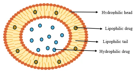
Fig: 1 Structure of proniosomes
COMPOSITION[18].
Table: 1 Composition of proniosomes
|
Components |
Importance |
Examples |
|
Non-ionic surfactants |
Serve as penetration boosters |
Span 20, 40, 60 Tween 20, 40, 60 |
|
Stabilizers |
Improve the stability of the formulation and reduce drug leakage. |
Cholesterol, Lecithin |
|
Solvents |
Enhance the solubility of the drug. |
Alcohol, Water, PBS 7.4, glycerol |
|
Carriers |
Increase the flexibility of the vesicular membrane. |
Maltodextrin or Sorbitol |
Non-ionic surfactants:
Non-ionic surfactants are the primary structural components used in the formulation of proniosomes. They consist of a polar hydrophilic head and a non-polar hydrophobic tail, making them uncharged and offering greater stability, compatibility, and lower toxicity than ionic surfactants. These surfactants enhance drug solubility and permeability through their wetting and emulsifying properties. The hydrophilic–lipophilic balance (HLB) is an important factor in surfactant selection, with values between 4 and 8 being optimal for proniosome vesicle formation. In contrast, highly hydrophilic surfactants are less suitable because their high solubility in aqueous media hinders aggregation and prevents the formation of the lamellar structures required for proniosomes[17].
Table: 2 Mostly used non-ionic surfactants with their HLB values[17].
|
Non-ionic surfactants |
Molecular weight (g/mol) |
HLB value |
Transition temperature |
|
Span 20 (sorbitan monolaurate) |
346.46 |
8.6 |
16°C |
|
Span 40 (sorbitan monopalmitate) |
402.57 |
6.7 |
42°C |
|
Span 60 (sorbitan monostearate) |
430.63 |
4.7 |
53°C |
|
Tween 20 (polyoxyethylene sorbitan monolaurate) |
522.68 |
16.7 |
- |
|
Tween 40 (polyoxyethylene sorbitan monostearate) |
648.92 |
14.9 |
- |
|
Tween 80 (polyoxyethylene sorbitan monooleate) |
604.82 |
15.0 |
- |
|
Tween 85 (sorbitan trioleate) |
957.49 |
1.8 |
- |
Cholesterol:
Cholesterol controls the structural and physical characteristics of proniosomes and can interact with non-ionic surfactants [19]. It regulates medication penetration through the membrane and increases the proniosomal membrane's stiffness and stability. The amount of cholesterol needed to prepare proniosomes depends on the HLB value of the surfactants. The amount of cholesterol should be raised to cover the larger groups when the HLB score is greater than 10[20]. However, over a specific cholesterol level, the produced formulation's entrapment efficiency (EE) decreases[21], potentially as a result of a drop in volume diameter[22].
Lecithin:
Proniosome formulation uses lecithin, a phospholipid, as a membrane stabilizer. Soya and egg lecithin are the most often utilized lecithins in the formulation. It has been claimed that hydrogenated-type lecithins offer advantages over non-hydrogenated lecithins, giving the cholesterol more stiffness and aiding in the creation of tight vesicles [23]. Non-hydrogenated lecithin's double bonds enable the molecular chains to bend (conformational rotation), preventing close contact with neighboring molecules during the formation of the niosomal membrane. As a result, the membrane has high permeability and low stiffness[21].
Solvents:
Phosphate buffer is typically employed as the hydration medium in proniosomes. The pH of the buffer is chosen based on the solubility of the medicine that is encapsulated[24]. Ruckmani and Sankar found that whereas EE increased when the hydration duration was extended from 20 to 45 minutes, medication leakage increased when the volume of hydration medium increased[25].
The solvent has the potential to improve penetration. It also has a significant impact on the vesicles' size. The type of alcohol used in a proniosomal formulation affects the drug's penetration rate and vesicle size[26].
The alcohol employed in Proniosomes has a substantial effect on vesicle size and drug penetration rate. The sizes of formatted vesicles are arranged as follows:
Ethanol > Propanol > Butanol > Isopropanol-due to its branched chain, ethanol forms the largest vesicles while isopropanol forms the smallest[27].
Carrier material:
Carriers play a crucial role in proniosome formulations by supporting drug loading and facilitating vesicle formation. An ideal carrier should be safe, non-toxic, free-flowing, sparingly soluble in the coating solution, and readily soluble in water to enable efficient hydration. In addition, carriers should provide a large surface area, allow flexibility in the surfactant-to-component ratio, and improve drug-loading capacity. Commonly used carriers include maltodextrin, sucrose stearate, sorbitol, glucose monohydrate, lactose monohydrate, and spray-dried lactose. Among these, maltodextrin is widely preferred because it offers greater flexibility in formulation and avoids the viscous slurry formation associated with more soluble carriers such as sorbitol, glucose monohydrate, and lactose monohydrate[23].
Types Of Proniosomes[28].
Dry Granular Proniososmes
Surfactant is used to cover water-soluble carriers, such as sorbitol and maltodextrin, in dry granular proniosomes. The end product of the coating process is a dry formulation with a thin layer of surfactant covering water-soluble particles. Vesicles must be prepared at a temperature higher than the non-ionic surfactant's transition temperature.
These are further divided into the following categories:
Sorbitol based Proniosomes: Sorbitol is used as the carrier in a dry formulation known as sorbitol-based proniosomes. After that, it is covered with a non-ionic surfactant and used as niosomes in a matter of minutes by stirring in water. They are usually made by spraying sorbitol powder with a combination of surfactants produced in an organic solvent, allowing the solvent to evaporate. Because the sorbitol carrier is soluble in organic solvent, the process must be repeated to obtain the necessary surfactant coating. The carrier has a very thin layer of surfactant coating, which hydrates and forms multilamellar vesicles as the carrier dissolves.
Maltodextrin based proniosomes: Recently, a proniosome formulation based on maltodextrin was developed that might be utilized to administer hydrophobic or amphiphilic medications. This formulation's more successful variant had hollow particles with enormous surface areas. Proniosomes with extremely high mass ratios of surfactant to carrier could be created with this formulation, and the amount of carrier required to maintain the surfactant could be easily changed.
Liquid Crystalline Proniosomes
Lipophilic chains of surfactants can change into a disordered, liquid state known as the lytorophic liquid crystalline state (neat phase) in three different ways when they are kept in contact with water. These three methods are,
In the lamellar (neat) phase, bilayers are arranged as stacked sheets separated by thin aqueous layers. When examined under a polarized microscope, these structures exhibit thread-like birefringence and produce characteristic X-ray diffraction patterns.
METHODS OF PREPARATION
MATERIALS AND TECHNIQUES
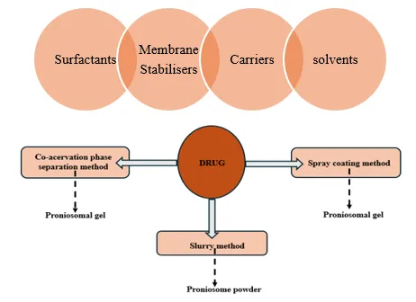
Fig: 2 Preparation methods of proniosomes
Slurry method
In this method, a slurry is prepared by mixing the carrier with a surfactant solution in a round-bottom flask. If required, additional organic solvent is added to achieve the desired slurry consistency. The slurry is then dried under vacuum to obtain a free-flowing proniosomal powder, which is stored in a sealed container at 4 °C. The preparation time is not affected by the carrier-to-surfactant ratio, making this method suitable for large-scale production[10,29,30].
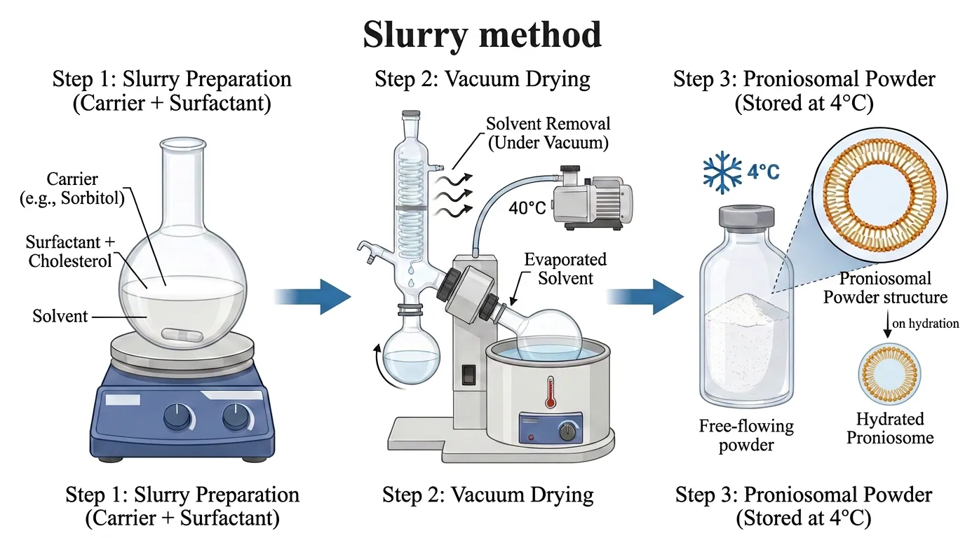
Fig: 3 Preparation of proniosomes by slurry method
Mohanty et al., (2022) prepared esomeprazole-encapsulated proniosomal formulation for the improved anti-ulcer activity by the slurry method placing maltodextrin as the carrier. Briefly, required amounts of drug, span 60, and cholesterol were dissolved in a chloroform and methanol to obtain a clear lipid solution. Maltodextrin gradually added under continuous stirring to produce a homogeneous slurry. The slurry was then subjected to solvent evaporation using a rotary evaporator at 40 °C under reduced pressure until the complete removal of solvent. The resulting proniosomal powder was further dried overnight in a vacuum oven, yielding a dry, free-flowing formulation[31].
Co-acervation phase separation method
The coacervation phase separation method is one of the most commonly used techniques for preparing proniosomal gels. In this method, accurately weighed amounts of the drug, cholesterol, and surfactants are placed in a dry, wide-mouthed glass beaker, followed by the addition of an appropriate organic solvent. The mixture is stirred and heated in a water bath at 60–70 °C until the surfactants dissolve completely, while minimizing solvent loss due to evaporation (close with lid). An aqueous phase is then added, and the mixture is further heated until a clear solution is obtained. Upon cooling and standing overnight, the solution converts into a proniosomal gel(creamy gel consistency)[32,33].
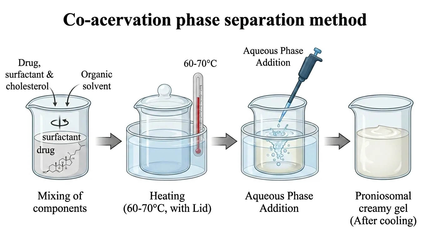
Fig: 4 Preparation of proniosomes by co-acervation phase technique
Sergio Liga et al., (2026) formulated puerarin-loaded proniosomal gel for the demal delivery and evaluated for in vitro antimelanoma potential and safety. Proniosomal gels were formulated using the coacervation-phase separation technique with a blend of nonionic surfactants and phospholipids. The lipid constituents, including Span 60, Tween 80, phosphatidylcholine, and cholesterol, were precisely weighed and dissolved in absolute ethanol in a glass vial. The mixture was maintained at 50 °C with continuous stirring until a clear and uniform solution was obtained. The vial was covered throughout the heating and stirring process to minimize ethanol loss due to evaporation. Meanwhile, puerarin was dissolved separately in preheated Milli-Q water and this aqueous solution was then added gradually to the lipid mixture with gentle stirring for 5 min. Upon cooling to room temperature, the formulation transformed into a yellowish, translucent, viscous creamy gel, confirming the successful formation of the proniosomal gel[34].
Spray-coating method
In this method, proniosomes are prepared by spraying a surfactant solution in an organic solvent onto a suitable carrier material, followed by solvent evaporation. This process deposits a thin surfactant film on the carrier surface, which upon hydration forms multilamellar vesicles[35,36].
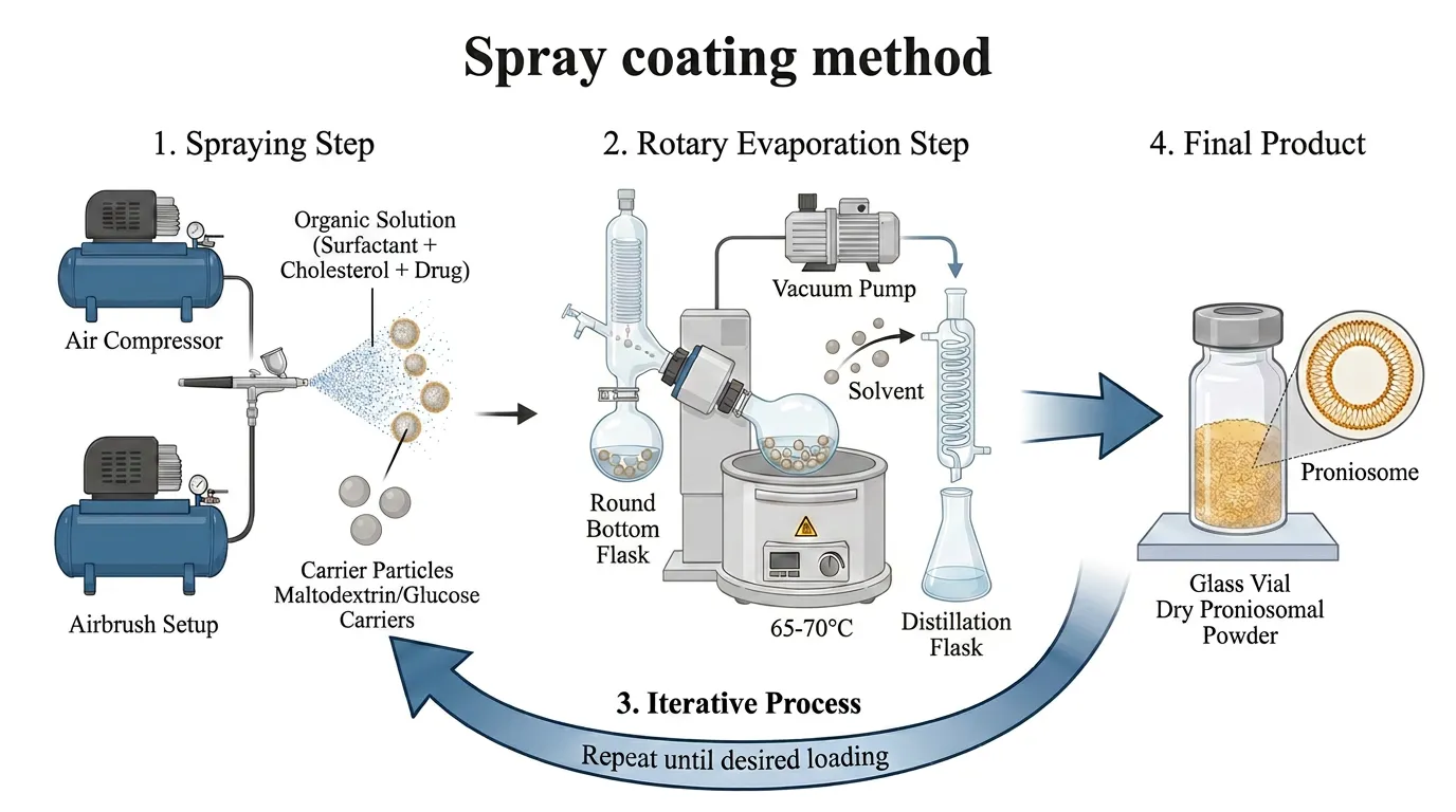
Fig: 5 Preparation of proniosomes by spray coating method
Niosome formation by hydration of proniosomes
Proniosomes can be hydrated to create niosomes by adding an aqueous phase containing the medication to them while briefly shaking them at a temperature higher than the surfactant's mean transition phase temperature[6].
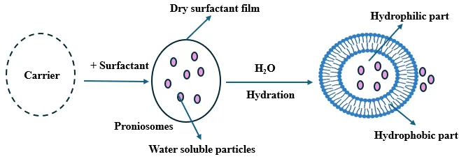
Fig: 6 Niosomes by hydration of proniosomes
Characterization of Proniosomes
Vesicular size and shape
Proniosomes become spherical or globular niosomal vesicles when they are hydrated. These vesicles' size, shape, and surface properties are significant factors that affect how well they carry drugs. Vesicle size and shape are assessed using a variety of analytical methods, including as optical microscopy, photon correlation spectroscopy, transmission electron microscopy (TEM), scanning electron microscopy (SEM), and freeze-fracture electron microscopy. These methods assure the stability and quality of the niosomal formulation by providing comprehensive data on vesicle size, surface shape, and structural integrity.[17]. The dynamic light scattering (DLS) method is used to measure the vesicular size distribution. DLS essentially determines the diffusion coefficient of the particles by measuring changes in scattered light intensity brought on by diffusing particles. The sizes of agitated and agitation-free noisome vesicles are measured. When hydration is done without stirring, the biggest size is produced[37].
Angle of repose
Using the funnel method, position a funnel over a level surface at a predetermined distance of 2 cm. Pour the powder into the funnel and let it pass slowly until the powder contacts the funnel's tip, creating a pile. After measuring the pile's diameter, the angle of repose was computed using the following formula:
tanθ=hr

Where, θ - angle of repose, h – the height of the pile, r – radius of the pile[38].
Zeta potential
Their extremely positive or highly negative surface charge, which keeps colloidal particles in the niosomes from aggregating due to repulsion between similarly charged particles, is responsible for the enhanced stability[39]. A proniosomal formulation is deemed physically stable if its zeta potential value is at least ± 30 mV. Thus, it is possible to prevent particle aggregation[40].
Drug content determination
By precisely measuring the necessary amount in a volumetric flask and dissolving it in 25 mL of methanol while being sonicated for 10 minutes in a sonication bath, the drug content of the proniosome gel was ascertained. After the solution was filtered through a 0.45-μm nylon filter and appropriately diluted, the drug content was ascertained using the HPLC technique[41].
Entrapment efficiency (EE %)
The EE% was found indirectly by spectrophotometric analysis of the unentrapped drug in the supernatant. In summary, the reconstituted formula was centrifuged for 1.5 hours at 4 °C and 22,000 rpm. The amount of unentrapped medication was measured using the supernatant[42,43].
% Entrapment= ED / TD X 100
Where, ED is the amount of entrapped drug and TD is the initial amount of drug.
Rate of spontaneity
The number of niosomes that form once proniosomes are hydrated is known as the rate of spontaneity. Proniosomal gel is moved and evenly distributed over the walls of the tiny stoppered glass tube container in order to calculate the rate of spontaneity. NaCl (0.154 M) was then carefully added and positioned to one side without causing any turbulence. The number of niosomes eluted from proniosomes is computed using Neubauer's chamber[17].
In vitro release studies
V.J. Mokale et al., performed in vitro release by USP dissolution apparatus II (Paddle Method), the drug-release rate investigation was conducted by comparing the produced famotidine proniosomes with the commercial tablet formulation[44]. Goyal et al., synthesised gugulipid incorporated proniosomes and modified Franz diffusion cell was used to measure the release of gugulipid from the proniosome formulation[45]. N.F. Younes et al., formulated ofloxacin loaded proniosomes for the enhanced trans-tympanic permeability. The dialysis method was used to determine the reconstituted optimal formula's in vitro release profile. First, the dialysis membrane was soaked overnight in the preferred medium, phosphate buffer saline solution (pH 7.4)[46]. In vitro skin permeation studies can be performed using flank skin, dorsal skin of albino rabbit (Alsarra et al)[9], female albino rat (Sprague-Dawley strain) (Vora et al)[32], Wistar rat skin (7–9 weeks old) (Fang et al)[47]. Drug release techniques such as drug diffusion from bilayered membranes, desorption from the vesicle surface, or a combination of diffusion and desorption mechanisms can be followed by niosomal vesicles generated from proniosomes[6].
Stability studies
The stability study was conducted to evaluate the ability of the proniosomal formulation to retain the drug under different storage conditions, including room temperature (25 ± 2 °C), oven temperature (32 ± 2 °C), and refrigerated conditions (4–8 °C). Samples were collected at predetermined time intervals over a period of eight weeks, and the drug content was determined using a spectrophotometric method. The percentage of drug retained was subsequently calculated to assess the stability of the formulation[44]. According to the ICH guidelines, dry proniosomal powder intended for reconstitution should undergo accelerated stability testing at 40 ± 2 °C and 75 ± 5% relative humidity. Long-term stability studies should be performed under storage conditions appropriate for the climatic zone where the product is intended to be marketed. For climatic zones I and II, the recommended conditions are 25 ± 2 °C and 60 ± 5% RH, whereas for zones III and IV, they are 30 ± 2 °C and 65 ± 5% RH. During stability studies, the formulation should be assessed for various quality attributes, including appearance, surface morphology, drug content, color, pH, particulate matter, assay, preservative content, pyrogenicity, and sterility[6].
Applications
Proniosomes as drug carriers
Table:3 Advancement in drug delivery by proniosomes as carrier
|
Route of drug administration |
Preparation techniques |
Status of evaluation |
Key findings |
References |
|
Terconazole loaded proniosomes (TCZ-PNS)-ocular |
Modified coacervation technique |
Comparative in-vitro release, corneal ex-vivo permeation, in-vivo permeation and microbiological studies. |
Compared to TCZ suspension, TCZ-PNS had a greater transcorneal ex-vivo penetration. Microbiological evaluations of optimal TCZ-PNS in comparison to TCZ suspension showed improved biofilm inhibition effectiveness at most tested doses, a roughly 50% bigger inhibition zone. |
[48] |
|
Glibenclamide pronisomes |
Slurry method |
In-vitro dissolution, in-vivo pharmacodynamics and histopathological studies. |
All formulations showed improved drug solubility since the dissolution rate was higher than that of the pure medication. According to pharmacodynamics data, animals treated with proniosomes had a significantly lower fasting blood glucose level than those given with pure medication, which had a 17.6% lower blood glucose level. No indication of liver damage. |
[38] |
|
Captopril-transdermal |
Coacervation-phase separation |
In vitro release and stability studies |
Extended release of entrapped captopril was seen in vitro. Better drug retention at refrigerated conditions. |
[33] |
|
Luliconazole-loaded proniosomal gel (LPG)-topical |
Coacervation-phase separation |
In-vitro release, ex-vivo skin permeation, and in-vivo antifungal activity studies. |
Due to improved vesicular dispersion and surfactant-mediated diffusion, LPG achieved 98.5% release within 24 hours. Ex vivo studies showed significantly higher permeation and good skin deposition. The in vivo experiments showed a superior antifungal activity with a minimum inhibitory concentration of 10.67 ng/mL compared to the commercial cream for fluconazole-resistant candida. |
[49] |
|
Valacyclovir-ocular |
Coacervation Phase Separation Method |
In vitro diffusion and kinetics of drug release |
For longer than ten hours, every formulation exhibited a sustained-release feature. The Korsmeyer-Peppas model of drug release kinetics was able to fit the drug release patterns from the in vitro studies. |
[50] |
|
Irbesartan-oral |
Slurry method |
In vitro drug release, solubility, stability and in vivo Pharmacokinetic Studies.
|
At 24 hours, the optimized batch displayed a CDR of 97.36 ± 1.13%. The proniosomal formulation was more soluble in phosphate buffer (pH 6.8) (2.65 ± 0.2 mg/mL) than in water (0.059 ± 0.02 mg/mL). The enhanced bioavailability of the medication in the optimized proniosomal formulation when compared to its pure drug suspension was validated by the pharmacokinetic investigation conducted in rats.
|
[39] |
|
Amphotericin B (Amp B)-topical |
Coacervation phase separation |
In vitro drug release, in vitro diffusion, ex-vivo permeability and anti-fungal studies. |
In vitro drug release of optimized formulation was 95.72%±0.30. Drug penetration was gradual and consistent, with 76.55%±0.27 of Amp B penetrating the skin in 24 hours from the optimized formulation. |
[51] |
|
Lornoxicam-topical |
Coacervation phase separation |
In vitro drug release, ex vivo, and the anti-inflammatory efficacy studies. |
Ex vivo studies indicated considerably increased skin permeability in the proniosomal gels. increased anti-inflammatory activity than standard formulation. |
[52] |
|
Ofloxacin- trans-tympanic |
Coacervation phase technique |
In vitro release, ex vivo permeation, microbiological, and in vivo tests. |
The optimal formula was found to have superior antibacterial activity, increased trans-tympanic permeability (3701.01 μg/cm2), and better otic tolerance than the ofloxacin solution (2118.72 μg/cm2). |
[46] |
|
Ketoprofen-oral(periodontitis) |
Coacervation phase technique |
Mucoadhesiveness, ex vivo drug permeation, retention and pharmacodynamic activity studies. |
Studies on viscosity and texture revealed smoothness and good adherence, which are needed for improved permeation. With the bone resorption process preserved, the illness status was improved.
|
[41] |
|
Clotrimazole-transdermal |
Coacervation technique |
In-vitro release, ex-vivo permeation and in-vitro antibacterial activity |
Comparative permeation and in vitro drug release studies demonstrated that the optimized proniosomal gel (F5) exhibited 1.75-fold higher skin penetration than the marketed gel and a higher anti-bacterial activity. |
[53] |
|
Silibinin-cutaneous
|
Coacervation technique |
In vitro drug release, skin permeation and cell viability assessment. |
The formulation demonstrated gradual in vitro drug release, sustained permeation, and a favorable safety profile, with no signs of cytotoxicity or irritation in the evaluated models. |
[54] |
|
Catechin-oral (food fortification) |
Thin-film hydration technique |
X-ray diffraction and in-vitro release studies. |
Studies on in vitro release showed that the proniosomes released catechins continuously. There were no chemical interactions between catechins and encapsulants, according to FTIR and X-ray diffraction spectra |
[55] |
|
Resveratrol- oral (food fortification) |
Thin-film hydration technique |
In-vitro release, and antioxidant activity. |
Resveratrol's bioavailability could be significantly increased by encapsulating it in proniosomes, according to studies on dynamic light scattering, FTIR, in-vitro release, and antioxidant activity. |
[56] |
Artificial Intelligence (AI) And Machine Learning (ML) In Proniosomes Development
In novel drug delivery systems (NDDS), integrating AI and ML into the development of proniosomes (and their hydrated form, niosomes) moves formulation design from conventional trial-and-error to advanced data-driven, predictive engineering. Proniosomes are dry, free-flowing formulations of surfactant-coated carrier particles that hydrate into niosomes upon contact with water or biological fluids. Developing them involves balancing many interdependent variables, which AI and ML excel at optimizing.
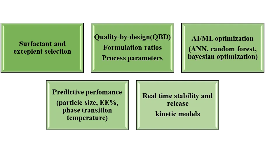
Surfactant and Excipient Selection
Choosing the appropriate non-ionic surfactant (such as Span 60, Tween 80) and membrane stabilizer (such as cholesterol) based on the drug's lipophilicity (log P), solubility, and molecular weight is crucial for proniosomal stability and vesicular shape. AI/ML's role is to assess the structural characteristics of active drug molecules and excipient libraries using machine learning algorithms like Support Vector Machines (SVM) and QSAR-based clustering. Before physical synthesis, they compute which surfactant-cholesterol combinations will result in the highest drug affinity and the best thermodynamic compatibility[57].
Quality-By-Design (QBD), Formulation Ratios, & Process Parameters
A systematic pharmaceutical methodology known as Quality-by-Design (QBD) determines the formulation's Critical Quality Attributes (CQAS) and how Critical Process Parameters (CPPS) impact them. The use of AI/ML: AI combines with QBD to model complicated, multi-factorial interactions, such as surfactant-to-cholesterol ratios, drug loading, hydration temperature, and carrier coating rates, rather than depending on conventional, labor-intensive Design of Experiments (DoE) that evaluate linear variables[58].
AI / ML Optimization (ANN, Random Forest, Bayesian Optimization)
This is the main computational engine that links the variables used in formulation with the results of experimental performance.
Utilized Algorithms:
ANNs, or artificial neural networks-are used to model extremely non-linear interactions between formulation inputs and output characteristics.
Random forest (RF) and gradient boosting- Assess the significance of features and forecast the success of categorical formulation.
Bayesian optimization:-Determines the best formulation formulas iteratively with the least amount of wet-lab testing[59].
Predictive Performance (Particle Size, EE%, Phase Transition Temperature)
Accurately predicting the physicochemical characteristics of hydrated vesicles prior to conducting laboratory experiments is the main objective of AI modeling.
Predicted Key Parameters:
Entrapment Efficiency (%EE): Indicates the proportion of medication that is effectively encapsulated within the bilayer or core.
Vesicle Size & Polydispersity Index (PDI): Guaranties post-hydration nanoscale size distribution (< 200 nm).
Phase Transition Temperature (Tc): Ensures spontaneous hydration at body temperature (37°C) by predicting the temperature at which surfactant bilayers change from a stiff gel state to a fluid liquid-crystalline state[60].
Real-Time Stability and Release Kinetic Models
Although proniosomes are designed to prevent liquid vesicular leakage, regulated medication release and long-term shelf life are still crucial evaluation criteria. AI/ML's role is to simulate sustained drug release over long periods of time by fitting in-vitro release profiles against mathematical models (Korsmeyer-Peppas, Higuchi, Zero-Order). Additionally, possible moisture sorption, drug crystallization, and long-term shelf stability under varying humidity and temperature are predicted by machine learning models based on thermal/diffraction data[61].
CONCLUSION
Proniosomes have become a flexible provesicular drug delivery vehicle with its enhanced stability, ease of storage, effective encapsulation of a variety of medicines, and controlled drug release. The kind of surfactant, cholesterol, lecithin, carrier materials, solvents, and preparation techniques all have a significant impact on their formulation properties. With advances in drug penetration, bioavailability, and therapeutic efficacy, recent studies show their promise across oral, topical, transdermal, ocular, and other administration routes. New possibilities for logical excipient selection, formulation optimization, critical quality attribute prediction, and medication release and stability modeling are presented by the growing integration of artificial intelligence and machine learning with Quality by Design. Even though the field of AI/ML-assisted proniosome development is still in its infancy, combining it with experimental methods may simplify formulations and increase the efficiency and predictability of drug delivery research. All things considered, merging proniosomal technology with data-driven formulation techniques is a promising approach to creating sophisticated and efficient drug delivery systems.
REFERENCES

Fathima Safa E. K., Archana O., Rifana C. K., Manoj K.*, Engineering Next-Generation Proniosomes: Advances in Formulation, Therapeutic Applications and AI/ML-Assisted Development, Int. J. Med. Pharm. Sci., 2026, 2 (9), 386-401. https://doi.org/10.5281/zenodo.22893335
 10.5281/zenodo.22893335