

We use cookies to ensure our website works properly and to personalise your experience. Cookies policy
1Research Scholar, Goenka College of Pharmacy, Laxmangarh, Rajasthan, India
2Associate Professor, Goenka college of Pharmacy, Gassu, Laxmangarh Sikar, Rajasthan
Inflammation is a protective physiological response to tissue injury and infection; however, persistent or excessive inflammation may contribute to chronic inflammatory disorders and require prolonged pharmacological intervention. Conventional anti-inflammatory agents are effective but may be associated with systemic and local adverse effects, creating a need for safer and more localized therapeutic approaches. Barbaloin, a bioactive anthraquinone glycoside associated with Aloe vera, has been reported to possess anti-inflammatory, antioxidant, and wound-healing properties. The present study aimed to formulate and evaluate a Barbaloin-loaded topical gel for localized anti-inflammatory therapy. Nine topical gel formulations (F1–F9) were prepared by the dispersion method using Carbopol 934 and hydroxypropyl methylcellulose (HPMC) at different concentrations. The formulations were evaluated for physical appearance, pH, homogeneity, spreadability, extrudability, viscosity, drug content, in-vitro drug release, release kinetics, stability, and in-vitro anti-inflammatory activity using the protein denaturation inhibition assay. Drug–excipient compatibility was investigated using Fourier transform infrared spectroscopy (FTIR) and differential scanning calorimetry (DSC). Barbaloin content was estimated using UV–Visible spectrophotometry. All formulations exhibited satisfactory physical characteristics and pH suitable for topical application. Drug content ranged from 97.45 ± 0.82% to 99.98 ± 0.54%, with F3 showing the highest drug content. Among the formulations, F3 containing 1.5% Carbopol 934 demonstrated the highest inhibition of protein denaturation (76.84 ± 0.88%) and was selected as the optimized formulation. F3 exhibited progressive drug release, reaching 98.21 ± 0.62% after 8 h. The release data showed the best fit with the Higuchi model (R² = 0.994), while the Korsmeyer–Peppas model showed an R² of 0.989 with an n value of 0.689, indicating anomalous release behaviour. Stability evaluation demonstrated no significant changes in the evaluated physicochemical characteristics during the study period. The developed Barbaloin-loaded topical gel, particularly F3, demonstrated satisfactory physicochemical characteristics, controlled drug release, stability, and promising in-vitro anti-inflammatory activity. The formulation may therefore serve as a potential topical approach for localized management of inflammatory skin conditions.
Inflammation is a complex protective biological response initiated in response to tissue injury, infection, or other harmful stimuli. It plays an essential role in eliminating injurious agents and initiating tissue repair. Although acute inflammation is generally beneficial and self-limiting, persistent or dysregulated inflammation may result in progressive tissue damage and contribute to the development of chronic inflammatory disorders, including dermatitis, psoriasis, rheumatoid arthritis, and osteoarthritis [1]. Non-steroidal anti-inflammatory drugs (NSAIDs) and corticosteroids are widely used for the management of inflammatory conditions because of their established therapeutic efficacy. However, prolonged or inappropriate use of these agents may be associated with adverse effects such as gastrointestinal irritation, renal impairment, cardiovascular complications, immunosuppression, and skin-related effects. These limitations have encouraged the investigation of alternative therapeutic agents with improved safety and the potential for localized drug delivery [2,3]. Natural products have attracted considerable interest in pharmaceutical research because of their diverse pharmacological activities and potential therapeutic benefits. Aloe vera (Aloe barbadensis Miller) is a medicinal plant widely investigated for its anti-inflammatory, antioxidant, antimicrobial, and wound-healing properties. Barbaloin (aloin), a major anthraquinone C-glycoside associated with Aloe vera, is one of its important bioactive constituents. Experimental studies have reported that Barbaloin can modulate inflammatory responses through effects on inflammatory mediators, oxidative stress, and cellular signalling pathways, supporting its potential as a natural anti-inflammatory agent [4–9]. Despite its pharmacological potential, the effective topical delivery of Barbaloin presents formulation challenges. The therapeutic performance of a topical drug depends not only on the intrinsic activity of the active compound but also on its physicochemical characteristics, stability, drug release, and ability to remain available at the site of application. Conventional administration may also result in inadequate drug concentration at the target site. Therefore, development of an appropriate topical delivery system may improve local drug availability and provide more controlled drug release [10]. Topical drug delivery is particularly advantageous for the treatment of localized skin disorders because it allows the active pharmaceutical ingredient to be delivered directly to the affected area while potentially reducing systemic exposure. Among various topical dosage forms, gels are widely used because of their ease of application, non-greasy nature, good patient acceptability, and ability to provide uniform drug distribution. Hydrophilic polymeric gels can also be designed to control drug release by modifying the viscosity and structure of the gel matrix [11–14]. The selection and concentration of gelling polymers are important determinants of the performance of topical formulations. Carbopol 934 and hydroxypropyl methylcellulose (HPMC) are commonly used hydrophilic polymers that can provide suitable viscosity, consistency, spreadability, and drug-release characteristics. Changes in polymer concentration can alter the microstructure of the gel matrix and consequently influence drug diffusion, drug availability, and overall formulation performance [15–18]. Hence, systematic optimization of polymer concentration is essential for developing a topical formulation with desirable physicochemical and therapeutic properties. The skin consists principally of the epidermis, dermis, and subcutaneous tissue and acts as an important barrier against external substances. While this barrier protects the body, it can also restrict the penetration of many therapeutic agents. For localized inflammatory skin conditions, an appropriately designed topical formulation may help maintain the active compound at or near the site of inflammation while minimizing unnecessary systemic exposure. Therefore, formulation strategies that provide satisfactory spreadability, skin compatibility, stability, and controlled drug release are important for successful topical therapy [19–21]. Although the pharmacological properties of Aloe vera and Barbaloin have been extensively investigated, comparatively limited work has focused on the systematic development and optimization of a Barbaloin-loaded topical gel using different concentrations of Carbopol 934 and HPMC, followed by comprehensive physicochemical, drug-release, kinetic, stability, and anti-inflammatory evaluation. In particular, establishing an appropriate polymer concentration that provides a balance between formulation characteristics and biological performance is important for the development of an effective topical dosage form [22–25]. Therefore, the present study was undertaken to formulate and evaluate Barbaloin-loaded topical gel formulations using Carbopol 934 and HPMC as gelling polymers. Nine formulations (F1–F9) were prepared with varying polymer concentrations and evaluated for their physicochemical properties, drug content, in-vitro drug release, release kinetics, stability, and in-vitro anti-inflammatory activity using the protein denaturation inhibition assay. Based on the overall evaluation, the formulation demonstrating the most desirable characteristics and anti-inflammatory performance was selected as the optimized formulation. The study was aimed at developing a stable and patient-acceptable topical gel capable of providing controlled Barbaloin delivery and promising localized anti-inflammatory activity.
2. MATERIALS AND METHODS
2.1 MATERIALS
Barbaloin was procured from Yucca Enterprises, Mumbai, India, and used as the active pharmaceutical ingredient. Carbopol 934 and hydroxypropyl methylcellulose (HPMC) were selected as gelling polymers for the development of topical gel formulations. Propylene glycol was incorporated as a humectant and penetration-enhancing agent [26], while methyl paraben served as the preservative. Triethanolamine was used as a neutralizing agent for pH adjustment and development of the desired gel consistency. Purified water was used as the aqueous vehicle. Methanol, phosphate buffer components, hydrochloric acid, and other reagents required for the analytical and experimental procedures were of analytical grade. Bovine serum albumin (BSA) was used for the in-vitro protein denaturation assay. All materials were used as received without further purification.
2.2 Preparation of Barbaloin Topical Gel
Barbaloin topical gel formulations were prepared by the dispersion method using Carbopol 934 or HPMC as the gelling polymer. The required quantity of polymer was gradually dispersed in purified water under continuous stirring and allowed to hydrate adequately. Propylene glycol and methyl paraben were subsequently incorporated into the hydrated polymer dispersion. Barbaloin was then added gradually with continuous stirring to obtain a uniform drug dispersion. Triethanolamine was added slowly to adjust the pH and obtain the required gel consistency. The prepared formulations were kept undisturbed to allow entrapped air to escape and were subsequently transferred into suitable containers for further evaluation. Nine formulations (F1–F9) were developed by varying the concentration of Carbopol 934 or HPMC, while the quantities of Barbaloin, propylene glycol, methyl paraben, and other formulation components were maintained constant. The composition of the prepared formulations is presented in Table 1.
Table 2.1Composition of Barbaloin topical gel formulations
|
Contents |
F1 |
F2 |
F3 |
F4 |
F5 |
F6 |
F7 |
F8 |
F9 |
|
Barbaloin (%w/w) |
1 |
1 |
1 |
1 |
1 |
1 |
1 |
1 |
1 |
|
Carbopol934(%w/w) |
0.5 |
1.0 |
1.5 |
2.0 |
_ |
_ |
_ |
_ |
_ |
|
HPMC(%w/w) |
_ |
_ |
_ |
_ |
0.5 |
1.0 |
1.5 |
2.0 |
2.5 |
|
Propylene glycol(%w/w) |
5 |
5 |
5 |
5 |
5 |
5 |
5 |
5 |
5 |
|
MethylParaben (%w/w) |
0.1 |
0.1 |
0.1 |
0.1 |
0.1 |
0.1 |
0.1 |
0.1 |
0.1 |
|
Triethanolamine |
q.s. |
q.s. |
q.s. |
q.s. |
q.s. |
q.s. |
q.s. |
q.s. |
q.s. |
|
Purified Water |
q.s. |
q.s. |
q.s. |
q.s. |
q.s. |
q.s. |
q.s. |
q.s. |
q.s |
The prepared formulations were subjected to physicochemical evaluation. Based on the overall evaluation results, the formulation exhibiting the most satisfactory characteristics was selected as the optimized formulation for subsequent in-vitro drug release, release kinetic, and stability studies.
2.4 Evaluation of Barbaloin Topical Gel
The developed formulations were evaluated for clarity, pH, homogeneity, drug content, spreadability, extrudability, viscosity, and in-vitro drug diffusion behaviour.
2.4.1 Clarity
The clarity of each gel formulation was examined visually against black and white backgrounds. The formulations were categorized according to their observed appearance as turbid, clear, or transparent/glassy.
2.4.2 PH
The PH of the formulations was measured using a calibrated digital pH meter. For measurement, 0.5 g of gel was dispersed in 50 mL of distilled water and allowed to equilibrate for 3 h at 25°C. The pH was then recorded.
2.4.3 Homogeneity
The prepared gels were examined visually and manually to assess their uniformity. Particular attention was given to the presence of lumps, aggregates, coarse particles, and phase separation.
2.4.4 Drug Content
Approximately 100 mg of gel was accurately weighed and transferred into 10 mL of methanol. The sample was thoroughly mixed to facilitate drug extraction and subsequently filtered through Whatman No. 42 filter paper. The filtrate was suitably diluted and analyzed using a UV–Visible spectrophotometer at the predetermined analytical wavelength. Drug content was calculated from the corresponding calibration curve.
2.4.5 Spreadability
Spreadability was determined using two clean glass plates. Approximately 0.5 g of gel was placed between the plates, followed by application of a 500 g load for 1 min. The diameter of the resulting spread was measured, and the spreadability was calculated.
2.4.6 Extrudability
Approximately 20 g of the prepared gel was filled into a collapsible aluminium tube. The filled tube was subjected to a pressure of 1 kg/cm² for 30 s using a Pfizer hardness tester. The amount of gel expelled from the tube was collected and weighed. The measurement was performed in triplicate.
2.4.7 Viscosity
The viscosity of the gel formulations was measured using a Brookfield viscometer equipped with spindle SC4-18/13R. Measurements were obtained within a torque range of 10–100%, and the corresponding viscosity values were recorded.
2.4.8 In-vitro Drug Diffusion Study
The in-vitro drug diffusion behaviour of the optimized Barbaloin gel was investigated using a Franz diffusion cell fitted with a dialysis membrane. The receptor compartment was filled with 60 mL of phosphate buffer (pH 7.4) and maintained at 37°C under continuous stirring at 150 rpm. A 0.5 g quantity of gel was applied uniformly to the donor compartment. At predetermined sampling intervals, 2 mL of receptor medium was withdrawn and immediately replaced with an equal volume of fresh phosphate buffer to maintain a constant receptor volume. The collected samples were suitably diluted, where necessary, and analyzed spectrophotometrically at the predetermined analytical wavelength. The cumulative percentage of drug released was calculated from the obtained absorbance values.
2.5 Drug Release Kinetic Analysis
The release data obtained from the optimized Barbaloin gel were subjected to mathematical modelling using zero-order, first-order, Higuchi, and Korsmeyer–Peppas equations [27]. The coefficient of determination (R²) was used to compare the suitability of the different kinetic models and to identify the model that best described the drug-release behaviour.
Zero-order model:
Qt = Q₀ + K₀t
where Qt is the amount of drug released at time t, Q₀ represents the initial amount of drug, and K₀ is the zero-order release constant.
First-order model:
log C = log C₀ − Kt/2.303
where C is the amount of drug remaining at time t, C₀ represents the initial drug concentration, and K is the first-order rate constant.
Higuchi model:
Q = Kᴴ√t
where Q represents the cumulative amount of drug released, Kᴴ is the Higuchi release constant, and t denotes the release time.
Korsmeyer–Peppas model:
Mₜ/M∞ = Ktⁿ
where Mₜ/M∞ represents the fraction of drugreleased at time t, K is the kinetic constant, and n is the release exponent used to characterize the drug-release mechanism.
2.6 In-Vitro Anti-Inflammatory Activity
The Anti-Inflammatory activity of the prepared Barbaloin gel formulations was evaluated by the protein denaturation inhibition method using bovine serum albumin (BSA). A 1% w/v BSA solution was prepared in phosphate buffer (pH 6.8). For each formulation, 1 mL of gel was mixed with 1 mL of the prepared BSA solution. The pH of the mixture was adjusted to 6.8 ± 0.2 using 1 N hydrochloric acid, wherever required. The reaction mixtures were incubated at 37 ± 2°C for 20 min and subsequently heated at 70 ± 2°C for 5 min in a thermostatically controlled water bath. After cooling to room temperature, the absorbance was measured at 660 nm using a UV–Visible spectrophotometer. A BSA solution without the test formulation was treated under identical experimental conditions and served as the control. All determinations were carried out in triplicate (n = 3), and the results were expressed as mean ± standard deviation (SD). The percentage inhibition of protein denaturation was calculated using the following equation:
% Inhibition = [(A₀ − A₁) / A₀] × 100
where A₀ represents the absorbance of the control and A₁ denotes the absorbance of the formulation-treated sample. A higher percentage inhibition indicated greater inhibition of protein denaturation.
2.7 Comparative Evaluation and Selection of Optimized Formulation
The nine Barbaloin gel formulations were compared on the basis of their percentage inhibition of protein denaturation along with their physicochemical characteristics. The formulation demonstrating the highest anti-inflammatory activity while maintaining satisfactory pH, viscosity, spreadability, homogeneity, and drug content was selected as the optimized formulation. The optimized batch was subsequently subjected to in-vitro drug diffusion, release kinetic, and stability studies.
3. RESULTS
3.1 Preparation of Barbaloin Topical Gel
Nine Barbaloin-loaded topical gel formulations (F1–F9) were prepared by the conventional dispersion method using Carbopol 934 and HPMC as gelling polymers. The polymers were dispersed in purified water and allowed to hydrate. Barbaloin was dissolved in ethanol containing propylene glycol and incorporated into the hydrated polymer dispersion with continuous stirring. Methyl paraben was added as preservative, and triethanolamine was added gradually to obtain the desired gel consistency. The formulations were allowed to stand to remove entrapped air before evaluation.
3.2 Evaluation of Barbaloin Topical Gel
3.2.1 Physical Appearance and Clarity
All Barbaloin-loaded gel formulations (F1–F9) exhibited clear to translucent appearance with a smooth and homogeneous texture. No visible particulate matter, grittiness, or phase separation was observed. The formulations showed satisfactory physical appearance and were considered suitable for further evaluation.
3.2.2 pH
The pH values of the formulations ranged from 6.10 ± 0.03 to 6.82 ± 0.04. All formulations exhibited pH values suitable for topical application. [28]
Table 3.1 PH of Various Batches Of Barbaloin Topical Gel
|
Formulation code |
PH (Mean ±SD) |
|
F1 |
6.10 ± 0.03 |
|
F2 |
6.18 ± 0.02 |
|
F3 |
6.24 ± 0.03 |
|
F4 |
6.31 ± 0.02 |
|
F5 |
6.45 ± 0.04 |
|
F6 |
6.52 ± 0.03 |
|
F7 |
6.60 ± 0.02 |
|
F8 |
6.71 ± 0.03 |
|
F9 |
6.82 ± 0.04 |
3.2.3 Spreadability
F1–F3 exhibited excellent (+++) spreadability, F4–F7 showed good (++) spreadability, while F8 and F9 demonstrated satisfactory (+) spreadability. The decrease in spreadability with increasing polymer concentration was associated with increased gel viscosity and polymer network density.
3.2.4 Viscosity
The viscosity of the Barbaloin-loaded gel formulations ranged from 8,900 to 10,180 cP. The Carbopol 934-based formulations (F1–F4) showed viscosity values ranging from 8,900 to 9,345 cP, whereas the HPMC-based formulations (F5–F9) exhibited values ranging from 9,498 to 10,180 cP. An overall increase in viscosity was observed with increasing polymer concentration, which may be attributed to the formation of a more structured polymeric network within the gel matrix [29].
3.7.5 Drug Content
The drug content of the formulations ranged from 97.45 ± 0.82% to 99.98 ± 0.54%, indicating satisfactory and uniform incorporation of Barbaloin.
Table 3.2 Drug content of Barbaloin-loaded topical gel formulations (F1–F9).
|
Formulation |
Drug Content (%±SD) |
|
F1 |
97.45±0.82 |
|
F2 |
98.12±0.76 |
|
F3 |
99.98±0.54 |
|
F4 |
99.21±0.61 |
|
F5 |
98.56±0.73 |
|
F6 |
98.84±0.68 |
|
F7 |
99.42±0.57 |
|
F8 |
99.69±0.48 |
|
F9 |
99.11±0.65 |
Among the formulations, F3 exhibited the highest drug content (99.98 ± 0.54%), demonstrating efficient drug incorporation and uniform distribution within the gel matrix.
3.7.6 HET-CAM Irritation Study
The irritation potential of the Barbaloin-loaded gels was evaluated using the HET-CAM assay. No visible hemorrhage, vascular lysis, or coagulation was observed for any formulation during the 5-min observation period. All formulations exhibited an irritation score of 0.0, corresponding to the non-irritant category (IS 0–0.9). These findings indicated a favourable acute irritation profile under the experimental conditions.
In-vitro Drug Release Study
The optimized Barbaloin-loaded topical gel (F3) was evaluated for in-vitro drug release using a Franz diffusion cell over a period of 8 h. The cumulative drug release increased progressively from 14.28 ± 0.42% at 0.5 h to 98.21 ± 0.62% at 8 h, indicating a gradual and sustained release profile. The release profile of F3 is presented in Table 3.3 and Figure 3.1,3.2 The release data were fitted to zero-order, first-order, Higuchi, and Korsmeyer–Peppas kinetic models to elucidate the release kinetics and mechanism. Among the models evaluated, the Higuchi model showed the highest regression coefficient (R² = 0.994), indicating that diffusion through the hydrated polymeric matrix was the predominant mechanism governing drug release. The Korsmeyer–Peppas model showed an R² value of 0.989 with a release exponent (n) of 0.689, indicating anomalous (non-Fickian) transport and suggesting the combined contribution of drug diffusion and polymer relaxation/swelling. The first-order and zero-order models showed R² values of 0.986 and 0.975, respectively. Overall, F3 exhibited a gradual and sustained release profile, suggesting that the polymeric gel matrix effectively regulated Barbaloin release and may provide prolonged drug availability following topical application.
Table3.3: In-vitro cumulative drug release profile of optimized Barbaloin-loaded topical gel formulation (F3)
|
Time (hr) |
Cumulative Drug release (%±SD) |
|
0.5 |
14.28±0.42 |
|
1 |
27.64±0.56 |
|
2 |
42.85±0.61 |
|
3 |
57.92±0.54 |
|
4 |
70.48±0.47 |
|
5 |
81.73±0.52 |
|
6 |
89.64±0.49 |
|
7 |
94.82±0.45 |
|
8 |
98.21±0.62 |
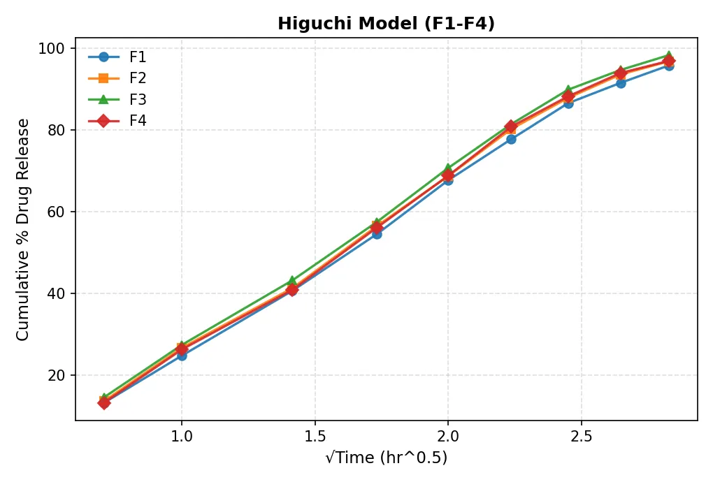
Figure 3.1 Higuchi modelrelease kinetic (F1-F4)
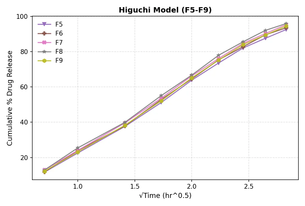
Figure 3.1Higuchi modelrelease kinetic (F5-F9)
.4.8 Drug Release Kinetic Study
The release data of the optimized Barbaloin gel (F3) were fitted to Zero-order, First-order, Higuchi, and Korsmeyer–Peppas kinetic models. The regression coefficients obtained from the respective plots are summarized in Table 6.23.
Table 3.4 Regression analysis of optimized Barbaloin gel (F3) using different kinetic models.
|
Kinetic model |
Regression coefficient (R²) |
|
Zero-order |
0.975 |
|
First-order |
0.986 |
|
Higuchi |
0.994 |
|
Korsmeyer–Peppas |
0.989 |
Figure 3.5 Drug release kinetic plots of optimized Barbaloin gel (F3): (A) Zero-order, (B) First-order, (C) Higuchi, and (D) Korsmeyer–Peppas model.
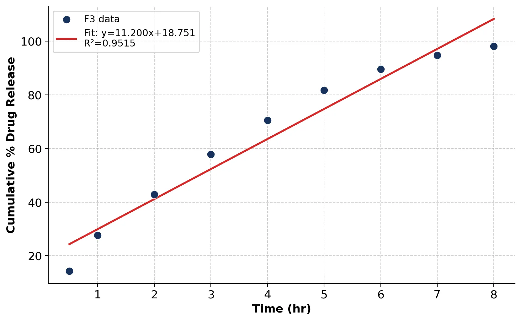
Figure 3.5(A) Zero-order Release Plot of Optimized Barbaloin Gel (F3)
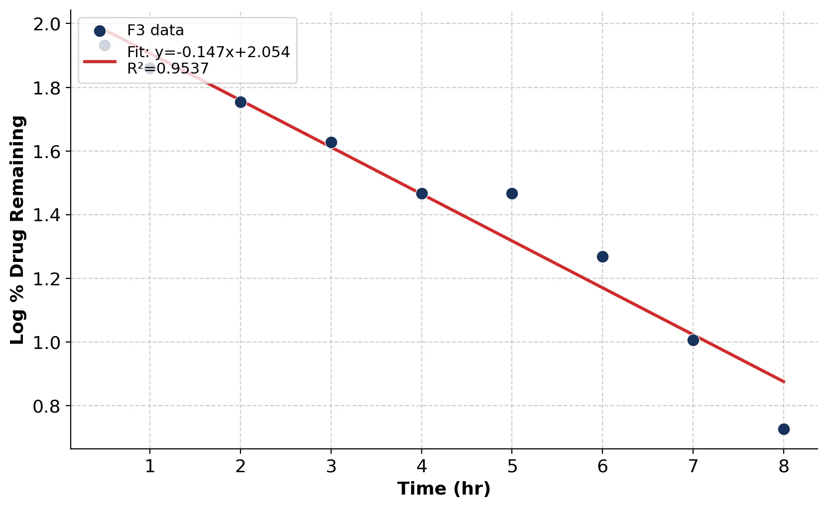
Figure 3.5(B) First-order Release Plot of Optimized Barbaloin Gel (F3)
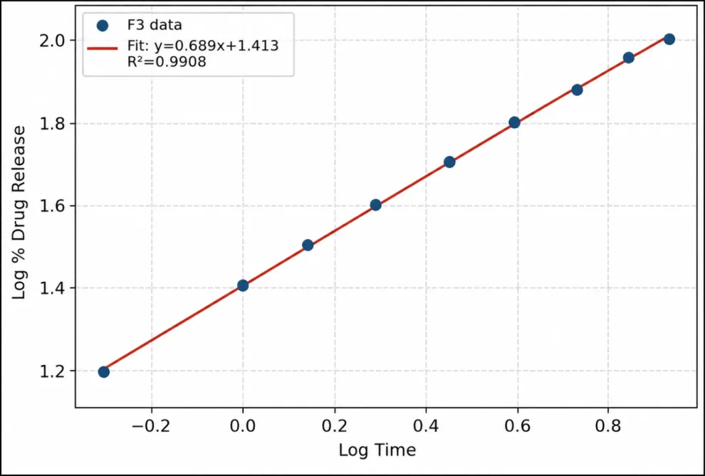
Figure 3.5(D) Korsmeyer–Peppas Release Plot of Optimized Barbaloin Gel (F3)
Discussion
The optimized Barbaloin-loaded topical gel (F3) exhibited the highest regression coefficient for the Higuchi model (R² = 0.994), followed by the Korsmeyer–Peppas (R² = 0.989), First-order (R² = 0.986), and Zero-order (R² = 0.975) models. The comparatively higher R² value obtained for the Higuchi model suggests that diffusion played a major role in the release of Barbaloin from the gel matrix. The good fit to the Korsmeyer–Peppas model further indicates that drug release may involve a combination of diffusion and polymer relaxation/swelling mechanisms. Overall, the kinetic analysis suggests that the release of Barbaloin from the optimized F3 gel was predominantly diffusion-associated, with possible contribution from polymeric matrix relaxation.
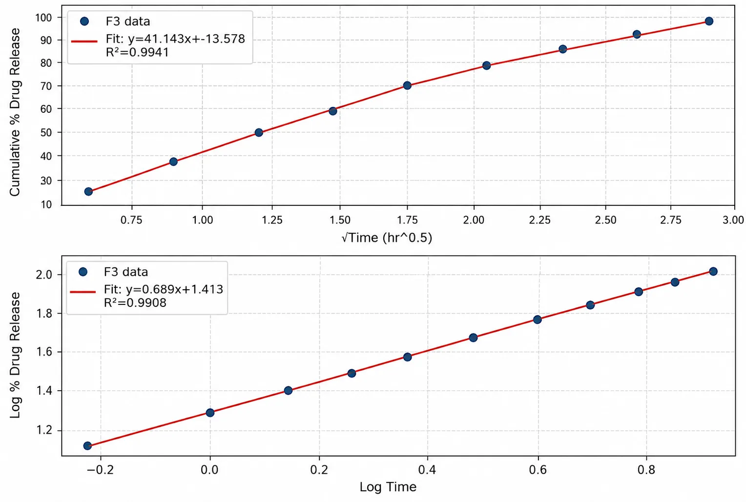
Figure3.6 Higuchi Model Release Plot
6.4.9 Comparative Drug Release Kinetic Study
The kinetic behaviour of the optimized Barbaloin gel (F3) was compared with that of standard Ketoprofen gel using the same mathematical models. The comparative regression coefficients are presented in Table 3.5
Table 3.5 Comparative regression analysis of optimized Barbaloin gel (F3) and standard Ketoprofen gel.
|
Parameter |
Optimized barbaloin gel (F3) |
Standard ketoperofen gel |
|
Zero order (R²) |
0.975 |
0.929 |
|
First order (R²) |
0.986 |
0.929 |
|
Higuchi (R²) |
0.994 |
0.971 |
|
Krosmeyer – peppas (R²) |
0.989 |
0.976 |
Discussion
The optimized Barbaloin gel (F3) showed higher regression coefficients than the standard Ketoprofen gel across all four evaluated kinetic models. For both formulations, the Higuchi model provided the highest correlation, with R² values of 0.994 for F3 and 0.971 for Ketoprofen gel. These findings support diffusion as an important mechanism governing drug release from the optimized Barbaloin gel
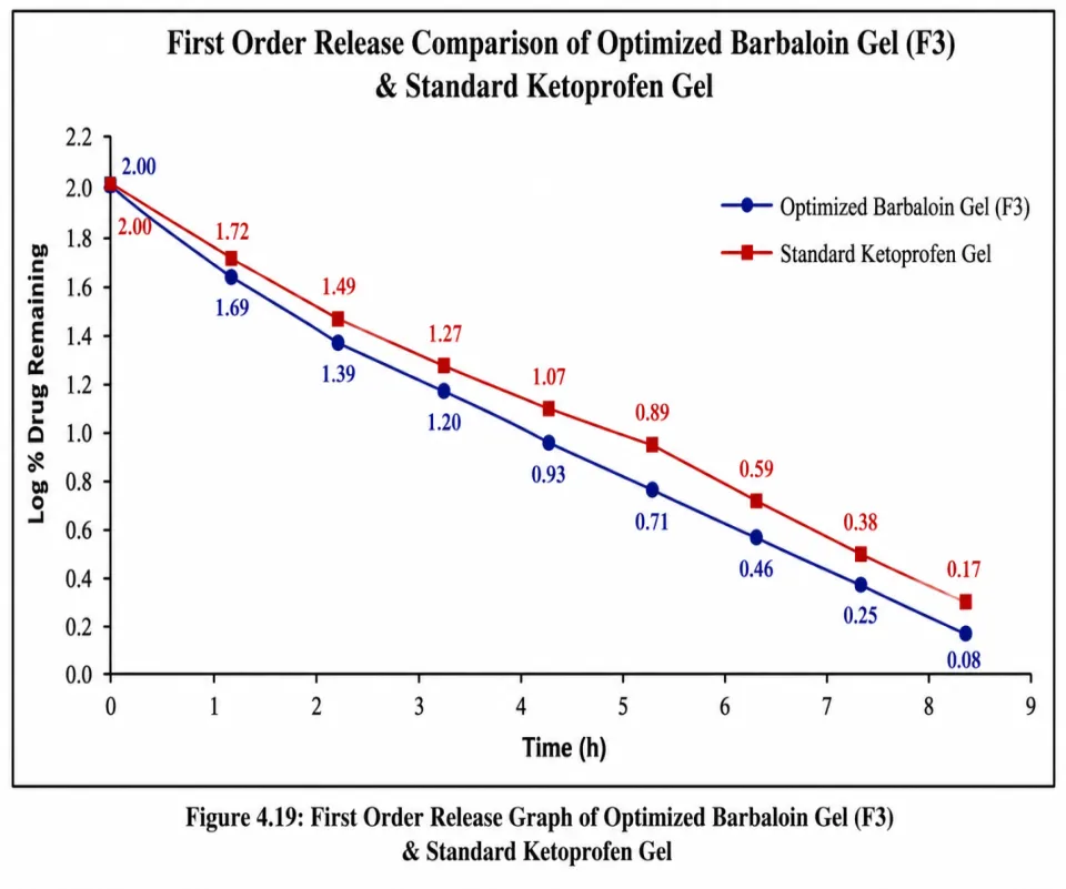
Figure 3.7-Higuchi Release Comparison of Optimized Barbaloin Gel (F3) and Standard Ketoprofen Gel.
6.4.10 Stability Study
The optimized formulation (F3) was subjected to stability studies under accelerated storage conditions. The formulation was evaluated for appearance, pH, viscosity, and drug content after storage.
Table 3.6 Stability Study of Optimized Barbaloin Topical Gel (F3)
|
Parameter |
Initial |
After 3 Months |
|
Appearance |
Clear, Homogeneous |
Clear, Homogenous |
|
Ph |
6.24 ± 0.03 |
6.22 ± 0.04 |
|
Viscosity (Cp) |
9188 |
9146 |
|
Drug Content (%) |
99.98 ± 0.54 |
99.36 ± 0.58 |
Discussion
No significant changes were observed in the appearance, pH, viscosity, or drug content after the storage period. The optimized formulation remained physically and chemically stable, indicating good stability under the selected storage conditions.
3.12 In-vitro Anti-inflammatory Activity
The in-vitro anti-inflammatory activity of the Barbaloin-loaded topical gel formulations (F1–F9) was evaluated using the protein denaturation inhibition assay. All formulations demonstrated inhibition of protein denaturation, with values ranging from 58.73 ± 1.26% to 76.84 ± 0.88%. The Carbopol 934-based formulations (F1–F4) generally exhibited higher inhibition than the HPMC-based formulations (F5–F9). Among the Carbopol 934-based formulations, the percentage inhibition increased from 63.42 ± 1.12% for F1 to 69.15 ± 0.94% for F2 and reached the maximum value of 76.84 ± 0.88% for F3 containing 1.5% Carbopol 934. A further increase in Carbopol 934 concentration to 2.0% resulted in a decrease in inhibition to 73.96 ± 1.05% for F4. In the HPMC-based formulations, inhibition increased from 58.73 ± 1.26% for F5 to 68.35 ± 1.14% for F8, followed by a decrease to 65.87 ± 1.19% for F9. The higher activity observed with F3 may be associated with an optimum balance between polymer concentration, gel structure, and drug availability. The reduction in activity at higher polymer concentrations may be related to increased viscosity and polymer network density, which can potentially restrict drug mobility and release. Based on its highest inhibition of protein denaturation (76.84 ± 0.88%) along with satisfactory physicochemical characteristics, F3 was selected as the optimized formulation.
Table 3.7. In-vitro anti-inflammatory activity of Barbaloin-loaded topical gel formulations (F1–F9) determined by the protein denaturation inhibition assay.
|
Formulation |
Gelling polymer |
Polymer concentration (% w/v) |
Inhibition of protein denaturation (%) |
|
F1 |
Carbopol 934 |
0.5 |
63.42 ± 1.12 |
|
F2 |
Carbopol 934 |
1.0 |
69.15 ± 0.94 |
|
F3 |
Carbopol 934 |
1.5 |
76.84 ± 0.88 |
|
F4 |
Carbopol 934 |
2.0 |
73.96 ± 1.05 |
|
F5 |
HPMC |
0.5 |
58.73 ± 1.26 |
|
F6 |
HPMC |
1.0 |
62.48 ± 1.08 |
|
F7 |
HPMC |
1.5 |
66.19 ± 0.97 |
|
F8 |
HPMC |
2.0 |
68.35 ± 1.14 |
|
F9 |
HPMC |
2.5 |
65.87 ± 1.19 |
Values are expressed as mean ± SD (n = 3).
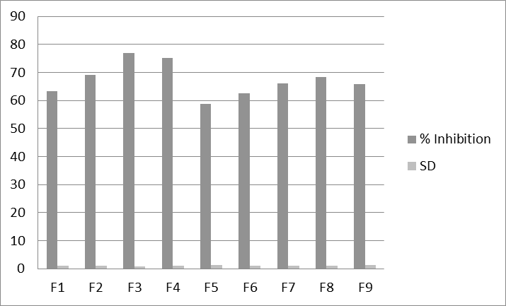
Figure 3.8 In-vitro anti -inflammatory acticity of barbaloin topical gel
Discussion
The variation in protein denaturation inhibition among the formulations indicates that the type and concentration of the gelling polymer influenced the anti-inflammatory performance of the Barbaloin-loaded gels. The comparatively higher inhibition observed with Carbopol 934-based formulations may be related to differences in gel structure and drug availability. F3, containing 1.5% Carbopol 934, showed the highest inhibition (76.84 ± 0.88%), whereas a slight reduction was observed following further increase in polymer concentration, as demonstrated by F4 (73.96 ± 1.05%). This reduction may be associated with increased polymer concentration and viscosity, which can restrict drug mobility and release. A similar trend was observed with HPMC, where activity increased up to F8 and subsequently decreased in F9. These findings suggest that an optimum polymer concentration is important for achieving desirable anti-inflammatory performance. Accordingly, F3 was selected as the optimized formulation for further evaluation.
DISCUSSION
The present study was focused on the formulation and evaluation of Barbaloin-loaded topical gel formulations using Carbopol 934 and HPMC as gelling polymers. Nine formulations (F1–F9) were prepared with varying polymer concentrations and evaluated for their physicochemical characteristics, drug content, drug-release behaviour, release kinetics, irritation potential, stability, and in-vitro anti-inflammatory activity. All prepared formulations exhibited satisfactory physical appearance, with clear to translucent appearance, smooth texture, and good homogeneity. No visible grittiness, particulate matter, or phase separation was observed. The pH values of the formulations ranged from 6.10 ± 0.03 to 6.82 ± 0.04, indicating that the prepared gels possessed pH values suitable for topical application [28]. A gradual increase in pH was observed with increasing polymer concentration. The spreadability of the formulations was also satisfactory, although formulations containing higher polymer concentrations showed comparatively lower spreadability. This behaviour may be attributed to the formation of a denser polymeric network and increased gel consistency. The viscosity of the formulations ranged from 8,900 to 10,180 cP. Carbopol 934-based formulations showed viscosity values ranging from 8,900 to 9,345 cP, whereas HPMC-based formulations exhibited values ranging from 9,498 to 10,180 cP. The overall increase in viscosity with increasing polymer concentration indicates that polymer concentration influenced the consistency and internal structure of the gel matrix. Higher viscosity may also affect drug mobility and consequently influence the release characteristics of the formulation [29]. The drug content of the prepared formulations ranged from 97.45 ± 0.82% to 99.98 ± 0.54%, demonstrating satisfactory and relatively uniform incorporation of Barbaloin into the gel formulations. Among the investigated formulations, F3 exhibited the highest drug content of 99.98 ± 0.54%. The high drug content observed across the formulations indicates that the preparation method was suitable for obtaining relatively uniform drug distribution within the gel matrix. The optimized F3 formulation was subjected to an in-vitro drug-release study using a Franz diffusion cell. The cumulative drug release increased progressively from 14.28 ± 0.42% at 0.5 h to 98.21 ± 0.62% at 8 h. The progressive release profile indicates that the gel matrix provided a controlled environment for Barbaloin release over the investigated period. The polymeric network may have influenced the movement of the drug through the hydrated gel matrix and thereby contributed to the observed release behaviour. The release data of F3 were fitted to Zero-order, First-order, Higuchi, and Korsmeyer–Peppas kinetic models. The Higuchi model exhibited the highest regression coefficient (R² = 0.994), followed by the Korsmeyer–Peppas model (R² = 0.989), First-order model (R² = 0.986), and Zero-order model (R² = 0.975). The comparatively higher R² value for the Higuchi model suggests that diffusion was an important contributor to Barbaloin release from the gel matrix. The good fit obtained with the Korsmeyer–Peppas model further indicates the possible contribution of both drug diffusion and polymer relaxation/swelling. The release exponent (n = 0.689), when interpreted using the appropriate release-data region and geometry, is consistent with anomalous or non-Fickian transport, suggesting the involvement of more than one release mechanism. The comparative kinetic analysis showed that F3 exhibited higher regression coefficients than the standard Ketoprofen gel for all the investigated models. The Higuchi model showed the highest R² for both F3 and Ketoprofen gel, with values of 0.994 and 0.971, respectively. These findings indicate that diffusion was an important component of drug release in both formulations. However, the difference in R² values represents model fit and should not be interpreted as evidence that F3 has greater clinical efficacy than Ketoprofen. The in-vitro anti-inflammatory activity of the formulations was evaluated using the protein denaturation inhibition assay. All formulations demonstrated inhibition of protein denaturation, with values ranging from 58.73 ± 1.26% to 76.84 ± 0.88%. Carbopol 934-based formulations generally showed higher inhibition values than HPMC-based formulations. Among the Carbopol formulations, the inhibition increased from 63.42 ± 1.12% in F1 to 69.15 ± 0.94% in F2 and reached the highest value of 76.84 ± 0.88% in F3 containing 1.5% Carbopol 934. Further increase in Carbopol 934 concentration to 2.0% resulted in a decrease in inhibition to 73.96 ± 1.05% in F4. Among the HPMC-based formulations, inhibition increased from 58.73 ± 1.26% in F5 to 68.35 ± 1.14% in F8, followed by a decrease to 65.87 ± 1.19% in F9. The observed variation indicates that both polymer type and concentration influenced the anti-inflammatory performance of the formulations. The higher activity observed with F3 may be associated with an optimum balance between polymer concentration, gel structure, and Barbaloin availability. The decrease observed at higher polymer concentrations may be related to increased viscosity and polymer network density, which can restrict drug mobility and release. The HET-CAM study showed an irritation score of 0.0 for the tested formulations, with no visible signs of hemorrhage, vascular lysis, or coagulation during the observation period. These findings indicate a favourable irritation profile under the experimental conditions. However, HET-CAM findings should be considered as an in-vitro preliminary assessment and should not be interpreted as confirmation of clinical skin safety. The optimized F3 formulation was further evaluated for stability. Only minor changes were observed in appearance, pH, viscosity, and drug content after three months of storage under the selected study conditions. The drug content decreased from 99.98 ± 0.54% to 99.36 ± 0.58%, while pH changed from 6.24 ± 0.03 to 6.22 ± 0.04 and viscosity changed from 9188 to 9146 cP. These observations indicate that F3 maintained its evaluated physicochemical characteristics during the investigated stability period. Overall, the findings demonstrate that the type and concentration of gelling polymer played an important role in determining the physicochemical properties, drug-release behaviour, and anti-inflammatory performance of the Barbaloin-loaded topical gels. Among the investigated formulations, F3 containing 1.5% Carbopol 934 provided the most favourable overall performance and was therefore selected as the optimized formulation.
CONCLUSION
The present study successfully developed and evaluated Barbaloin-loaded topical gel formulations using Carbopol 934 and HPMC as gelling polymers. Nine formulations (F1–F9) were prepared and evaluated for their physicochemical properties, drug content, in-vitro drug release, release kinetics, irritation potential, stability, and anti-inflammatory activity. Among the prepared formulations, F3 containing 1.5% Carbopol 934 demonstrated the most favourable overall performance. It exhibited the highest drug content (99.98 ± 0.54%) and the highest inhibition of protein denaturation (76.84 ± 0.88%). The optimized F3 formulation showed progressive drug release, reaching 98.21 ± 0.62% after 8 h. The release data showed the best fit with the Higuchi model (R² = 0.994), indicating a major contribution of diffusion to the release process, while the Korsmeyer–Peppas model (R² = 0.989; n = 0.689) suggested the possible contribution of diffusion along with polymer relaxation/swelling. The optimized formulation also exhibited a favourable in-vitro irritation profile with an HET-CAM irritation score of 0.0 and maintained satisfactory physicochemical characteristics during the three-month stability study. The findings demonstrate that appropriate selection and concentration of the gelling polymer are important for achieving desirable formulation performance. In conclusion, the developed Barbaloin-loaded topical gel, particularly formulation F3, demonstrated satisfactory physicochemical characteristics, favourable drug-release behaviour, stability, and promising in-vitro anti-inflammatory activity. The formulation may therefore be considered a potential candidate for further investigation as a topical delivery system for Barbaloin. Further studies, including detailed skin permeation, in-vivo efficacy, and comprehensive safety evaluation, would be required to establish its therapeutic potential for topical inflammatory conditions.
REFERENCES


Vishakha Bhati*, Deepika Chaudhary, Formulation, Characterization and Anti-Inflammatory Evaluation of Barbaloin Loaded Topical Gel, Int. J. Med. Pharm. Sci., 2026, 2 (9), 518-532. https://doi.org/10.5281/zenodo.22972154
 10.5281/zenodo.22972154