

We use cookies to ensure our website works properly and to personalise your experience. Cookies policy
Department of Pharmaceutics, Krishna School of Pharmacy and Research, Drs. Kiran and Pallavi Global University, Varnama, Vadodara, Gujarat 391243, India
Cardiovascular diseases (CVDs) remain the leading cause of mortality worldwide because adult cardiomyocytes have a limited capacity to recover following ischemic injury. Unfavorable ventricular remodeling, long-term myocardial injury, and progressive loss of cardiac function are associated with heart failure and myocardial infarction. Although current pharmacological and interventional therapies reduce acute mortality, they are often limited by systemic toxicity, inadequate targeting, and poor retention at the disease site. By facilitating tailored drug delivery, prolonged release, better biodistribution, and increased therapeutic effectiveness, nanotechnology has emerged as a viable approach to get around these restrictions. In addition to helping with enhanced diagnostics and cardiac regeneration, designed nanocarriers such nanoparticles, nanogels, and bioengineered nanopatches have shown promise in the treatment of atherosclerosis, myocardial infarction, and heart failure. The mechanisms of action of nanotechnology-based therapeutic and diagnostic techniques for cardiovascular illnesses are highlighted in this study, along with their existing obstacles and potential for clinical translation.
Cardiovascular diseases (CVDs) continue to be a major cause of death and morbidity despite the recent tremendous growth of the global healthcare sector. Earlier this year, the American Heart Association reported that 17.6 million deaths globally were attributed to CVDs in 2016. [1]. Among the several conditions that fall under the category of CVDs, ischemic heart disease is the leading cause of death. When an artery becomes blocked, ischemia occurs. This blockage damages a specific area or tissue in the body by preventing blood flow and oxygen supply. A clot resulting from atherosclerosis, thrombosis, or embolism may be the source of this blockage. An ischaemic event occurring in the heart deprives it of its blood and oxygen supply leading to myocardial infarction (MI) or heart attack. In such cases, the usual treatment is to relieve the occlusion as soon as possible and restore the blood flow. However, paradoxically, the restoration of blood flow (also called reperfusion) that accompanies this may cause further damage [2]. The current treatment for MI is reperfusion as soon as possible following arterial obstruction. Pharmacological or invasive methods such as coronary artery bypass grafting (CABG) or percutaneous coronary intervention (PCI) can be used to achieve this [3]. Fatigue, peripheral edema, and dyspnea are symptoms of heart failure, a clinical disease condition caused by reduced cardiac functioning [4]. When the heart muscle is unable to adequately pump blood to meet the body's metabolic needs, this kind of cardiomyopathy develops [3]. About 25% of the heart's cardiomyocytes (CMs) are lost after a MI. The heart cannot regenerate new CMs to replace lost ones because adult CMs have a limited capacity to multiply. As a consequence, it repairs itself by creating fibrotic scar tissue that unlike CMs cannot beat. This healing process, called ventricular remodeling, thus increases the burden that the heart has to cope with and thus slowly weakens its muscles. The heart will then slowly deteriorate, losing contractility and muscle strength and, in the long term, this may lead to congestive heart failure and, ultimately, death [5]. A variety of cardiac symptoms, including irregular pulse, valvular, pericardial, or endocardial abnormalities, and left ventricular dysfunction, are together referred to as heart failure [4]. roughly 80 million Americans, or roughly one-third of the population, suffer from cardiovascular disease. [6]. The majority of CVD treatments rely on invasive surgery or oral drugs, and we have not yet been able to halt or reverse this silent pandemic [7]. The current incidence figures show that novel medicines and technological advancements are needed to address CVDs.
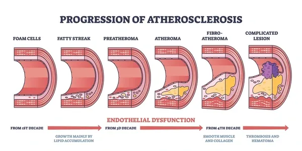
Figure 1: Atherosclerotic plaque progression indicating blood vessel narrowing
Atherosclerosis precedes a number of vascular diseases. Calcium, fat, cholesterol, and macrophage cells are accumulated in the vasculature and eventually solidify to form atherosclerotic plaques. This results in a reduction in blood flow, a shrinkage of the blood vessels, a rupture, and ultimately ischaemia [8]. Lifestyle decisions are one of the main causes of atheromatous plaques. It is well known that consuming meals heavy in sugar, salt, and saturated fat greatly increases the risk of getting a CVD in later life. A high low-density lipoprotein (LDL)-to-high-density lipoprotein (HDL) ratio is especially dangerous because LDLs lodge on blood vessel walls and contribute to the development of plaques. The current treatments for CVDs focus on restoring normal blood flow through or around the injured vasculature and preventing further cardiovascular shocks. Statin therapy affects fibrous and dense calcium volumes, external elastic membranes, and the formation and thickness of atherosclerotic plaques [9]. Dual antiplatelet drugs, which employ cyclooxygenase inhibitors like aspirin and P2Y12 inhibitors like clopidogrel to reduce platelet aggregation and clot formation, are first-line therapies for avoiding CVDs [10]. Antiplatelet therapy has risks that lead to extremely poor side effect profiles and low patient compliance, which is the main reason these therapies need to be improved [11]. Furthermore, some patients have poor responses to antiplatelet medication, which worsens their prognosis over the long run.
Nanoparticles
Nanoparticles for Therapeutic Delivery
There are several cardiac therapies available following an ischemic incident.To ensure that these medicines reach the myocardium, intracoronary catheterization or direct intramyocardial injection could be required [12]. Functionalized poly(lactic-co-glycolic acid) (PLGA) nanoparticles containing insulin-like growth factor (IGF)-1 injected intramyocardially into the heart have been shown to have effective cardioprotective effects [13]. Even though intramyocardial injection has shown encouraging outcomes, it is a somewhat intrusive procedure that may cause further harm to the heart. In contrast, an intracoronary injection entails a direct inside injection. Catheterization may not be suitable for every medicine and may result in embolization. Because intravenous infusion is less intrusive, less dangerous, and safer, all of these qualities make it more desirable. Therapeutics will go through the circulatory system when administered via systemic intravenous injection. We previously used this phenomenon to show that heparin-conjugated intravenous infusion might reduce thrombus development in a mouse model of acute hind limb thrombosis. Because carbon nanocapsules are administered intravenously, they are more biocompatible than other types of carbon nanomaterials, which is why they were used. Compared to heparin or carbon nanocapsules alone, heparin-conjugated carbon nanocapsules have been demonstrated to exhibit antithrombotic effect and to lengthen the thrombus formation time [14]. Following intravenous injection, the medications are removed and tolerated by various organs as they travel through the bloodstream. Non-functionalized treatments will mostly collect in organs of the reticuloendothelial system, such as the liver and spleen, and will never be able to target the cardiovascular region particularly [12]. The current idea is to "protect" these treatments by encasing them in nanoparticles. Depending on the physicochemical characteristics of the carrier, the medicines may take a varied path of retention to their free form after being engulfed in nanoparticles. Drug encapsulation in nanoparticles may be advantageous as it may extend the medications' half-lives [12,13,15]. The nanoparticles can target the disease areas by passive targeting even if they don't have active targeting groups. The EPR effect is thought to be the phenomena that underlies passive targeting, in which drug-loaded nanoparticles were able to enter because the vasculatures of diseased and injured regions became unusually porous [16]. Because the EPR effect lasts for a very long period, it has been demonstrated that it has been exploited as a targeting method for malignancies. The increased permeability of the disease vasculature has been linked to the production of the vascular permeability factors [17] and a number of other compounds, including those involved in the enhancement of this effect, like nitric oxide, peroxynitrite, and bradykinin [18]. The capacity of the nanoparticles to be maintained inside the tumors has been linked to both the absence of lymphatic capillaries within the tumors [18,19] and the one-way extravasation within the vasculatures [20].
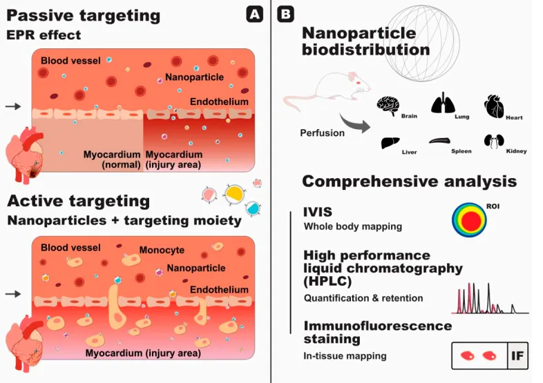
Figure 2: Application of functionalized nanoparticles for treatment of ischemic injury in cardiac tissues: (A) Passive targeting based on the enhanced permeability and retention (EPR) effect in comparison to active targeting,using nanoparticles decorated with targeting moieties. Nanoparticles with monocyte-targeting abilities have can actively bind to the surface of monocytes and then migrate to the damaged area; (B) Nanoparticle biodistribution experiments play an essential part in assessing the fate of nanoparticles in the body. in vivo. Perfusion is carried out to eliminate freely circulating nanoparticles found in the blood prior to In vivo. Perfusion is carried out to eliminate freely circulating nanoparticles that are found organ collection. In summary, the biodistribution study outlines the spread of the nanoparticles in within the body and within the tissue. It also shows the amount retained within each tissue, giving researchers with an overall view of the ultimate disposal of their nanoparticles.
A research has also demonstrated the EPR effect in the myocardium after an ischemia event [21]. In order to promote angiogenesis and replenish oxygen in the damaged myocardium, there is an increase in vascular endothelial growth factor production during the post-MI phase [22, 23]. Additionally, prior research has demonstrated that VEGF increases vascular permeability in this setting [22, 24, 25], which in turn causes the myocardium to experience the post-MI EPR effect [22]. However, in contrast to previous tumor cell-related instances, the post-MI EPR impact only takes a short time to manifest, peaking 24–48 hours after the MI and exhibiting some functional cardiac recovery after two weeks [21]. In order to prevent depressing left ventricular remodeling in this case, therapy and myocardial recovery from an ischemia event take a long time [26]. However, this time frame is still insufficient for administering medication in this situation. However, recent research has shown that the EPR effect may be used to passively target the heart after an ischemic episode [26–29]. One study reported the usage of nanoparticles that can target the heart using the EPR effect. When stimulated by matrix metaloproteases (MMPs), which include the enzymes MMP-2 and MMP-9, the particles can self-aggregate. The aggregate matures to form a scaffold that is able to retain itself within a maximum of 28 days post-MI [26]. Although the study has successfully increased the retention time, the nanoparticles would still have to be done before the EPR effect is subdued. Consequently, elimination. The use of the EPR effect remains the best approach. This can be realized through the use of nanoparticles decorated with targeting modules actively. The PLPs were engineered to leverage the platelet and monocyte binding, imitate the binding, and utilize monocytes as a shuttle bus that could carry the drugs directly to the heart. The drug cobalt protoporphyrin (CoPP) has been chosen for use in the study. This compound has the ability to inhibit the inflammatory function of macrophages through the induction of heme oxygenase-1 (HO-1) protein synthesis, but as a harmful effect, the compound causes hepatotoxicity and nephrotoxicity. The cobalt protoporphyrin-encapsulated PLPs successfully bound themselves to the monocytes and piggybacked their way towards the infarcted hearts without using the EPR effect. After the extravasation into the myocardium, the PLPs were phagocytosed by the macrophages, and the subsequent release of cobalt protoporphyrin directly inside the macrophages enhanced the treatment outcomes. The cobalt protoporphyrin-encapsulated PLPs showed a dramatically increased targeting potential and successful improvement in heart function with less side effects compared to the free cobalt protoporphyrin or the cobalt protoporphyrin-encapsulated liposomes. Although a dramatic increase in targeting potential showed the success of the study, a measly 5% of the injected PLPs could be found in the heart, the majority instead getting stuck in the spleen and the liver. Although higher than previously reported in other studies, this number should improve so that more nanoparticles could reach the heart and thus increase the therapeutic outcome.
Nanogel
Diseases have been treated by protein and cell treatments. Since the existing treatments simply stop further harm to the afflicted region, the illnesses are intended to be cured via tissue repair regeneration. However, human trials using protein or cell therapy to treat cardiovascular illnesses have not been able to surpass the current therapies. The limited retention of the active components within the damaged site is notably responsible for this. Hyaluronic acid hydrogels [28], alginate hydrogels [29], and self-assembling peptide nanofibers [27] are examples of nanomaterials that have been designed to function as a matrix in trapping medicines and an environment favorable to heart healing in order to overcome this problem. We demonstrated the effectiveness of a combination of autologous bone marrow mononuclear cells and self-assembling degradable peptide nanofibers for the treatment of pigs with experimentally induced MI using a different model of the disease, and we discovered that this combination was successful in improving both systolic and diastolic function parameters following injury [28]. Additionally, we showed that in both murine and pig model animals, a combination of nanofibers and vascular endothelial growth factors may stimulate arteriogenesis, improving heart systolic performance and reducing infarction size within four weeks following MI [30]. Biodegradable peptide nanofibers maintained the area of injury for three months; this created a favorable environment for cardiac repair and added mechanical strength. Worth mentioning is the fact that the efficacy of cell therapy is time-dependent; the procedure must be applied within four days after MI [31]. In conclusion, it is demonstrated that nanofibers and nanogels play a crucial role in improving the efficacy of cell/protein therapy.
Nanopatch
Pharmacologic therapy may not be sufficient in situations when myocardial cell damage is severe; thus, stem cell therapy and transplantation may be necessary. For cell-based regeneration of the injured myocardial cells in MI, ESC and iPSC-derived CM cells have shown promise. It has been shown that injecting murine ESCs or hiPSCs directly into post-MI rat hearts can restore cardiac functioning; nevertheless, one of the key obstacles need to be addressed is the implanted CM cells' retention, survival, and capacity to correctly integrate with the host cells [32, 33]. Bioengineered cardiac grafts have been developed to aid in post-MI myocardial regeneration in order to overcome the difficulties related to cell survival and retention. In order to produce three-dimensional cell sheets, Sekine et al. [34,35] cultivated neonatal rat CMs either by themselves or in conjunction with endothelial cells in temperature-responsive culture pans. The effectiveness of the method in enhancing the retention of the transplanted CMs in the infarct area was next demonstrated by transplanting these cell sheets into the infarcted heart of a rat model of MI [34, 35]. Thus, the left ventricular dysfunction could be quantified by decreased left ventricle wall thickness and fractional shortening, which indicates percentage change in the left ventricular cavity during systolic contraction. Consequently, the hearts that were subjected to cell sheet transplantation showed improved fractional shortening, left ventricle wall thickness, and neovascularisation [34]. By using the same temperature-sensitive cell sheet technology, Kawamura et al. developed hiPSC-derived CM sheets, combined with the omental flap, and finally transplanted the cell sheet into a porcine ischemic cardiomyopathy model. Originating from the peritoneal layers, the omental flap supplies blood to the cell sheet. Similarly, they demonstrated that the hiPSC-derived transplanted CMs had excellent long-term survival on the cell sheets and markedly enhanced cardiac functions, including as LVEF and the post-MI porcine heart's end-diastolic and systolic volumes [36].
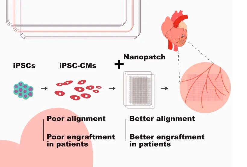
Figure 3: The integration of cardiomyocytes produced from pluripotent stem cells would improve when these cells are attached to a cell sheet. The shortcomings of these cells are misalignment and the inability to integrate. When attached to a nanopatch, the cardiomyocytes will be better aligned, ultimately leading to better integration for the patient.
Prior to the use of cardiac patch technology for post-MI therapy, some issues had to be resolved. In order for the cells to survive, the cardiac patch must first work correctly on the host heart, starting with the vascular connection. When arrhythmia arises following a heart transplant, the relationship between the cardiac patch and the heart should also be taken into account.
Diagnostics
Nanotechnology holds the key to significantly speeding up the implementation of personalized medicine within the cardiovascular community, enabling the rapid, multiplex, point-of-care detection of single nucleotide polymorphisms (SNPs). The former will, in turn, provide information relative to the risks of developing specific cardiovascular diseases, and pharmacogenetic guidance on the most appropriate therapeutic approach for the patient. A useful system will also have to be quick, accurate, capable of simultaneous measurement for multiple genotypes, and able to complete the entire analysis without requiring assistance. There appear to be a great many competing technologies emerging. A recent review describes a great many systems, some of these nano-technologies. [37] Besides genotyping, nanotechnology also enables the ultr-sensitive detection of proteins and other biomarkers. The ‘bio-barcode’ approach, for example, has a sensitivity for protein detection down to 100 nM, and has been used by the Mirkin group to show that diffusible ligands derived from amyloid, when present in cerebrospinal fluid, could be a marker for Alzheimer’s disease. [38] The test involves magnetic particles that bind a primary antibody for capturing a protein target, followed by a binding reaction for protein targets and hundreds of ‘identical’ oligonucleotides tagged with antibodies for gold particles. The target is then magnetically captured, washed, and eluted with a heating solution for release of the bound oligonucleotides, followed by analysis as described above for methods that employ genomic probes tagged with gold particles can also be multiplexed in various combinations of antibodies and oligonucleotides in a multiplex microarray. It also has an on-chip format that can easily read results by use of the Verigene ID System. [39]
Imaging
One of the main goals of using nanotechnology in the cardiovascular system has been to characterize atherosclerotic plaque. In the past, medical professionals have assessed plaque using contrast agents in conjunction with plaque X-rays or, more recently, computed tomography (CT) to look for vascular constriction. However, because the artery remodels while plaque accumulates, a significant constriction of the coronary artery does not happen until a plaque has fully matured. Moreover, anatomical imaging cannot predict the probability of plaque rupture, which is the cause of most MIs and strokes. Figure 4 [40] displays a range of markers pertinent to each stage of plaque development. Using targeted imaging agents to identify the expression of indicators specific to various stages of plaque progression is the aim of molecular imaging. Plaque imaging can also target blood monocytes that move into the artery intima and develop into macrophages. Macrophages are believed to play a crucial role in destabilizing and rupturing plaque by releasing proteases that degrade and harm the extracellular matrix and the fibrous cap that protects the plaque. A thrombus may develop and block the artery when plaque ruptures, releasing thrombogenic plaque components. Hyafil et al. used N1177, a macrophage-avid iodinated nanoparticulate contrast agent, to detect macrophage-rich atherosclerotic plaques that were thought to be at high risk of rupturing.8. The agent allowed for the rapid (2-hour) identification of atherosclerotic plaque in rabbits using CT. The capacity to image with CT may be especially useful for the identification of plaque in coronary arteries as spatial and time constraints might limit the significance of other imaging techniques like MRI and positron emission tomography. [41]
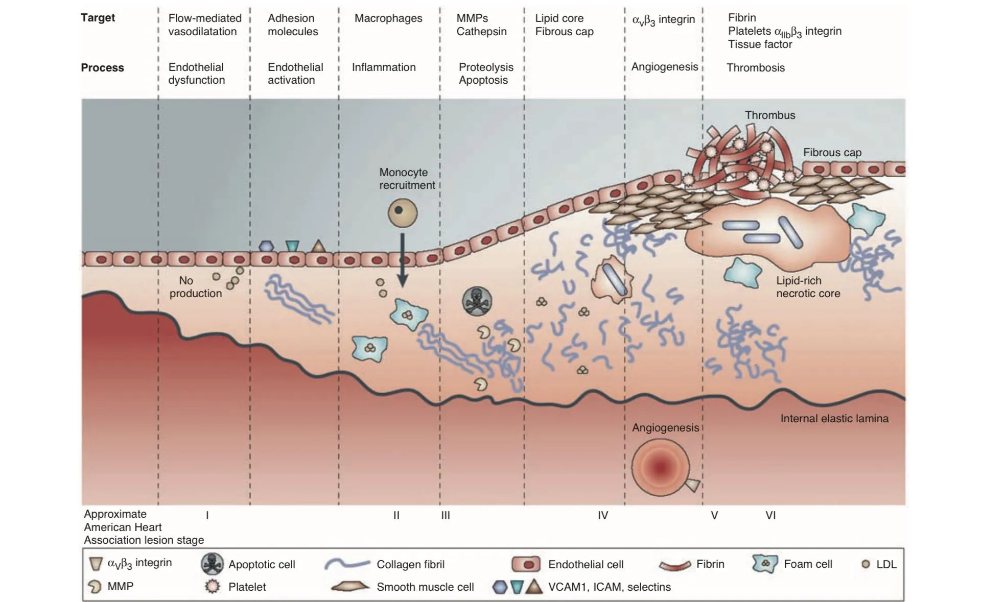
Figure 4: Illustration of processes of atherogenesis ranging from prelesional endothelial dysfunction (a) through monocyte recruitment to the development of advanced plaque complicated by thrombosis (b). The mechanisms are grossly simplified but focus on components (for example, cell adhesion molecules, macrophages, connective tissue elements, lipid core, and fibrin) and processes (for example, apoptosis, proteolysis, angiogenesis, and thrombosis) in plaques that have been imaged or that present useful potential imaging targets. ICAM, intercellular cell adhesion molecule; LDL, low-density lipoprotein; MMP, matrix metalloproteinase; NO, nitric oxide; VCAM, vascular cell adhesion molecule.
Therapeutics
Distribution of drugs to atherosclerotic plaque can be targeted using the same techniques used for molecular imaging. The expansion of the vasa vasorum, the blood supply to the artery wall, coincides with the development of atherosclerotic plaque and the thickening of the vascular intima. [42] In animals, it has been demonstrated that blocking the thrombotic expansion of the vasa vasorum inhibits the development of plaque; nevertheless, high dosages of anti-angiogenic drugs have negative neuropsychological effects in people. Winter et al., however, showed a reduction in microvessels in atherosclerotic rabbit aortas by encapsulating the anti-angiogenic medication fumagillin in paramagnetic nanoparticles and directing them to the neovasculature using a peptide that binds to the αvβ3 integrin. [43] The paramagnetic properties of the nanoparticles also allowed for MRI imaging of αvβ3 integrin on the plaque, revealing a corresponding decrease in integrin expression due to the drug. Naturally, cardiovascular health is impacted by the function of other organs, including the liver, and nanotechnology may help target hepatocytes. By employing a N-acetylgalactosamine targeting ligand to deliver a novel siRNA-containing nanoparticle formulation to the hepatocytes, Rozema et al. prevented Kupffer cell uptake. [44] The nanoparticles are transported by endocytosis into the endosomal compartment, where the reduced pH causes ligand breakdown to reveal positively charged groups. The siRNA is released into the cytoplasm as a result of endosomal instability. Release of siRNA has been utilized to downregulate apoB expression, lowering plasma cholesterol, in a proof-of-concept study in mice. By reducing the cardiotoxicity of drugs used to treat ailments other than heart disease, nanotechnology will also enhance cardiovascular health. Halofantrine is an antimalarial medicine that might be useful in treating multidrug-resistant malaria. However, the medication's usage is restricted and can result in bradycardia and hypotension if the QT interval—the space between the Q wave and the conclusion of the T wave in the heart's electrical cycle—is extended. By encasing the medication in polycaprolactone nanocapsules, Leite et al. were able to successfully cure malaria in mice while lessening the drug's cardiotoxic effects. In [44] The cancer medication doxorubicin, which exhibits similar efficacy but less cardiotoxicity when packed into 100-nm pegylated liposomes (Doxil), is one example of a medicine whose cardiotoxic effects may be reduced via nanoparticle encapsulation. [45]
Repair and Regeneration of Tissues
Blood Vessels
Because present methods are plagued by thrombosis and restenosis, the development of tiny diameter blood arteries is a major unmet need for cardiovascular repair. Nanofibrous scaffolds show great promise as the base for vascular grafts. Stankus et al. simultaneously electrosprayed smooth muscle cells and electrospun a biodegradable elastomer polymer to construct a small diameter conduit with uniform cell integration. [46] Although the resultant vasculature was strong and adaptable and shared traits with native arteries, thrombogenicity is still an issue that has to be resolved. This problem could be resolved by another study that makes use of nanofibrous scaffolds. Electrospun biodegradable scaffolds filled with mesenchymal stem cells showed exceptional long-term patency when inserted into rat carotid arteries during a bypass procedure [47]. The scaffold promoted efficient cellular infiltration and matrix remodeling similar to that of native arteries. Another top aim is the creation of long-lasting biosynthetic heart valves that have dynamic characteristics, a structure, and a function similar to the native valve. Bacterial cellulose and polyvinyl alcohol nanocomposite hydrogels have demonstrated potential as a material for biosynthetic heart valves with characteristics akin to those of the natural valve [48]. By using nanotechnology to examine the nanoscale topography of the valve basement membrane, the issue is also being addressed. This could help with the logical design of scaffolds with the required characteristics [49].
Cardiac Restoration
Another challenge is preventing ventricular remodeling following MI. In individuals with bigger infarcts (transmural infarcts), which result in damage across the whole width of the heart wall, the dead heart tissue is replaced by a noncontracting fibrotic scar. Over time, the heart chamber expands and the ventricle wall thins, leading to heart failure. Both in vitro tissue engineering and in vivo regeneration methods are being investigated to solve this problem. Furthermore, the in vitro development of scaffolds to support cardiomyocytes is showing promise via electrospinning. [50,51] Similar to the extracellular matrix, the scaffolds offer external signals for both isotropic and anisotropic development, enabling cells to proliferate into and pull on the fibers. Ishii et al. grew neonatal rat cardiomyocyte constructions up to five layers thick using nanofibrous poly (-caprolactone) meshes. The layers were connected morphologically and electrically, and they beat vigorously. The implementation of a vascularization plan will be necessary for future development. Nanotechnology has also been used for in vivo cardiac regeneration by infusing RADA16 self-assembling nanofibers (NF) into the heart. Together, the peptides form intramyocardial microenvironments that attract endothelial cells and promote their survival. [52] Furthermore, the peptide NF has the ability to modulate the transit of proteins to the heart, which may have therapeutic benefits.[53, 54, 55] Co-injection of platelet-derived growth factor (P-BB) reduced cardiomyocyte mortality and infarct size following MI in rats (Figure 4). The NF protected against ischemia/reperfusion injury and preserved systolic function after MI.[53] They have also been used in combination with cell therapy to provide insulin-like growth factor, which has enhanced function following experimental MI.[54] When protease-resistant stromal cell derived factor-1 was attached to the injected fibers, natural stem cells were more likely to be drawn to the site of injury to improve heart function.[55] Injectable NF can thereby promote extended protein transport to the myocardium, preserving healthy heart muscle and postponing the development of heart failure.
Additional developments in nanotechnology
In addition to the systemic administration of tailored nanoparticles, there are a number of other nanotechnological developments that might greatly enhance the sector. In addition to intravenous injection, technical improvements have made it feasible to provide medications and cells to the heart intramyocardially and intrapericardially. [56] One example of a therapeutic device is the implanted epicardial reservoir called "Therepi," developed by Whyte et al. for the repeated infusion of drugs or cells. By changing the porosity of the semipermeable membrane, the researchers modified their treatment rate around the infarct border zone. In a mouse MI study, the researchers showed that the device may be utilized for recurring cell administration to improve heart function. [57] A similar strategy to customizable drug release is shown by implantable nanochannel membrane devices, which may be passively [58,59] or actively [60,61] managed to deliver cardiovascular drugs for scheduled treatments. In a primate artery graft model, an extended polytetrafluoroethylene (ePTFE)-based drug delivery device locally infused heparin and improved graft patency without systemic anticoagulation to evaluate its application in people. [62, 63] Furthermore, therapeutic heart regeneration has showed promise when drugs and stem cells are included into microneedle patches that might be placed to a damaged heart. [64. Last but not least, functionalizing endovascular intervention devices with magnetic nanoparticles such as superparamagnetic iron oxide can result in magnetic resonance imaging (MRI) visible devices suitable for interventional cardiovascular magnetic resonance (iCMR), [65] which can guide interventions with real-time imaging in multiple planes for treatments of the heart and peripheral arteries. When combined, these approaches—highlighted in Figure 5—show that nanotechnology may be used in a number of innovative ways to enhance cardiovascular care, outside the main region of payload distribution from a focused nanocarrier.
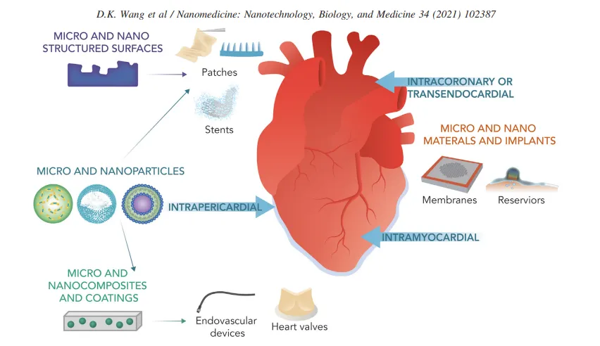
Figure 5: Diagram illustrating the applications of nanotechnology in cardiovascular disease.
Medical Devices
Surface modification is one area that will likely be quite significant for cardiovascular devices. Among the goals are thrombosis and infection control, cellular adhesion modification, and medicine delivery regulation. Although late thrombotic events caused by delayed endothelialization have raised concerns, drug-eluting stents (DES) are helpful in reducing neointimal thickness and restenosis. One important sector market for technical advancement is stents. In the current generation of DES, the drug is adsorbed onto a nondegradable polymer that remains in continuous contact with the vessel wall. It is believed that inflammatory responses to the polymer matrix have an impact on late thrombotic events. Using a biocompatible porous composite matrix made of carbon nanoparticles embedded in glassy polymeric carbon is one way to solve this issue. In terms of endothelialization, neointimal hyperplasia, inflammation, and fibrin deposition, cobalt chromium stents with this carbon-carbon coating and low and medium dosages of paclitaxel worked well in pigs. [66] Clinical trials are now being conducted for the stents, which are sold under the brand name Corel-C. Another area with great potential is ventricular assist devices (VADs). The groundbreaking randomized evaluation of mechanical assistance for the treatment of congestive heart failure trial found that the implantation of left VADs doubled one-year survival compared to optimal medical therapy and also improved quality of life in patients with advanced heart failure who were not candidates for heart transplantation. [67]
CONCLUSION:
Cardiovascular diseases continue to be a major global health burden, and conventional treatment approaches are often limited by poor target specificity, systemic adverse effects, and inadequate tissue regeneration. Nanotechnology has emerged as a promising interdisciplinary approach that addresses many of these limitations through targeted drug delivery, controlled release systems, enhanced imaging, and regenerative therapies. Various nanotechnology-based platforms, including nanoparticles, nanogels, nanofibers, nano-patches, and nanostructure medical devices, have demonstrated significant potential in the prevention, diagnosis, treatment, and repair of cardiovascular disorders such as atherosclerosis, myocardial infarction, and heart failure. Advances in nano medicine have improved drug bioavailability, reduced toxicity, enhanced therapeutic efficacy, and enabled early disease detection through sensitive diagnostic and imaging techniques. Furthermore, nanotechnology-supported tissue engineering and stem cell therapies offer new possibilities for cardiac regeneration and functional recovery following myocardial injury. Despite these promising developments, challenges related to long-term safety, biocompatibility, large-scale manufacturing, regulatory approval, and clinical translation remain to be fully addressed. Overall, nanotechnology represents a transformative strategy in cardiovascular medicine with the potential to revolutionize disease management and improve patient outcomes. Continued research, multidisciplinary collaboration, and well-designed clinical studies are essential to translate these innovative technologies from laboratory research into routine clinical practice.
REFERENCES


Zeel Desai*, Reinn Patel, Astha Patel, Pankti Patel, Role of Nanotechnology in Cardiovascular System, Int. J. Med. Pharm. Sci., 2026, 2 (8), 783-796. https://doi.org/10.5281/zenodo.22170675
 10.5281/zenodo.22170675