

We use cookies to ensure our website works properly and to personalise your experience. Cookies policy
1Research Associate, R&D, Ami Polymer Pvt Ltd
2Senior Executive, R&D, Ami Polymer Pvt Ltd
3CEO, Ami Polymer Pvt Ltd
4Associate Manager, R&D, Ami Polymer Pvt Ltd
5Executive. R&D, Ami Polymer Pvt Ltd
6Research Trainee. R&D, Ami Polymer Pvt Ltd
Articular cartilage exhibits negligible intrinsic regenerative capacity following trauma or degenerative affliction, frequently culminating in chronic arthrosis. Contemporary tissue engineering paradigm leverages biopolymer-based scaffolds to engineer biomimetic microenvironments conducive to chondrogenesis. This investigation explores the efficacy and physicochemical optimization of composite hydrogel matrices constructed from naturally derived macromolecules—specifically hyaluronic acid, chitosan, and gelatin—for articular cartilage restoration. Novel porous scaffold architectures were synthesized via lyophilization and enzymatic cross-linking techniques. The resulting biopolymeric constructs underwent rigorous physicochemical characterization, evaluating porosity, swelling kinetics, and compressive modulus. In vitro cytocompatibility and chondrogenic differentiation potential were subsequently appraised through primary human chondrocyte encapsulation, assessing extracellular matrix (ECM) deposition and pertinent gene upregulation via quantitative reverse transcription polymerase chain reaction (qRT-PCR). Empirical evaluation revealed that the optimized composite scaffolds possessed an interconnected trabecular architecture with high porosity exceeding eighty-five percent, thereby facilitating efficacious nutrient diffusion and cellular infiltration. The mechanical stiffness closely approximated native subchondral cartilage parameters. Furthermore, biological assays substantiated enhanced cellular proliferation and significant upregulation of chondrogenic phenotypic markers, notably aggrecan and type II collagen, alongside diminished fibroblastic dedifferentiation. The synthesized biopolymer-based systems demonstrate exceptional biocompatibility and biomechanical resilience, offering a propitious therapeutic modality for osteochondral defect remediation and regenerative orthopedics.
Articular cartilage is a unique type of connective tissue found in joints like the knee. It allows smooth movement and helps spread pressure across the joint with very little resistance. However, this tissue does not have blood vessels, nerves, or lymphatic channels, making it difficult to repair. This leads to slow recovery and impacts how we treat joint injuries and illnesses. Traditional ways to fix cartilage issues include methods like microfracture, abrasion arthroplasty, and using cartilage from one part of the body to another. These approaches often lead to the growth of fibrocartilage, which is not as strong or durable as natural hyaline cartilage. Over time, this can cause joint problems that require more invasive treatments, such as joint replacement. Tissue engineering uses cells, growth factors, and biomaterials to create alternatives for damaged tissues. Biopolymer scaffolds are a key part of this field. These materials are biocompatible and resemble the natural structure of tissue. This paper explores the latest developments in biopolymer scaffolds for knee cartilage repair, including the types of biopolymers, fabrication techniques, mechanical properties, and future prospects. [1]
2. Overview of Natural Biopolymers
Natural biopolymers are widely used in tissue engineering scaffolds because they are readily available and closely resemble the body's natural extracellular matrix. These materials are mainly classified into polysaccharides and proteins. Hyaluronic acid helps with shock absorption and lubrication within joints. Chitosan, derived from chitin, possesses antimicrobial properties and can improve scaffold strength when blended with other materials. Sodium alginate, extracted from seaweed, forms hydrogels through calcium-ion crosslinking and is commonly used in scaffold fabrication. Among protein-based biomaterials, collagen is the major structural component of cartilage and provides an excellent environment for cell growth. Gelatin, obtained by hydrolysing collagen, has a lower melting point and is easier to process for tissue engineering applications. [2]
3. Fabrication Techniques for Cartilage Scaffolds
The fabrication of biomimetic scaffolds for articular cartilage restoration necessitates sophisticated manufacturing paradigms capable of recapitulating the intricate architectural anisotropy of native extracellular matrix. Contemporary biofabrication methodologies—encompassing advanced electrospinning, thermally induced phase separation, precision 3D bioprinting, and supercritical fluid foaming—facilitate meticulous spatial deposition of biopolymeric matrices. These sophisticated engineering modalities engender interconnected porous networks that optimize cellular infiltration, nutrient permeation, and mechanical competence, thereby engendering an auspicious microenvironment for orchestrated chondrogenesis and osteochondral defect amelioration. Several methods are used to fabricate biopolymer-based scaffolds. Injectable hydrogels are prepared by mixing a biopolymer solution with cells and growth factors before injection into the damaged site, where the material gels in situ, providing a minimally invasive treatment option. Three-dimensional (3D) bioprinting enables the fabrication of complex scaffold structures using bio-inks composed of biopolymers, cells, and growth factors. Porous scaffolds contain interconnected pores that facilitate nutrient transport, cell migration, and tissue regeneration. [3]
4. Current Challenges in Biopolymer Research
Despite their advantages, biopolymer scaffolds face several challenges. Their mechanical strength is often insufficient for load-bearing applications. Controlling the degradation rate remains difficult, and additional research is required to translate laboratory findings into successful clinical treatments. Despite profound advancements in regenerative medicine, contemporary biopolymer research encounters multifaceted bottlenecks that impede the seamless clinical translation of engineered cartilage substitutes. Paramount among these impediments is the persistent dichotomy between biological affinity and mechanical robustness; naturally derived macromolecular matrices frequently exhibit superlative biocompatibility yet lack the requisite load-bearing competence mandatory for articular joint environments. Furthermore, achieving precise spatiotemporal regulation over bioactive molecule liberation, preventing undesirable chondrocyte dedifferentiation, and mitigating interfacial immunogenicity remain formidable obstacles. Overcoming these interdependent physicochemical and physiological hurdles necessitates innovative cross-linking strategies and composite material configurations to ensure long-term structural integrity and functional neotissue integration. [4]
5. Biopolymeric Constituents and Material Classification in Cartilage Tissue Engineering [5]
5.1. Polysaccharide-Derived Matrices
Glycan-based macromolecules—predominantly chitosan, hyaluronic acid, and sodium alginate—constitute a fundamental class of biopolymers extensively exploited for constructing articular cartilage scaffolds. Chitosan, a cationic deacetylated derivative of chitin, possesses intrinsic antimicrobial attributes alongside structural similarities to glycosaminoglycans, thereby engendering a propitious microenvironment for chondrocytic attachment. Hyaluronic acid, a ubiquitous non-sulfated glycosaminoglycan within native synovial fluid and articular cartilage, actively engages CD44 cellular receptors to orchestrate cell signaling, proliferation, and extracellular matrix synthesis. Meanwhile, sodium alginate, an anionic polysaccharide extracted from brown algae, facilitates rapid ionic cross-linking in the presence of divalent cations (such as calcium), yielding highly tunable viscoelastic hydrogels that effectively cushion compressive intra-articular forces while maintaining superlative cytocompatibility. [Figure:1]
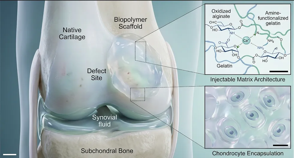
Figure 1: Schematic Representation of Biopolymer Scaffold
Composite network structure showing the interaction between polymer chain. And encapsulated chondrocytes and sol-to-gel transition mechanism biomimetic matrix
5.2. Protein-Based Scaffolds
Structural scleroproteins and fibrous macromolecules—specifically collagen, gelatin, and silk fibroin—offer exceptional biological mimicry owing to their inherent amino acid sequences and conserved bioactive motifs. Type I and type II collagens provide structural scaffolding mirroring the native fibrillar network, presenting specialized integrin-binding domains (such as the ubiquitous arginine-glycine-aspartic acid or RGD sequences) that profoundly augment cellular adhesion, migration, and cytoskeletal organization. Gelatin, a hydrolyzed derivative of collagen retaining critical cell-adhesive ligands, is frequently leveraged for its exceptional water-binding capacity and enzymatic degradability. Concurrently, silk fibroin—derived from silkworm cocoons—exhibits remarkable tensile resilience, slow enzymatic degradation kinetics, and versatile processability, rendering it an indispensable biomaterial for fabricating load-bearing osteochondral constructs. [Figure: 2]
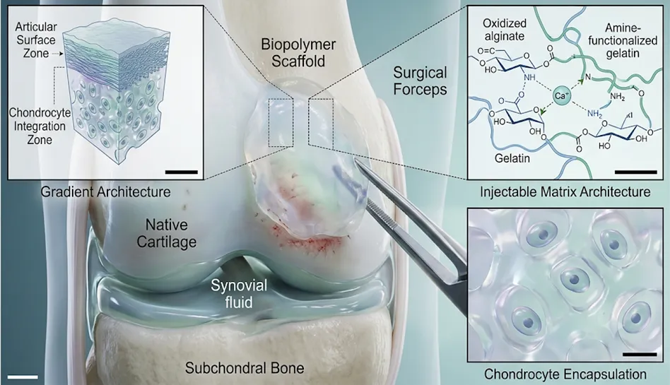
Figure: 2 Implantation of pre-formed, gradient biopolymer scaffold
5.3. Composite and Synthetic Hybrids
To circumvent the intrinsic limitations associated with single-component biomaterials—such as the rapid degradation of polysaccharides or the inadequate mechanical stiffness of soft protein hydrogels—contemporary regenerative strategies increasingly rely on composite and synthetic hybrids. By synergistically amalgamating naturally derived macromolecules with high-performance synthetic polyesters (such as polycaprolactone, polylactic acid, or poly(lactic-co-glycolic acid)), bioengineers can meticulously tailor degradation kinetics and amplify structural fortitude. These hybrid architectures successfully reconcile the perennial dichotomy between biological affinity and mechanical robustness, engendering robust, multi-phasic scaffolding systems capable of withstanding physiological biomechanical loads while orchestrating harmonious neo-tissue regeneration.
CONCLUSION
Biopolymer hydrogels and scaffolds represent promising strategies for treating cartilage damage and osteoarthritis. Continued research is needed to improve their mechanical performance, optimise degradation rates, and expand their clinical applications.
REFERENCES


Roshan Sambar, Pallab Mandal*, Priyabrata Pattnaik, Prasenjit Swain, Maahi Bisht, Ramavath Arun, Biopolymer- Based Systems Knee Cartilage Repair, Int. J. Med. Pharm. Sci., 2026, 2 (8), 515-518. https://doi.org/10.5281/zenodo.21927855
 10.5281/zenodo.21927855