

We use cookies to ensure our website works properly and to personalise your experience. Cookies policy
1Gawande College of Pharmacy, Sakharkherda
2Shree Sant Gajanan Maharaj College of Pharmacy
3Dr. R. G. Bhoyar, Institute of Pharmaceutical Education and Research, Wardha
4Mahatma Phule Pharmacy College, Akola
5Datta Meghe College of Pharmacy, S. Alod Wardha
Transdermal delivery offers a non-invasive, patient-friendly alternative to oral and parenteral administration, but the barrier function of the stratum corneum restricts conventional patches to small, lipophilic, potent molecules. Microneedles (MNs) are micron-scale projections, typically 150 to 1,500 µm long, that create transient microchannels through the stratum corneum and deliver drugs to the epidermis and upper dermis with minimal pain and little to no bleeding. Since the first demonstrations in the 1990s, MN technology has expanded from solid silicon arrays to coated, dissolving, hydrogel-forming, and hollow designs, enabling delivery of vaccines, peptides, proteins, nucleic acids, and small molecules. This review summarizes MN types, materials, and fabrication; delivery mechanisms and pharmacokinetics; applications in vaccination, biologic therapy, and chronic disease management; and the translational, regulatory, and manufacturing challenges that remain. We also outline emerging directions, including closed-loop and smart systems, MN-based diagnostics, and integration with wearable electronics.
The skin is the body's largest organ and an attractive route for drug administration. It avoids first-pass hepatic metabolism, gastrointestinal degradation, and the pain, needle-phobia, sharps waste, and trained-personnel requirements associated with hypodermic injection. Despite this appeal, only a limited number of drugs are marketed as transdermal products, such as nicotine, fentanyl, nitroglycerin, estradiol, and scopolamine. The reason is the stratum corneum, a 10 to 20 µm layer of corneocytes embedded in a lipid matrix that excludes most molecules above roughly 500 Da, as well as hydrophilic and charged compounds [1]. Strategies to overcome this barrier fall into three generations: (1) conventional passive patches, (2) chemical enhancers, iontophoresis, and sonophoresis, and (3) methods that physically disrupt the stratum corneum, including thermal ablation, electroporation, jet injection, and microneedles. Microneedles occupy a unique position in this third generation because they combine a simple, patch-like format with the ability to deliver macromolecules, particulates, and even live-attenuated or nucleic-acid vaccines [2,3]. The concept was proposed in the 1970s and first experimentally realized with silicon microfabrication in the late 1990s [4]. Over the following two decades, advances in polymer science, micromolding, and 3D printing shifted the field from proof-of-concept devices toward clinical-grade products [5,6]. This review surveys that progress with emphasis on three areas of greatest clinical interest: vaccines, biologics, and chronic disease management.
2. Skin Anatomy and Rationale for Microneedle Delivery
Human skin comprises the epidermis (including the stratum corneum, 10 to 20 µm, and viable epidermis, roughly 50 to 150 µm), the dermis (1 to 3 mm), and the hypodermis. The dermis is richly vascularized and contains lymphatic vessels, while the epidermis and dermis host a dense network of antigen-presenting cells, including Langerhans cells and dermal dendritic cells. Microneedles of 250 to 1,000 µm typically penetrate the stratum corneum and reach the viable epidermis and papillary dermis, but are too short to stimulate the deeper nerve endings that mediate most pain. Clinical studies report that MN insertion is perceived as considerably less painful than hypodermic needles, and often as barely perceptible [7].
Two features of this anatomy underpin the major application areas:
Penetration is not trivial. Skin is viscoelastic and deforms under the needle tip, so effective insertion depends on needle geometry (tip radius, aspect ratio, base width), array density (the "bed of nails" effect), insertion speed, and application force. Applicator devices that deliver controlled velocity or impact generally improve reproducibility [8]. skin layers and microneedle penetration depth
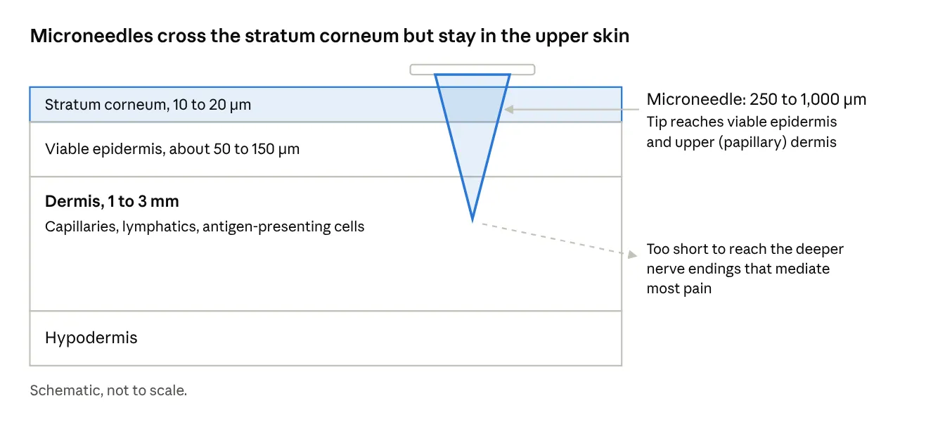
Figure 1. Schematic skin cross-section showing microneedle penetration relative to skin layers.
3. Types of Microneedles
Microneedles are commonly categorized by their delivery mechanism, and each category has distinct trade-offs summarized below.
3.1 Solid microneedles ("poke and patch")
Solid MNs, made from silicon, metal, or ceramic, are used to pretreat skin and create microchannels, after which a conventional patch, gel, or cream is applied. They are simple and mechanically robust, but require two steps, and microchannels reseal within hours (faster without occlusion), which makes dose control difficult. Dermarollers and fractional devices used in cosmetic practice follow the same principle [9].
3.2 Coated microneedles ("coat and poke")
A drug formulation is coated onto the surface of solid needles and dissolves in the skin after insertion. Coated MNs support rapid delivery of potent drugs, vaccines, and peptides. Their key limitation is payload: coatings typically carry tens of micrograms per patch, so they suit highly potent molecules [10,11]. Coating formulations require careful control of viscosity, surface tension, and excipients (for example, carboxymethylcellulose, sugars, surfactants) to achieve uniform, stable layers. Zosano Pharma's coated titanium MN patch for the migraine drug zolmitriptan (ZP-Zolmitriptan, M207) progressed to pivotal clinical trials, demonstrating that coated systems can reach late-stage development [12].
3.3 Dissolving microneedles ("poke and release")
Dissolving MNs are fabricated entirely from water-soluble or biodegradable polymers and sugars, such as hyaluronic acid, polyvinylpyrrolidone (PVP), polyvinyl alcohol (PVA), carboxymethylcellulose, trehalose, maltose, chitosan, or PLGA, with the drug encapsulated in the matrix. After insertion, needles dissolve or degrade and release the payload, leaving no sharps waste. Advantages include high loading relative to coated systems, one-step application, safety, and suitability for low-cost micromolding. Challenges include preserving the stability of biologics through casting, drying, and storage; achieving adequate mechanical strength without sacrificing dissolution rate; and controlling release kinetics, which can range from minutes (fast-dissolving) to weeks (biodegradable depot needles) [13,14].
3.4 Hydrogel-forming microneedles
Hydrogel-forming (or swellable) MNs are made from crosslinked polymers that absorb interstitial fluid and swell, forming continuous aqueous channels through which drug diffuses from an attached reservoir [15]. They remain intact on removal, avoiding polymer deposition in skin, and can deliver larger doses (milligram scale) than dissolving MNs. They have also been explored for minimally invasive sampling of interstitial fluid for therapeutic drug monitoring [16].
3.5 Hollow microneedles
Hollow MNs contain a bore through which liquid formulations flow, analogous to a miniature hypodermic needle [17]. They enable high-volume, controllable infusion and can handle conventional liquid biologic formulations, including insulin and monoclonal antibody solutions. Challenges include tip clogging, back-pressure from dermal tissue, fabrication cost, and device complexity. Intradermal delivery systems based on hollow microneedles have reached the market for influenza vaccination.
3.6 Emerging designs
Recent innovations include:
Table 1. Comparison of the main microneedle types.
|
Type |
Material |
Mechanism |
Typical dose capacity |
Main advantage |
Main limitation |
|
Solid |
Silicon, metal, ceramic |
Poke, then apply patch or gel |
Depends on topical formulation |
Strong, simple |
Two steps; channels reseal; variable dose |
|
Coated |
Metal or silicon with drug film |
Coating dissolves in skin |
Micrograms |
Fast release of potent drugs |
Very low payload |
|
Dissolving |
Hyaluronic acid, PVP, PVA, sugars, PLGA |
Needle matrix dissolves and releases drug |
Micrograms to low milligrams |
One step; no sharps; scalable micromolding |
Biologic stability in matrix; strength vs dissolution |
|
Hydrogel-forming |
Crosslinked swellable polymers |
Swollen needles form diffusion channels from a reservoir |
Milligrams |
Larger doses; intact removal |
Slower onset; reservoir needed |
|
Hollow |
Metal, silicon, glass, polymer |
Liquid flows through a bore |
Microlitres to millilitres |
Liquid biologics; controllable infusion |
Clogging; back-pressure; complexity |
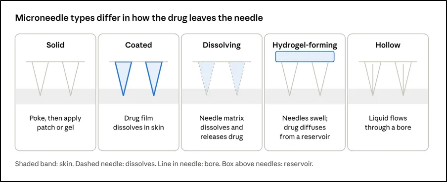
Figure 2. Release mechanisms of the five main microneedle types (schematic).
4. Materials and Fabrication
Material choice determines mechanical performance, biocompatibility, drug compatibility, and cost.
Inorganic materials. Silicon was the first MN material owing to mature semiconductor microfabrication, but brittleness and cost limit translational use [20]. Stainless steel, titanium, and nickel provide mechanical strength and are used for solid and coated MNs. Glass and ceramics have been used for specialized hollow or solid needles.
Polymers. Water-soluble and biodegradable polymers dominate current research because they allow low-temperature, aqueous processing compatible with biologics. Common choices include hyaluronic acid, PVP, PVA, carboxymethylcellulose, gelatin, silk fibroin, chitosan, PLGA, and polylactic acid [21,22].
Fabrication methods. Principal approaches are:
Manufacturing at scale requires attention to drug loading in the needle tips (to minimize waste of expensive biologics), aseptic or low-bioburden processing, reproducible drying, and packaging with appropriate moisture barriers.
5. Delivery Mechanisms and Pharmacokinetics
Drug release and absorption from MN patches depend on several interacting factors: the needle type, depth of deposition, dissolution or swelling rate, drug physicochemical properties (molecular weight, solubility, charge), and skin site. Dissolving MNs typically achieve rapid release, with partial dose delivery within minutes, while longer wear times or depot-forming materials produce prolonged release. Pharmacokinetic profiles for MN-delivered drugs often show a rapid onset similar to subcutaneous injection and can be tuned toward sustained exposure through formulation. Bioavailability can vary because of incomplete insertion, skin hydration, hair, subcutaneous fat thickness, and the loss of drug into the patch backing. Quantifying delivered dose, for example by measuring residual drug on removed patches or by dye-based insertion assays, is an important quality attribute. Variability between individuals and anatomical sites remains a key consideration for regulatory acceptance of MN products [23].
6. Vaccine Delivery
6.1 Rationale
Skin is an immunologically privileged site for vaccination. Delivering antigens to epidermal and dermal antigen-presenting cells can produce robust humoral and cellular responses, with dose-sparing benefits demonstrated for several antigens [25]. MN patches also address practical barriers, including cold-chain dependence, trained-personnel requirements, and sharps disposal, which are especially significant for mass campaigns in low-resource settings [24].
6.2 Influenza
Influenza has been the lead model. Preclinical studies in mice and other species showed that MN delivery of inactivated influenza vaccine induced immune responses at least comparable to intramuscular injection, with improved lung viral clearance and durable memory [26-28]. A first-in-human, randomized, partially blinded Phase 1 trial of an inactivated influenza vaccine dissolving MN patch reported that patches were well tolerated, produced mostly mild and transient local reactions, and induced antibody responses similar to those from intramuscular injection; participants also strongly favored the patch, and self-administration was feasible [29,30].
6.3 Measles, rubella, and other childhood vaccines
MN patches for measles and rubella vaccine have been developed for use in low- and middle-income countries, where live-attenuated vaccines require reconstitution and cold chain. Studies in animals, and early-stage clinical work, indicate that patch formulations can improve thermostability and simplify administration by lay health workers or caregivers [31,32]. Patches for polio, hepatitis B, and other antigens are in preclinical or early clinical evaluation.
6.4 Nucleic-acid and next-generation vaccines
Following the success of mRNA vaccines, MN delivery of mRNA, DNA, and self-amplifying RNA has attracted considerable attention. Skin cells and resident immune cells can take up and express nucleic acids, and MN patches may offer improved thermostability relative to lipid-nanoparticle suspensions [34]. Challenges include maintaining RNA integrity during fabrication and storage, protecting cargo from nucleases, and achieving efficient intracellular delivery, which has led to designs incorporating lipid nanoparticles, polyplexes, or other carriers within dissolving needles. During the COVID-19 pandemic, preclinical studies demonstrated MN-delivered recombinant spike protein and DNA vaccines that elicited neutralizing antibodies in animals [33].
6.5 Adjuvants, immunomodulation, and cancer vaccines
MNs can co-deliver antigens with adjuvants such as TLR agonists, enabling local immune activation while limiting systemic exposure. Cancer vaccine and immunotherapy applications, including delivery of tumor antigens, checkpoint inhibitors, and immune-modulating nanoparticles to skin or tumors, have shown promising results in animal models, with some approaches using MNs for intratumoral or peritumoral delivery [35,36].
7. Biologics: Peptides, Proteins, and Antibodies
Biologics are administered almost exclusively by injection because of poor oral bioavailability and inability to cross intact skin. MN patches are attractive for pain-free and potentially self-administered delivery.
Peptides. Parathyroid hormone analogues are among the most advanced examples. Abaloparatide delivered by a coated MN system (abaloparatide-sMTS) was studied as an alternative to daily subcutaneous injection for osteoporosis, with early studies showing comparable pharmacokinetics [37] and subsequent clinical development. Other peptides, such as exenatide, desmopressin, leuprolide, and octreotide, have been explored in MN formats.
Proteins and hormones. Insulin, growth hormone, interferons, erythropoietin, and enzymes have been delivered by MN patches in animal models. Silk fibroin and sugar glass matrices can stabilize proteins during storage [38].
Monoclonal antibodies. Antibodies present a particular challenge because therapeutic doses are in the milligram range, exceeding the capacity of most dissolving or coated MNs. Hollow MNs, hydrogel-forming MNs with reservoirs, and high-loading dissolving formulations are approaches under investigation. Intradermal and lymphatic uptake of large antibodies is also slower and more variable than for small peptides, so formulation and device design must be tailored.
Stability considerations. A distinguishing advantage of solid-state MN formulations is that dried biologics can be stable at ambient temperature for extended periods, reducing cold-chain requirements. However, processing steps such as heating, drying, shear, and exposure to organic solvents or interfaces can denature proteins, so stabilizing excipients (trehalose, sucrose, amino acids, surfactants) and gentle processing are essential.
8. Chronic Disease Management
8.1 Diabetes
Diabetes is the most extensively studied chronic disease application. MN patches can deliver insulin without needles, and a major research thrust is glucose-responsive (closed-loop) delivery. In a widely cited approach, MN arrays loaded with glucose-sensitive vesicles released insulin in response to elevated glucose and controlled blood glucose in diabetic mice for hours [39]. Other designs use glucose oxidase, phenylboronic acid, or concanavalin A chemistries [40,41]. Hollow MN pumps and wearable systems combine continuous glucose monitoring with microneedle-based sensing and delivery. Key translational questions include the reliability and safety margin of glucose responsiveness (avoiding hypoglycemia), reproducibility across skin types, and scale-up.
8.2 Osteoporosis and endocrine disorders
In addition to abaloparatide, MN formats for teriparatide and other bone anabolic agents have been explored, as have MN systems for growth hormone deficiency, aiming at weekly or daily dosing without injections.
8.3 Cardiovascular and metabolic disease
MN patches have been investigated for antihypertensive agents, anticoagulants such as heparin and enoxaparin, lipid-lowering biologics, and GLP-1 receptor agonists for obesity and type 2 diabetes. Long-acting depot MNs that leave polymer microdepots in the skin could support weekly or monthly dosing for conditions where adherence is a major challenge.
8.4 Neurological and pain conditions
Coated MN patches for acute migraine therapy (zolmitriptan) demonstrated rapid absorption and efficacy in clinical studies [12]. MN delivery of local anesthetics, opioids, naloxone for overdose reversal, and drugs for Parkinson's disease and Alzheimer's disease has been explored, mostly in preclinical stages.
8.5 Dermatological and local conditions
MNs are used clinically for cosmetic skin remodeling and are being evaluated for delivery of agents in psoriasis, alopecia, keloids, hyperhidrosis, and skin infections. In these cases, localized delivery maximizes drug concentration at the target and reduces systemic exposure. MN-mediated delivery of botulinum toxin, corticosteroids, and antifungals has been studied.
8.6 Contraception and long-acting therapies
Separable MN patches that embed biodegradable levonorgestrel-loaded tips for sustained release over weeks to months have been reported in animal studies, with the aim of giving users a self-administered long-acting method [42,43]. Similar depot approaches are being considered for HIV pre-exposure prophylaxis and antiretroviral therapy.
8.7 Ocular and other non-skin sites
MN technology has also been adapted for suprachoroidal and intrascleral ocular delivery, oral mucosal delivery, and delivery to other tissues such as heart or gastrointestinal surfaces, indicating that the platform's utility extends beyond skin.
Table 2. Representative microneedle applications and the stage of evidence typically reported
|
Application |
Typical MN format |
Evidence stage |
Ref. |
|
Inactivated influenza vaccine |
Dissolving |
Phase 1 human trial |
[29,30] |
|
Measles and rubella vaccine |
Dissolving |
Preclinical to early clinical |
[31,32] |
|
Recombinant coronavirus vaccine |
Dissolving or coated |
Animal studies |
[33] |
|
Osteoporosis (PTH analogues) |
Coated |
Human PK and later-phase trials |
[37] |
|
Acute migraine (zolmitriptan) |
Coated |
Pivotal clinical trials |
[12] |
|
Glucose-responsive insulin |
Dissolving, depot |
Animal studies (mice, minipigs) |
[39,41] |
|
Long-acting contraception |
Separable, effervescent |
Animal studies |
[42,43] |
|
Cancer immunotherapy |
Dissolving, depot |
Animal studies |
[35,36] |
9. Smart and Integrated Microneedle Systems
A growing body of work integrates MNs with sensing, electronics, and responsive materials.
10. Safety, Tolerability, and Patient Acceptance
Safety data to date are broadly favorable. Typical adverse events are transient erythema, mild edema, and occasional pruritus at the application site, resolving within hours to days. Microchannels generally close within 24 to 72 hours, depending on occlusion. Theoretical concerns include infection through microchannels, residual polymer or needle fragments, immune sensitization to excipients, and scarring or pigmentary change with repeated application. Studies of properly manufactured dissolving MNs have not shown clinically significant infection, but sterility or low bioburden and quality control are essential, and long-term data in repeated-use scenarios are still limited. Patient surveys consistently show a preference for MN patches over injections, largely owing to reduced pain and the possibility of self-administration. [40]
11. Translational, Regulatory, and Manufacturing Challenges
Drug loading and dose capacity. The small volume of needle tips restricts dose, especially for antibodies. Strategies include tip-loading, hollow designs, reservoirs, and long-acting depots.
Reproducibility of delivery. Variability in insertion, skin properties, and wear conditions affects delivered dose. Applicators, insertion indicators, and standardized wear instructions are being developed.
Stability and shelf life. Biologics and nucleic acids must remain active through fabrication and storage. Packaging, desiccants, and excipient screening are key.
Scale-up and cost. Roll-to-roll micromolding, automated filling, and continuous manufacturing are needed for mass production at costs competitive with syringes, particularly for vaccines in low-income markets.
Regulation. MN products are typically regulated as drug-device combination products. Regulatory agencies have issued guidance on microneedle systems and related transdermal products, but standards for characterization (mechanical strength, penetration, residual drug, sterility, and needle dissolution) are still evolving. Defining critical quality attributes and bioequivalence approaches for MN products remains an active area of discussion.
Environmental and disposal considerations. Although dissolving MNs eliminate sharps, backing materials and packaging need sustainable design. Non-dissolving MNs retain sharps-waste considerations.
Clinical evidence. Many MN technologies still lack large, late-stage clinical trials. Positive Phase 1 and 2 results must be translated into pivotal studies, with attention to diverse populations, skin types, ages, and anatomical sites.
12. FUTURE PERSPECTIVES
Several trends are likely to shape the next decade of MN research and commercialization:
CONCLUSION
Microneedle patches have matured from laboratory curiosities into a versatile platform capable of delivering vaccines, peptides, proteins, nucleic acids, and small molecules through the skin with minimal pain and potential for self-administration. Their greatest promise lies in vaccination, where dose sparing, thermostability, and ease of use could transform global immunization, and in chronic disease management, where painless, patient-controlled, and possibly glucose-responsive or long-acting systems could improve adherence and outcomes. Realizing this potential requires solving practical issues of dose capacity, reproducible delivery, stability, scalable manufacturing, and regulatory clarity. With continuing advances in materials, device engineering, and clinical evidence, microneedles are well positioned to become a mainstream mode of drug delivery.
REFERENCES

Aniket Chandurkar*, Pranali Mangle, Yamini Bhagat, Vaishnavi Anpan, Aaliya Tabassum Shaikh Jameel, Prachi Raut, Krutika Bhisekar, Microneedle-Based Transdermal Drug Delivery: Innovations in Vaccines, Biologics, and Chronic Disease Management, Int. J. Med. Pharm. Sci., 2026, 2 (11), 1-10. https://doi.org/10.5281/zenodo.23935885
 10.5281/zenodo.23935885