

We use cookies to ensure our website works properly and to personalise your experience. Cookies policy
College of Pharmaceutical Sciences, Govt. Medical College, Kozhikode
Vulvovaginal candidiasis (VVC) ranks among the most widespread fungal infections in women globally, with Candida albicans responsible for the majority of cases. Yet treatment has grown increasingly difficult, driven by a rise in infections from non-albicans Candida species and by recurrent VVC (RVVC) — a trend fueled in part by growing antifungal resistance Although topical vaginal formulations (creams, gels, tablets, suppositories) and oral antifungals remain effective for uncomplicated infections, their clinical utility is often constrained by poor vaginal retention, leakage, need for repeated dosing, suboptimal drug concentrations at the target site, systemic side effects, and poor patient compliance. To overcome these limitations, advanced vaginal drug delivery systems have been developed to enhance localized distribution, prolong retention, and sustain therapeutic action. This review critically examines recent advances in such systems, including vaginal films, hydrogels, intravaginal rings, microneedle-based platforms, and nanoparticle carriers (liposomes, solid lipid nanoparticles, and nanostructured lipid carriers), as well as the emerging role of AI/ML in vaginal drug delivery. It also discusses the pathophysiology of vulvovaginal candidiasis (VVC), current treatment strategies, and the shortcomings of conventional dosage forms, with particular focus on biodegradable and stimuli-responsive systems—covering their formulation approaches, therapeutic benefits, and recent progress. Collectively, these innovations offer promising strategies to improve treatment efficacy, patient adherence, and reduce VVC recurrence.
“Vulvovaginal candidiasis” is a fungal infection which occurs mostly in the women of reproductive age. It occurs due to the overabundance of candida species in the vagina. It has become a major global health concern. Most of the studies states that almost three out of four women will experience at least one episode in their lifetime and about half of the women will experience recurrent infection. The prevalence of VVC puts a significant burden on women's health as well as healthcare systems in general.[1–4]. Candida albicans is the main causative agent of VVC, which accounts for around 80–90% of cases and is still more common when compared with other candida species. Several non-albicans Candida (NAC) species — including C. glabrata, C. tropicalis, C. krusei, and C. parapsilosis — have been identified as causing infections in recent years. Rising rates of infection with these species have been tied to the growing number of resistant strains, often driven by repeated or long-term antifungal use. Compared to C. albicans, many NAC species show lower susceptibility to standard antifungal drugs—especially azoles—making treatment failure and recurrent or persistent infections more likely[1,4–6]. Major symptoms associated with VVC includes severe itching, burning sensation, irritation, redness and swelling and also pain during urination and intercourse. A thick white cheese like discharge is an indication of the condition. These symptoms can be moderate to severe, and they frequently get worse with recurrent or persistent infections. Even though it is not a life threatening condition, its recurring and persistent nature often disrupt the physical comfort, sexual health, and quality of daily life The major predisposing factors associated with the infection includes antibiotic use, increased estrogen levels during pregnancy or hormonal therapy, uncontrolled diabetes mellitus, and tight-fitting clothing etc.[1–3,7,8]. Many women of reproductive age suffer greatly from recurrent vulvovaginal candidiasis (RVVC), a difficult-to-treat fungal infection that is characterized by three or more symptomatic episodes in a 12-month period. Even when Candida strains test as susceptible to standard first-line antifungals, treatment for recurrent vulvovaginal candidiasis (RVVC) frequently fails — a pattern that points to something beyond simple drug resistance at play. One explanation that has gained traction is biofilm formation, long considered a key virulence mechanism in Candida species and a likely contributor to both the persistence of RVVC and its poor response to treatment.[1–3,8,9]. There are generally two categories of RVVC, primary and secondary. Primary RVVC occurs in healthy and immunocompetent women, without any noticeable risk factors and therefore it considered as idiopathic in nature. On the other hand, a number of triggering events, such as excessive estrogen levels, hormone replacement treatment, diabetes, pregnancy, allergic diseases, or the use of corticosteroids and antibiotics, can cause secondary RVVC. Recurrences are also more common in women living with HIV, mostly because it is more difficult for them to control Candida due to a compromised immune system.[9]. VVC is generally treated with antifungals — most often azoles (fluconazole, clotrimazole, miconazole, terconazole, ketoconazole) or polyenes like nystatin — delivered orally or through topical vaginal forms such as creams, gels, tablets, and suppositories. RVVC management follows two stages: induction (7–14 days of topical or oral antifungals to clear the infection) and maintenance (typically weekly oral fluconazole for about six months to prevent recurrence). Treatment choice isn't universal, though — it depends on the specific Candida species involved and its susceptibility to available drugs.[2,9,10]. Standard antifungal treatments are generally effective for the treatment of uncomplicated VVC, but they are associated with several drawbacks such as the chances of systemic side effects and possible drug interactions on using oral antifungal agents, and also poor retention, leakage, frequent dosing and inconsistent drug delivery are associated with other conventional therapies which reduces the patient compliance. Antifungal resistance, the suboptimal drug concentration at the site of infections and the persistent biofilm formation are the other reasons which contribute to the treatment failure.[6,11]. In order to get around these problems, scientists have developed more sophisticated vaginal drug delivery systems that limit the amount of drug that enters systemic circulation while keeping the drug in place longer, adhering better to the mucosal surface, and releasing the active compound gradually. New techniques that offer better treatment results, increased patient compliance, and a lower risk of infection recurrence include intravaginal rings, nanoparticles, liposomes, nanofibers, hydrogels, microneedle-based systems, and stimuli-responsive formulations.[9,11–14].
The pathophysiology of vulvovaginal candidiasis is intricate, resulting from a dynamic interaction between the virulence characteristics of Candida species, disruptions of the normal vaginal microbiota, and host immune systems.[5].
Candida species are present in the vagina as part of the normal microbial community and do not cause any symptoms in healthy women. there are many factors that disrupt the balance between the host immune response and the the vaginal microbial environment, which includes pregnancy, diabetes mellitus, prolonged broad-spectrum antibiotic use, hormonal fluctuations, immunosuppression, and elevated estrogen levels etc. Vulvovaginal candidiasis arises from a disruption of this microbial balance, which permits the proliferation of Candida species.[4–6,15,16].
The pathogenesis of vulvovaginal candidiasis is initiated by the adhesion of Candida cells to the vaginal epithelium, representing the first critical step in disease establishment. Key fungal surface proteins drive this process—mainly the agglutinin-like sequence (ALS) family and hyphal wall protein 1 (Hwp1), which act as adhesins. Once Candida effectively latch onto the vaginal mucosa, it is able to colonize the tissue—and that sets the stage for everything that follows, from invading deeper tissue to forming biofilms and driving a full-blown infection. [4–6,16,17].
A key part of what makes Candida albicans harmful is its shape-shifting ability—it can switch from a single-celled yeast form into thread-like hyphae and pseudo-hyphae. Candida penetrates the vaginal lining using these invasive structures in two ways: it gets taken up by host cells through endocytosis, and it actively pushes its way in on its own. The transition from yeast to hyphal morphology is a key virulence mechanism underlying tissue invasion, and is widely regarded as a prerequisite for the onset and progression of symptomatic VVC.[4–6,16,18].
Beyond its ability to grow as hyphae, Candida albicans relies on several other virulence factors to cause disease. These include the capacity to create candida lysin, a cytolytic peptide toxin that damages epithelial cells, build biofilms, avoid host immunological responses, and secrete hydrolytic enzymes such secreted aspartyl proteinases (SAPs), phospholipases, lipases, and hemolysins. Together, these virulence factors enable the fungus to adhere to and colonize host tissue, invade it, persist over time, and resist clearance by the immune system.[4–6,16,19,20].
Candida has the ability to form a microbial community emebdded within a self-produced extracellular matrix called as biofilms on both the medicl devices and vaginal mucosa. These biofilms play an important role in the pathogenesis of RVVC. They have the ability to enhance the fungal survival, shield the organism from the host's immune system defense and thereby reducing its sensitivity to antifungal treatment. [4–6,8,16,21].
Vulvovaginal candidiasis symptoms arise mainly from an overactive immune response in the host, rather than from direct tissue damage caused by the fungus itself. When vaginal epithelial cells detect the presence of Candida, they respond by releasing cytokines and chemokines—signaling molecules that in turn recruit neutrophils to the site. Yet these immune cells frequently fall short of clearing the fungus entirely, leading to ongoing inflammation and symptoms like itching, burning, redness, swelling, and vaginal discharge.[4–6,16].
Recurrent vulvovaginal candidiasis (RVVC) is diagnosed when a woman experiences three or more symptomatic episodes within a single year. The condition is thought to arise from multiple contributing factors, including persistent fungal colonization, biofilm development, genetic predisposition of the host, dysregulated immune responses, and incomplete elimination of Candida from vaginal reservoirs, which together promote repeated episodes of infection[3–6,8,9,16].
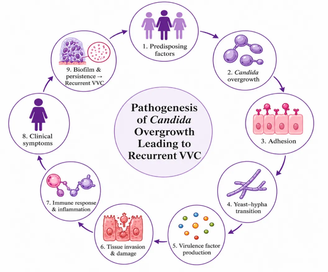
Figure 1: Pathogenesis of VVC
Vaginal creams are one of the standard first line dosage form for treating uncomplicated VVC. These are usually water-in-oil or oil-in-water type of emulsions which contains antifungal drugs. These types of semisolid preparations are generally applied intravaginally using an applicator. These formulations helps to reduce the chances of systemic adverse effects and possible drug interactions by delivering drugs to the site of infection.[1,22,23]. VVC is usually treated with creams containing azole antifungal agents such as clotrimazole, miconazole, terconazole, butoconazole or tioconazole. Among these clotrimazole and miconazole is mostly acceptable and frequently prescribed due to their safety profiles, established clinical history and broad-spectrum action. These formulations are available in a variety of dose regimens and strengths, ranging from conventional 7-day courses to shorter 1- to 3-day treatments. For example, clotrimazole is typically marketed as a 1% cream for a seven-day regimen or as a 2% formulation for a shorter three-day course. Miconazole follows a similar pattern — a 2% cream for seven days of use, or a 4% cream when a more concentrated three-day course is needed. That said, conventional vaginal creams aren't without their drawbacks. Issues like leakage, messiness, inconsistent retention within the vaginal cavity, and the need for repeated dosing can all make them less convenient for patients, potentially affecting how well they stick to their treatment regimen[22–24].
Gels are semisolid preparations formed by a three-dimensional polymer network. This network gives the gel its solid-like structural integrity and shape retention, while still allowing it to hold a significant volume of liquid within its matrix [25]. These formulations also deliver the drugs to the target site and helps minimize the possible systemic toxicity. Examples for gelling agents: carbomers, poloxamers, hydroxypropyl methyl cellulose (HPMC), hydroxy ethyl cellulose (HEC), chitosan, pectin, etc. And the frequently used antifungal agents include clotrimazole, miconazole, fluconazole, amphotericin B, and terbinafine.[25–28].
Another kind of semisolid dosage form is vaginal ointments, which are made by mixing antifungal medications with an oleaginous or water-miscible base to be applied to the vaginal mucosa. Although they can be highly effective in managing vulvovaginal candidiasis (VVC)—especially in cases involving significant vulvar symptoms—ointments are used less frequently than creams and gels in routine clinical practice. Clotrimazole, miconazole, and nystatin are common antifungal drugs utilized in these formulations; they restrict systemic drug exposure while acting locally on the afflicted tissue. However, the semisolid formulation has several disadvantages, including greasiness, leaking, and less comfortable application, despite its benefit of extending contact time with the mucosal surface.[24,27].
These are solid dosage forms formulated for local delivery to the vaginal tract, most commonly used to treat gynecological conditions such as vaginal infections. They are manufactured using the same compression techniques employed in conventional oral tablet production and typically contain a comparable range of excipients. When inserted, these tablets absorb moisture from vaginal fluid, as a result they swell and adhere to the vaginal mucosa. This adhesion keeps the drug in place and allows for gradual release, improving local retention and therapeutic effectiveness. Surfactants can enhance drug penetration, but mucoadhesive polymers—such as hydroxypropyl methylcellulose, carbopol, chitosan, and polycarbophil—play the larger role in prolonging retention. Antifungal vaginal tablets, most commonly containing clotrimazole, miconazole, or dequalinium chloride, are widely used, with clotrimazole the most popular choice. Available in several strengths and treatment regimens, clotrimazole vaginal tablets have shown strong clinical outcomes, with mycological cure rates comparable to oral azole therapy for uncomplicated vulvovaginal candidiasis.[27,29–31].
Vaginal suppositories—also known as ovules—are solid dosage forms generally prepared with water-soluble bases, such as glycerol-gelatin, or water-miscible bases, such as polyethylene glycol (PEG)[22,27,29,32]. Unlike rectal suppositories, which melt when inserted, vaginal suppositories dissolve instead, allowing for targeted, localized medication release due to their hygroscopic nature. To improve stability and boost therapeutic efficacy, formulations frequently include excipients like surfactants and preservatives. [29,32,34]. Research has shown that a single 1200 mg dose of miconazole nitrate, delivered as an ovule, offers quicker symptom relief and is generally preferred by patients over the traditional 7-day course of miconazole cream for treating VVC[29,33]. Fenticonazole ovules have also proven effective in treating vaginal trichomoniasis, with patients generally tolerating the treatment well — even at higher doses, side effects reported were typically mild to moderate[29,32,34].
Table 1: Advantages and limitations of conventional vaginal dosage forms
|
Conventional Vaginal Dosage Form |
Advantages |
Limitations |
References |
|
Creams |
|
|
[1,3,9,22,27,29,35] |
|
Gels |
|
|
[13,22,25–27,29] |
|
Vaginal ointments |
|
|
[3,9,22,27,29] |
|
Vaginal tablets |
|
|
[13,22,25–27,29] |
|
Vaginal suppositories |
|
|
[1,22,24,27,29] |
Advanced vaginal drug delivery system
Vaginal films are thin, solid polymer-based dosage forms designed to dissolve quickly once they come into contact with vaginal fluids, releasing the drug right at the site of infection. Compared to gels and creams, they're much less messy, cause less leakage, and tend to be better accepted by patients[27,29,36]. Their compact, pliable shape makes them easy to insert intravaginally without needing an applicator. Because they're dry rather than semisolid, they hold up well during storage and aren't easily affected by changes in temperature or humidity[27,29,36].Vaginal films were originally designed for contraceptive and microbicidal purposes, but they're now gaining attention as potential carriers for antifungal drugs in treating vulvovaginal candidiasis. This growing interest stems from their ability to improve drug stability, extend contact time with the mucosal surface, and make the treatment easier for patients to stick with[27,29,36] Kumar et al. developed fluconazole-loaded vaginal films using HPMC via the solvent casting method. These films showed consistent drug content, good mechanical strength, and a sustained release profile[37]. They also demonstrated strong antifungal activity against Candida albicans, suggesting they could serve as a promising alternative to conventional vaginal dosage forms for treating localized VVC[37]. Conte et al. developed vaginal films made from biopolymers, loaded with fluconazole and thymol — and among the variations tested, the chitosan/HPMC films stood out for their flexibility, swelling behavior, and mechanical strength[38]. Pairing fluconazole with thymol boosted the formulation's antifungal potential against resistant Candida strains, making it a promising strategy for tackling recurrent and drug-resistant VVC[38].
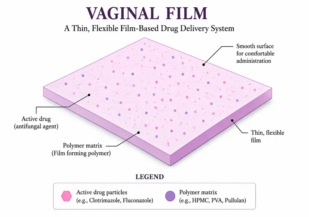
Figure 2: Vaginal film
Table 2: Advantages and limitations of vaginal films
|
Advantages |
Limitations
|
References |
|
|
[25,27,29,36]
|
Table 3: Components of vaginal films
|
Component |
Function |
Example |
References |
|
Film forming polymers |
Form the structural matrix |
Polyvinyl alcohol (PVA), Hydroxypropyl methylcellulose (HPMC), Hydroxyethyl cellulose (HEC), Sodium alginate |
[22,27,29,36,39] |
|
Mucoadhesive polymers |
Improve adhesion to vaginal mucosa |
Chitosan, Carbopol, Sodium carboxymethyl cellulose, Hyaluronic acid |
[22,27,29,36,40] |
|
Plasticizers |
Increase flexibility and prevent brittleness |
Glycerol, Polyethylene glycol (PEG), Propylene glycol |
[22,27,29,36,40] |
|
Surfactants/Penetration enhancers |
Improve wetting and drug permeation |
Tween 80, Poloxamers |
[22,27,29,36,40] |
Vaginal hydrogels are three-dimensional, cross-linked polymer networks capable of soaking up large amounts of water without losing their structural integrity. Due to their strong biocompatibility, mucoadhesive properties, extended residence time in the vaginal cavity, and ability to release drugs gradually, they've emerged as promising delivery systems for treating vulvovaginal candidiasis locally — offering better drug retention, improved bioavailability, and greater patient compliance than conventional vaginal formulations[41]. One of the advancements in the hydrogel based vaginal drug delivery systems include embedding nanocarriers—like liposomes, nanoparticles, nanoemulsions, and microsuspensions—within the hydrogel matrix. by integrating both of these systems the formulation will have the ability to stay in place for extended periods a property which is derived from hydrogel paired with the nanocarriers' improved tissue penetration and greater capacity to carry drug payloads. [41]. Vaginal hydrogels loaded with terbinafine and formulated using natural polymers—such as chitosan, sodium carboxymethylcellulose, and Carbopol—have shown sustained drug release along with stronger antifungal activity against Candida species compared to commercially available products. The optimized hydrogel formulations were well-tolerated, causing no irritation, and remained physically stable throughout a three-month stability study[42,43]. Researchers have developed caspofungin-loaded vaginal hydrogels as a promising topical approach for treating vulvovaginal candidiasis (VVC). Formulated with chitosan and thermosensitive Poloxamer 407, these hydrogels demonstrated sustained release of caspofungin, minimal permeation across the vaginal mucosa, broad-spectrum activity against various Candida species, and good biocompatibility, with no signs of tissue irritation.[44].
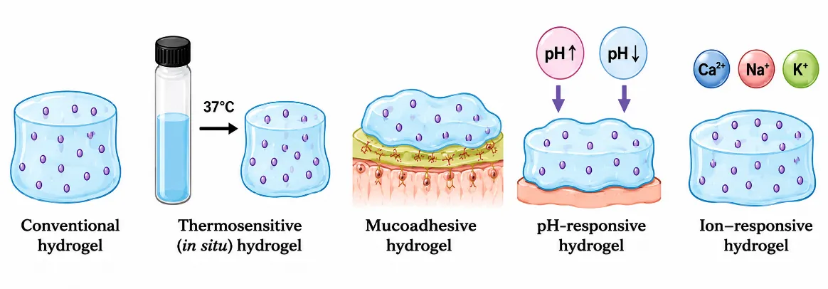
Figure 3: Types of hydrogels
Table 4: Advantages and limitations of hydrogels
|
Advantages |
Limitations |
References |
|
Increased residence time and mucoadhesive property |
High water content may lead to microbial infection. |
[27,29,41,45] |
|
Enhanced bioavailability of poorly soluble antifungal drugs |
Lower physical stability compared with solid dosage forms. |
[27,29,41,45] |
|
Protection of drugs from degradation and minimum systemic absorption. |
Drug loading may be limited for poorly soluble compounds. |
[27,29,41,45]
|
Table 5: Polymers for preparing hydrogels
|
Polymer |
Category |
Advantages |
References |
|
Chitosan |
Natural |
Mucoadhesive, intrinsic antimicrobial activity. |
[7,22,27,29,42] |
|
Sodium alginate |
Natural |
Biocompatible, gel-forming. |
[7,22,27,29,42] |
|
Hyaluronic acid |
Natural |
Tissue healing, hydration. |
[7,22,27,29,42] |
|
Carbopol |
Synthetic |
High viscosity, mucoadhesion.
|
[7,22,27,29,42] |
|
Poloxamer 407 |
Synthetic |
Thermosensitive gelation |
[7,22,27,29,42] |
|
Polyvinyl alcohol (PVA) |
Synthetic |
Mechanical strength |
[7,22,27,29,42] |
|
HPMC |
Semi-synthetic |
Film-forming and viscosity modifier |
[7,22,27,29,42] |
Table 6: Classification of hydrogels
|
Type |
Mechanism |
References |
|
Conventional hydrogels |
Preformed cross-linked polymeric network that hydrates, swells and releases drug by diffusion and polymer relaxation. |
[22,41,42] |
|
Mucoadhesive hydrogels |
Form hydrogen bonding and electrostatic interactions with vaginal mucin, increasing adhesion and residence time while sustaining drug release. |
[42,46,47] |
|
Thermosensitive (in situ) hydrogels |
Liquid at room temperature and undergo sol-to-gel transition at vaginal temperature (≈37 °C), minimizing leakage after administration. |
[42,48–50] |
|
pH-responsive hydrogels |
Swell or alter network structure in response to changes in vaginal pH during infection, triggering controlled drug release. |
[22,41,42,45,51] |
|
Ion-sensitive hydrogels |
Gelation occurs in the presence of physiological ions (Ca²⁺, Na⁺, K⁺) present in vaginal fluid, producing an in situ gel depot. |
[41,42,45,48] |
|
Multi-stimuli-responsive hydrogels |
Respond simultaneously to two or more stimuli (e.g., temperature + pH, temperature + ions) for intelligent and controlled drug release. |
[41,42,45,48] |
Vaginal rings (VRs), also called intravaginal rings (IVRs), are flexible, polymer-based devices built to deliver drugs in a sustained, controlled way directly within the vaginal cavity. They're usually made from biocompatible materials like silicone elastomers, polyurethane (PU), or thermoplastic polyurethanes (TPU), and once inserted into the upper vagina, they can steadily release therapeutic agents for weeks or even months at a time[29,42,52–55]. Compared to traditional vaginal dosage forms, vaginal rings provide a number of benefits, including longer retention times, more regulated medication release, and fewer doses—all of which contribute to improved patient adherence. Vaginal rings are a very promising treatment for recurrent vulvovaginal candidiasis (RVVC) because of these characteristics [29,42,52–55]. Mattia Tiboni and colleagues developed a 3D-printed intravaginal ring loaded with clotrimazole for treating recurrent vaginal candidiasis. Using fused deposition modeling, they created a device capable of releasing the drug steadily over several days, by retaining its antifungal potency against Candida albicans[11]. Unlike conventional creams or tablets that need to be applied repeatedly, the ring provided extended local drug exposure from just a single insertion — a feature that could boost patient compliance and lead to better treatment outcomes[11]. To treat vulvovaginal candidiasis, researchers created a 3D-printed intravaginal ring (IVR) constructed of ethylene-vinyl acetate (EVA) and loaded with the antifungal medications clotrimazole and bifonazole. The ring exhibited good drug stability, sustained antifungal release for up to one week, appropriate mechanical strength, and complete inhibition of Candida albicans growth within four days, demonstrating its potential as an effective single-application therapy for recurrent VVC[52,56]. A recent study by Suresh et al. explored a novel approach to managing vulvovaginal candidiasis using an intravaginal ring coated with a quinazoline-derived copper(I) complex. The coated ring proved highly effective against Candida albicans, eliminating around 70% of established biofilms and blocking the fungus's transition into its invasive hyphal form under simulated vaginal conditions[57].
Table 7: Types of intravaginal rings and their characteristics
|
Type |
Structure |
Mechanism |
References |
|
Matrix ring |
Drug dispersed throughout polymer matrix. |
Diffusion through polymer. |
[29,42,52,58,59] |
|
Reservoir ring |
Drug core surrounded by rate-controlling membrane. |
Membrane-controlled diffusion. |
[22,42,52,58,59] |
|
Sandwich ring |
Drug layer between polymer layers. |
Controlled diffusion. |
[52,60,61] |
|
Pod ring |
Drug-containing pods embedded in ring. |
Individual pod-controlled release. |
[14,62,63] |
|
Biodegradable ring |
Drug dispersed within degradable polymer. |
Polymer degradation and diffusion |
[64–67] |
Table 8: Polymers used for the preparation of IVRs
|
Polymer |
Type |
Examples / Applications |
References |
|
Silicone elastomer (Polydimethylsiloxane) |
Non-biodegradable |
Estring, Femring, Annovera, Dapivirine IVR |
[14,68–70] |
|
Ethylene-vinyl acetate (EVA) |
Non-biodegradable thermoplastic |
NuvaRing, Ornibel membrane |
[42,52,56,56,71–73] |
|
Thermoplastic polyurethane (TPU) |
Non-biodegradable thermoplastic |
Ornibel core |
[14,54,71,74,75] |
|
Polylactic acid (PLA) |
Biodegradable polyester |
Experimental biodegradable IVRs |
[42,71] |
|
Styrene-butadiene block copolymer |
Biodegradable/erodible experimental polymer |
Estradiol vaginal rings |
[14,62] |
|
Polycaprolactone |
Biodegradable polyester |
Experimental antifungal, antiviral and contraceptive IVRs |
[64,65,67,76,77] |
Microneedle arrays
Microneedle arrays (MNs) have gained attention as a novel platform for delivering drugs directly to the vaginal site, offering a minimally invasive alternative to traditional dosage forms[23,42]. Microneedles are tiny, microscopic projections—typically ranging from 25 to 1000μm—that painlessly pierce the outermost layer of the vaginal epithelium. In doing so, they create temporary microchannels that boost drug permeation, all while leaving deeper tissue layers and nerve endings unharmed[23,42]. One study focused specifically on VVC took a different approach, using rapidly dissolving microneedles loaded with lipid nanocarriers containing clotrimazole[78]. The microneedles rapidly dissolved following vaginal insertion, releasing the antifungal-loaded nanoparticles into the mucosa. Compared with blank microneedles and lipid nanocarriers alone, the microneedle formulation produced significantly greater inhibition of both Candida albicans and Candida glabrata.[78] Enggi et al. developed dissolving microneedles incorporating multilayer microcapsules containing Lactobacillus plantarum as a probiotic therapy for VVC. Following vaginal administration, the microneedles dissolved rapidly, releasing viable probiotic microcapsules that inhibited the growth of Candida albicans up to three-fold more effectively than conventional hydrogel and patch formulations in an ex vivo vaginal model[79]. In another study, Aziz et al. developed fluconazole nanocrystal-loaded dissolving microneedles for localized vaginal delivery. Nanocrystal incorporation significantly improved the dissolution characteristics of fluconazole, achieving an in vitro drug release of 89.51%, while the dissolving microneedle system demonstrated a drug recovery of 96.45% following administration[80]. Microneedle arrays appear to be a promising novel treatment for vulvovaginal candidiasis. Their ability to administer probiotics, antifungal drugs, and other therapeutic agents directly into the vaginal mucosa has several advantages, including increased drug penetration, higher bioavailability, maintained local drug concentrations, reduced dose frequency, and enhanced patient compliance[27,42,81].
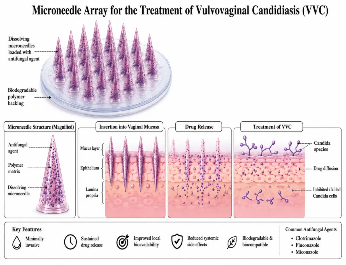
Fig 4: Microneedle array
Table 9: Types of Microneedles used for Vaginal Drug Delivery
|
Type |
Materials |
Mechanism |
References |
|
Solid |
Silicon, stainless steel |
Creates microchannels followed by topical drug application |
[23,42,81] |
|
Coated |
Metal/polymer coated with drug |
Rapid dissolution of coating |
[22,23,42,81] |
|
Dissolving |
PVA, PVP, hyaluronic acid |
Needle dissolves after insertion |
[23,42,79,80] |
|
Hydrogel-forming |
Cross-linked polymers |
Swelling-controlled release |
[23,42,82] |
|
Biodegradable |
PLA, PLGA, PCL |
Polymer degradation |
[23,42,83] |
Liposomes are spherical vesicles made up of one or more phospholipid bilayers surrounding an aqueous core, allowing them to encapsulate both hydrophilic and lipophilic drugs. They are a desirable carrier choice for vaginal drug delivery due to their great biocompatibility, capacity for regulated drug release, and ability to prolong drug retention. When their surface is modified with mucoadhesive polymers, vaginal residence time and local therapeutic efficacy against vulvovaginal candidiasis (VVC) are further enhanced[27,42,84].
Table 10: Applications of liposomes for vaginal drug delivery
|
Formulation |
Drug |
Key findings |
Reference |
|
Chitosan-coated liposomes. |
Clotrimazole |
Chitosan coating (0.1–0.6%) enhanced mucoadhesion, prolonged drug release, increased vaginal retention, and reduced tissue penetration. Lower chitosan concentrations exhibited better mucoadhesive performance. |
[42,85] |
|
Deformable propylene glycol-containing liposomes (DPGLs) in Carbopol hydrogel |
Clotrimazole / Metronidazole |
Showed high drug entrapment, sustained diffusion-controlled release, rapid penetration into the hydrogel matrix, and maintained suitable mechanical properties for vaginal application. |
[86] |
Solid lipid nanoparticles are nanoscale, lipid-based drug carriers featuring a solid lipid core that is stabilized by surfactants.
Table 11: Applications of SLNs in vaginal drug delivery
|
Formulation |
Drug |
Key finding |
References |
|
Polymer-coated SLNs |
Ketoconazole, Clotrimazole |
Controlled drug release and enhanced antifungal activity against C. albicans. |
[42,87] |
|
Cationic SLNs |
Clotrimazole + α-Lipoic acid |
Enhanced stability, prolonged release, and improved activity against resistant Candida. |
[42,88] |
|
SLNs |
Fluconazole |
High drug encapsulation and improved efficacy against fluconazole-resistant Candida |
[42,89] |
These are a combination of liquid and solid lipids, a second-generation lipid nanocarrier system. When compared to solid lipid nanoparticles (SLNs), they have several advantages because of their structure, which include increased drug-loading capacity, increased stability, and longer-lasting drug release. And also due to their strong mucoadhesive properties and higher absorption rate NLCs are considered as a promising delivery system for vulvovaginal candidiasis (VVC) [42,90].
Table 12: Applications of NLCs in vaginal drug delivery
|
Formulation |
Drug |
Key findings |
References |
|
Mucoadhesive NLC hydrogel |
Hypericin |
Sustained release and improved photodynamic antifungal activity. |
[42,91] |
|
Thermosensitive NLC gel |
Clotrimazole |
Sustained release, minimal systemic absorption, and enhanced anti-Candida activity. |
[42,92] |
|
NLCs |
Voriconazole |
Improved activity against Candida biofilms. |
[42,93] |
Electrospinning stands out as a versatile and cost-effective technique for producing fibers on the nano- to micro-scale. these electrospun nanofiber mats are one the most promising targeted drug delivery system within the vaginal environment due to their unique structural and physicochemical properties[42,94]. Electrospinning works by applying a strong electric field to a polymer solution or melt as it's pushed through a syringe needle. This field draws the charged liquid out into a thin jet, which is pulled toward a grounded collector plate. As the jet travels through the air, the solvent evaporates, leaving behind continuous, ultra-fine fibers that pile up on the collector to form a nonwoven, porous mat. Because of this porous structure, the resulting mats are well-suited for loading and delivering drugs or other medicinal compounds.[94].
One or more of the following methods control the release of drugs from electrospun nanofibers[42,94]:
The pronounced hydrophilicity and consequent aqueous solubility of polymers such as PVP and PVA typically result in rapid, near-instantaneous drug release profiles, often characterized by an initial burst effect[94]. In contrast, hydrophobic polymers such as PCL restrict the ingress of aqueous media into the polymer matrix, thereby retarding drug diffusion kinetics and facilitating a more controlled, sustained release profile over an extended duration.[94]. Several studies have provided support for the application of nanofibers in managing VVC. For instance, Souza et al. employed electrospinning to develop amphotericin B-loaded poly(lactic-co-glycolic acid) (PLGA) nanofibers intended for localized vaginal drug delivery, demonstrating sustained release of the drug over a continuous eight-day period.[95]. In a related investigation, Sharma and colleagues formulated fluconazole-loaded PVA nanofibers exhibiting a uniform, bead-free structure, with fiber diameters spanning 150–180 nm. Relative to the free drug, these nanofibers exhibited enhanced antifungal efficacy against Candida species and sustained drug release over a 6-hour period[96]. Nematpour et al. developed clotrimazole-loaded vaginal formulations using polymeric materials such as polyvinyl alcohol (PVA), sodium alginate, and dextran, fabricating both electrospun nanofibrous mats and conventional films for comparison. Evaluation of the two formulations revealed marked differences in Young's modulus, mucoadhesive strength, and antifungal efficacy. Notably, the electrospun nanofibrous mats demonstrated superior vaginal mucoadhesion and enhanced antifungal activity relative to their film-based counterparts. [97]. Mishra et al. reported that incorporating a eucalyptol/β-cyclodextrin inclusion complex into electrospun gellan/polyvinyl alcohol nanofibers (EPNF) achieved a sustained release profile, resulting in approximately 70% inhibition of biofilm formation by both Candida albicans and C. glabrata[98].
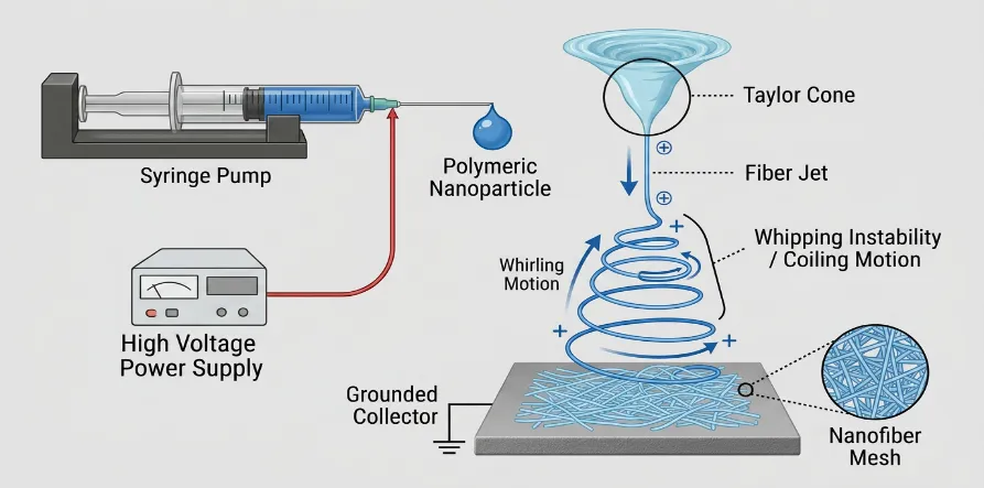
Figure 4: Electrospinning
Vaginal formulation design and development using AI and ML techniques
Molecular dynamics simulations and empirical mathematical models have improved our comprehension of the behavior of vaginal medication delivery devices. By combining data from many variables, machine learning-based methods go one step further and offer more effective and flexible formulation development tools. For instance, researchers linked the composition and thickness of ethylene–vinyl acetate (EVA) polymer membranes to estradiol permeation behavior using an artificial neural network (ANN)-based optimization framework. They then used constrained optimization to identify formulation parameters that could reach particular target doses. More generally, ML models are excellent at integrating diverse data sources and capturing the intricate, multifaceted connections between formulation characteristics and vaginal physiology[99]. Pałkowski et al. investigated the relationship between formulation composition and processing parameters and post-application vaginal pH in tablets and pessaries using dominance-based rough set theory, a rule-based artificial intelligence technique. In order to optimize formulations that quickly disintegrate or deform while maintaining physiological pH, they examined the effects of excipients such methylcellulose, glycerol, lactic acid, chitosan, and Eudragit E-100 on vaginal acidity[99,100]. Ndesendo et al. integrated artificial neural networks (ANN) with molecular modeling to optimize the bioadhesive and erosion properties of intravaginal polymeric devices. They developed caplet-shaped devices containing zidovudine and polystyrene sulfonate, using an ANN-based analysis to guide rational polymer selection—maximizing bioadhesion while preserving matrix integrity under simulated vaginal conditions. This work illustrated how molecular mechanics simulations can be combined with ANN modeling to fine-tune polymer composition, contributing to more rational, data-driven approaches for enhancing formulation performance[99,101]. An ANN model can be used to investigate the effects of polymer ratio, mucoadhesive strength, and delivery system type (tablet versus gel) on drug release, mucoadhesive force, formulation viscosity, etc. In a similar vein, data from enzyme assays and simulated vaginal fluid (SVF) dissolution tests can be processed by AI/ML tools to identify underlying patterns and forecast the formulation's likely behavior under physiological conditions. Furthermore, explainable AI (XAI) methods can provide deeper scientific understanding of the underlying mechanisms by identifying which particular factors—such as polymer hydrophobicity or enzyme affinity—have the biggest influence on product performance. Vaginal formulation development could become a more predictable, efficient, and repeatable process by integrating AI and ML, hastening the shift from lab design to clinically practical drug delivery systems [99,102-104].
CONCLUSION
The benefits of the vagina as a potential drug administration site have been acknowledged and utilized for many years, despite the fact that the entire potential of the vaginal route appears to be overstated, as indicated in multiple extensive reviews related to this topic. Extending the residence period in the vaginal canal is one of the most crucial areas of research for polymers used in vaginal medication delivery. For this reason, mucoadhesive and smart polymers—which enhance viscosity when they interact with the physiological environment—are frequently investigated. Now a days AI and ML is also used for designing, optimization and characterization of vaginal drug delivery systems. pharmaceutical research. Future management of VVC and RVVC will likely depend on the combination of biofilm-targeted treatments, data-driven formulation design, and more sophisticated material science Experts from various fields such as microbiologists, computer researchers and pharmaceutical industry should work in collaboration to make further advancements in this field.
REFERENCES

Archana O.*, Fathima Safa E. K., Rifana C. K., Manoj K., Vaginal Drug Delivery Systems for Vulvovaginal Candidiasis: Current Status AI & ML Enabled Innovations and Future Perspectives, Int. J. Med. Pharm. Sci., 2026, 2 (9), 287-307. https://doi.org/10.5281/zenodo.22671319
 10.5281/zenodo.22671319