
We use cookies to ensure our website works properly and to personalise your experience. Cookies policy
Department of Pharmacology, BVV Sangha’s HSK College of Pharmacy Bagalkot
Background: Nephrotoxicity is a major cause of acute kidney injury (AKI) due to toxic drug exposure. Gentamicin, a widely used aminoglycoside antibiotic, induces renal damage through oxidative stress, inflammation, and lipid peroxidation. Herbal drugs with antioxidant properties are being explored as safer alternatives to NSAIDs. Manilkara zapota (Sapota) is rich in flavonoids, tannins, and polyphenols, with proven antioxidant and anti-inflammatory effects. Objective: This study aimed to evaluate the nephron-protective effect of the hydroalcoholic extract of Manilkara zapota (HAMZ) and its ethanolic nanosuspension (EENs) formulation against gentamicin-induced nephrotoxicity in Wistar rats by assessing biochemical, antioxidant, and histopathological parameters. Materials and methods: Gentamicin (100 mg/kg, i.p.) was administered for eight days to induce nephrotoxicity. The rats were divided into five groups: normal control, disease control, standard (selenium 2 mg/kg), EENs (200 mg/kg), and HAMZ (400 mg/kg). Serum and urine samples were analyzed for creatinine, BUN, uric acid, calcium, phosphorus, and magnesium levels. Antioxidant markers (SOD, CAT, GSH, and LPO) and kidney histology were evaluated. Results: Gentamicin caused significant elevation of serum creatinine, BUN, uric acid, and LPO levels with a decline in antioxidant enzymes. Treatment with HAMZ and EENs significantly restored renal function and antioxidant status, reducing tubular necrosis and inflammation in the kidneys. The 400 mg/kg HAMZ dose showed results comparable to those of selenium. Conclusions: The Manilkara zapota extract and its nano-suspension exhibited strong nephron-protective effects against gentamicin-induced toxicity, mainly through antioxidant and anti-inflammatory mechanisms.
The kidneys are the most essential organs of excretion that lie within the upper compartment of the belly on both sides of the vertebral column. The nephron is the functional and morphological unit of the kidney. Each kidney contains approximately 1 million nephrons within each kidney1. Nephrotoxicity may be described as acute worsening of renal failure occurring as an immediate or indirect consequence of drug, environmental, or occupational exposure to toxins. Similarly, multiple pathologies, such as diabetes, lead to diabetic nephropathy or chronic kidney disease (CKD). Nephropathies are not limited to one form of renal injury. Some chemical toxins act on a discrete anatomical area of the kidney and affect only one category of cell2. Heavy metals are well established to be nephrotoxic to the kidney by their deposition, leading to the formation of a wide range of morphological and functional alterations in the kidney. Several drugs and antibiotics, such as penicillin, cephalosporins, tetracycline, sulphone-amides, antiviral agents, and aminoglycosides, have also been proven to be potential nephrotoxins 3,4. Nephroprotection is greatly advised if cancer and bacterial infections are to be treated. Some strategies have been attempted to constrain nephrotoxicity using xenobiotics and man-made drugs, but none have been successful, and most are unsafe5. Therefore, scientists worldwide are turning to natural sources or alternative or traditional medical systems to explore the potential of natural origin drugs. Ayurveda is a traditional system of medicine that proposes the use of numerous herbs for nephrotoxicity management6. Medicinal plants are a rich source of potentially useful compounds with curative action because they contain multiple complex chemical compounds that can be used to synthesize effective therapies against a wide range of kidney diseases. Numerous herbs are established nephron-protective agents, but few herbs do not have any scientific background to support such assertions. The formulation of an appropriate herbal medicine to cure advanced kidney disease calls for a thorough exploration of characteristics such as nephritic syndrome, acute kidney failure, and chronic intestinal nephritis7. Herbal medicines have curative value because they retain their chemical components, such as flavanols, polyphenols, glycosides, carotenoids, saponins, and tannins.
Mechanism of gentamicin-induced renal toxicity:
Gentamicin (GM), a highly important member of the aminoglycoside class of bactericidal antibiotics, was discovered for the first time in 1963 and is widely used for the treatment of severe gram-negative infections. These infections can lead to endocarditis, sepsis, pneumonia, pelvic inflammatory disease, meningitis, urinary tract infection, and osteomyelitis8. Among the most prominent side effects of GM are nephrotoxicity and ototoxicity, that is, vestibular and cochlear damage. Nephrotoxicity is the leading dose-limiting side effect, in fact9, the mechanisms through which GM can cause nephrotoxicity are unknown 10% to 25% of patients exposed to GM develop AKI. Due to its effectiveness and cost, GM remains the most widely used treatment for ITP, despite its side effects. The key pathophysiology of GM-induced nephrotoxicity encompasses the generation of ROS in glomerular, tubular, and vascular tissues. ROS comprise superoxide anions, hydroxyl radicals, and hydrogen peroxide, and in the kidney, RNS cause a severe reduction in antioxidant defense mechanisms. Acid hydrolases are also secreted along with GM, causing the inhibition of mitochondrial respiration. Induction of acute tubular necrosis, apoptosis, overexpression of transforming growth factor β (TGF-β), intracellular edema, elevation of endothelin I, monocyte/macrophage infiltration, basal membrane damage, and glomerular congestion have all been attributed to GM-induced nephrotoxicity, which decreases the glomerular filtration rate (GFR) and causes renal impairment 10, 11.
Fig.1: Pathological mechanisms of gentamicin-induced nephrotoxicity
Gentamicin is a potent aminoglycoside antibiotic used to treat infections caused by gram-negative bacteria. The chief adverse effects of aminoglycosides are nephrotoxicity and ototoxicity. The nephrotoxicity of aminoglycosides is in the following decreasing order: neomycin > gentamicin ≥ tobramycin ≥ amikacin ≥ netilmicin > streptomycin. The clinical manifestations of aminoglycoside-induced NPT include non-oliguric kidney failure and a gradual increase in serum creatinine levels. The accumulation of aminoglycosides in the PCT of nephrons is the major cause of NPT12. A histopathological report from a research paper revealed that tubular necrosis is the prime practical toxicity of aminoglycosides. Stephan et al, have reviewed about the NPT caused by aminoglycosides in children with its mechanism. Only 5 % of the administered drug accumulates in the lysosomes, Golgi apparatus, and endoplasmic reticulum of the epithelial cells of the PCT. When the concentration threshold increases, aminoglycosides leak into the cytosol and bind to the mitochondria, resulting in the activation of the apoptosis pathway, leading to the production of reactive oxygen species or free radicals13.
Brief review of literature:
Plant profile of Manilkara Zapota14.
|
Synonyms: |
Achradelpha mammosa L, Achras sapota L Achras zapota L |
|
Phylum: |
Magnoliophyta |
|
Kingdom: |
Plantae |
|
Class: |
Magnoliopsida (Dicotylidanae) |
|
Order: |
Ericales |
|
Family: |
Sapotaceae |
|
Genus: |
Manilkara |
|
Species: |
M. zapota |
|
Parts used: |
Fruit and Seeds |
|
Distribution: |
America, Mexico, India, Pakistan, Thailand, Malaysia, Indonesia |
|
Common Name: |
Sapodilla, Sapote, Chicozapote, chicoo |
|
Kannada: |
Chikku, Sapota |
|
Telugu: |
Sima ippacettu |
|
Tamil: |
Chappotta |
Fruits
Seeds
Chemical Constituents: The Manilkara zapota plant contains various phytochemicals, such as flavonoids, tannins, phenolic acids, hydroxybenzoic acid (p-hydroxybenzoic, gallic, and ellagic), flavanols (catechin and epicatechin), flavanols (quercetin), and hydroxycinnamic acid15.
Traditional Use: The traditional literature mentions the Use: The Manilkara zapota. It plays an important role in curing diseases such as diabetes, kidney stones, and hepatotoxicity16.
Pharmacological activity: Biological screening of Manilkara zapota for anti-tumor activity, anti-lipidemic activity17, anti-diarrheal activity18, anti-inflammatory activity19, anti-diabetic activity17, anti-oxidant activity, anti-microbial activity, and anthelmintic activity20.
MATERIALS AND METHODS:
Drugs and chemicals: Selenium, 5,5’ Dithio-bis-(nitro benzoic acid) (DTNB), trichloroacetic acid, 2-Thio barbituric acid (TBA), and epinephrine were purchased from Sigma-Aldrich Co. Gentamicin. All other chemicals used were of analytical grade.
Diagnostic kits: creatinine, BUN, uric acid, Phosphorous, Magnesium, calcium.
Instruments: Refrigerator centrifuge (MPW-350R), UV-Spectrophotometer (UV-1900i, Shimadzu Corporation, Kyoto, Japan), research centrifuge (Remi Industries, Mumbai), and homogenizer (Remi Motors, Mumbai). Hot air oven (Lab Hosp, KW-800W, Mumbai, India).
Fruit material:
Fresh ripe fruits of Manilkara zapota were collected from Kaladagi village, Bagalkot district, Karnataka, in February 2024. The fruit was identified and authenticated at the Department of Botany, Akkamahadevi Women’s Arts, Science and Commerce College, Bagalkote, Karnataka, and the fruit specimen (BVVS/AMWASCC/PG-Bot/15) was deposited in the herbarium of the same college for future reference.
Preparation of fruit extract by maceration procedure:
The collected fruits and seeds were cleaned, and the fruits were cut into small pieces. Small pieces of fruit and seeds were shade-dried and then pulverized into a coarse powder using a grinding machine. The powdered fruits and seeds were macerated in hydroalcohol (30% water and 70% ethanol). The extracts were filtered through a filter paper (Whatman No.1). The filtrate was then evaporated to dryness. Dried extracts were used in the present study21.
Fig.2: Preparation of fruit extract by maceration procedure
Experimental Animals:
Wistar albino rats (200 – 250 g) were obtained from the central animal house of H.S.K. College of Pharmacy and Research Centre in Bagalkot. The animals were housed at room temperature (22-28 ºC) for 12 h. dark and 12 hr. light cycles and were given standard laboratory feed and water ad libitum. The study was approved and conducted according to the norms of the Institutional Animal Ethics Committee will be provided.
Preparation of nano-suspension formulation: (Solvent evaporation method)
MZ nano-suspension was prepared using solvent evaporation technique. The organic phase solution containing MZ extract was dissolved in 3-5 ml absolute ethanol was injected into 0.5, 1 or 3% of stabilizer (SLS) dissolved in water (100 ml) by using a syringe with needle. The mixture was then stirred with a magnetic stirrer at 500 rpm for 60 min. The organic solvent in the solution was evaporated by placing the solution in an open container for 1 hour. At last, nano-suspension is formed.
Evaluation of gentamicin-induced nephrotoxicity in experimental animals:
Experimental Design:
Group I: Normal control (Drinking Water) for 8 days (n=6).
Group II: Control group, received gentamicin (100 mg/kg) intra-peritoneally for 8 days (n=6).
Group III: Gentamicin (100 mg/kg) + selenium (2 mg/kg) intra-peritoneally for 8 days (n=6).
Group IV: Gentamicin (100 mg/kg) + MMN’s Manilkara zapota nano-suspension (200 mg/kg) i.p for 8 days.
Group IV: Gentamicin (100 mg/kg) + HAMZ (400 mg/kg) i.p for 8 days.
Assessment of gentamicin-induced nephrotoxicity
Collection and analysis of urine sample:
All animals were kept in individual metabolic cages and urine samples of 24h were collected on the 8th day of Gentamicin injection. Animals had free access to drinking water during the urine collection period. Urine was analyzed for creatinine, uric acid, BUN, Oxalate, Phosphorus, Magnesium, calcium.
Collection and analysis of serum sample:
After the experimental period, blood samples were collected from the retro-orbital plexus centrifuged at 3000 x g for 10 minutes to obtain serum, the resulting serum was collected in properly labeled, clean and dry micro-centrifuge tubes analyzed immediately for creatinine, uric acid, BUN, Calcium, Phosphorous, Magnesium by using kits (Manufactured by ERBA diagnostics Biosystem) using star-21 plus semi auto analyzer.
Kidney homogenate analysis:
On the 8th day, animals were sacrificed by cervical decapitation or chloroform. The abdomen was opened to remove both kidneys and washed in 0.9% cooled saline, kept once and one kidney is stored in 10% formalin for histopathology. Another one was homogenized in cold phosphate buffer (0.1M, pH 7.4). The homogenates were centrifuged at 10000rpm for 10min at 4ºC (MPW-350R, Korea). Another part of the homogenized supernatant was centrifuged at 17000 rpm for 1 hr at 4ºC. The supernatant obtained was used for further estimation of SOD, CAT, GSH and LPO22 ,23.
Biochemical estimation
Effect of MZNs and HAMZ on serum Creatinine levels in gentamicin induced nephrotoxicity in rat model.
Serum creatinine of the Normal Control group of rats is 0. 0.4381±0.05 mg/dl maintained in the normal range. In Disease control rats are 4.203±0.23 mg/dl significantly (p<0.001) increase from the normal rats. Next comes to standard drug gentamicin treated rats along with MZNs Manilkara zapota nano suspension at 200mg/kg and HAMZ- Hydro Alcoholic Manilkara zapota at 400mg/kg was reduced significantly (p?0.001) 22.864±0.18 mg/dl and 2.167±0.43 mg/dl respectively as compared against the disease control group. The serum creatinine level of the standard group treated with gentamicin induced rats is significantly (p<0.001) reduced to 1.778±0.32 mg/dl serum creatinine when compared to the disease control group.
Effect of MZNs and HAMZ on serum BUN levels in gentamicin induced nephrotoxicity in rat model.
The serum BUN of the Normal Control group of rats is 39.32±16.4 mg/dl maintained in the normal range. In disease control rats is 161.5±5.80 mg/dl significantly (p<0.001) increase from the normal rats. Next comes to standard drug gentamicin treated rats along with MZNs Manilkara zapota nano suspension at 200mg/kg and HAMZ- Hydro Alcoholic Manilkara zapota at 400mg/kg was reduced significantly (p?0.001) 70.61±3.16mg/dl, 60.47±2.60 mg/dl respectively as compared against the Disease control group. The serum BUN level of the standard group treated with Gentamicin-induced rats is significantly (p<0.001) reduced to 56.47±8.20 mg/dl serum BUN when compared to the disease control group.
Fig: 4 Serum BUN
Effect of MZNs and HAMZ on serum Phosphorus, levels in gentamicin induced nephrotoxicity in rat model.
The serum Phosphorus (mg/dl) of the Normal Control group of rats is 12.32 ± 0.74 mg/dl maintained in the normal range. The Serum Phosphorus levels of Disease control rats is 2.920 ± 0.20 mg/dl significantly (p<0.001) increase from the normal rats. The serum Phosphorus level in Gentamicin treated rats along with MZNs Manilkara zapota nano suspension at 200mg/kg and HAMZ- Hydro Alcoholic Manilkara zapota at 400mg/kg was reduced significantly (p?0.001) 4.972 ± 0.44 mg/dl, 5.393 ± 0.55 mg/dl respectively as compared against the Disease control group. The serum Phosphorus level of the standard group treated with Gentamicin-induced rats is significantly (p<0.001) reduced to 6.178 ± 0.48 mg/dl serum Phosphorus when compared to the Disease control group.
Effect of MZNs and HAMZ on serum Magnesium levels in gentamicin induced nephrotoxicity in rat model
The serum Magnesium (mg/dl) levels of the Normal Control group of rats is 1.327 ± 0.20 mg/dl maintained in the normal range. The Serum Magnesium levels of Disease control rats is 9.283 ± 0.32 mg/dl significantly (p<0.001) increase from the normal rats. The serum Magnesium level in Gentamicin treated rats along with MZNs Manilkara zapota nano suspension at 200mg/kg and HAMZ- Hydro Alcoholic Manilkara zapota at 400mg/kg was reduced significantly (p?0.001) 2.390 ± 0.30 mg/dl, 2.138 ± 0.36 mg/dl respectively as compared against the Disease control group. The serum Magnesium level of the standard group treated with Gentamicin-induced rats is significantly (p<0.001) reduced to 1.508 ± 0.18 mg/dl serum Magnesium when compared to the Disease control group.
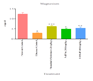
Fig: 6 Serum Magnesium
Effect of MZNs and HAMZ on serum Calcium levels in gentamicin induced nephrotoxicity in rat model.
The serum Calcium (mg/dl) levels of the Normal Control group of rats is 1.908 ± 0.24 mg/dl maintained in the normal range. The serum Calcium levels of Disease control rats is 14.84 ± 0.53 mg/dl significantly (p<0.001) increase from the normal rats. The serum Calcium level in gentamicin treated rats along with MZNs Manilkara zapota nano suspension at 200mg/kg and HAMZ- Hydro Alcoholic Manilkara zapota at 400mg/kg was reduced significantly (p?0.001) 4.327 ± 0.96 mg/dl, 4.277 ± 0.77 mg/dl respectively as compared against the Disease control group. The serum Calcium level of the standard group treated with gentamicin-induced rats is significantly (p<0.001) reduced to 3.875 ± 0.28mg/dl serum Calcium when compared to the Disease control group.
Effect of MZNs and HAMZ on serum Uric acid levels in gentamicin induced nephrotoxicity in rat model.
The serum uric acid (mg/dl) levels of the Normal Control group of rats is 1.210 ± 0.10 mg/dl maintained in the normal range. The serum uric acid levels of Disease control rats are 4.080 ± 0.06 mg/dl significantly (p<0.001) increase from the normal rats. The serum uric acid level in gentamicin treated rats along with MZNs Manilkara zapota nano suspension at 200mg/kg and HAMZ- Hydro Alcoholic Manilkara zapota at 400mg/kg was reduced significantly (p?0.001) 2.213 ± 0.24 mg/dl, 2.153 ± 0.31 mg/dl respectively as compared against the Disease control group. The serum uric acid level of the standard group treated with gentamicin-induced rats is significantly (p<0.001) reduced to 2.002 ± 0.15 mg/dl serum uric acid when compared to the Disease control group.
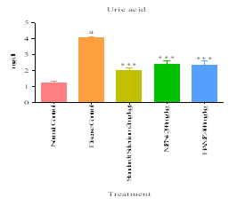
Fig: 8 Serum Uric acid
Urine analysis
Effect of MZNs and HAMZ on Urine Calcium levels against gentamicin induced Nephrotoxicity
The urine Calcium (mg/dl) levels of the Normal Control group of rats is 8.362 ± 0.32 mg/dl maintained in the normal range. The urine Calcium levels of Disease control rats is 27.42 ± 0.65 mg/dl significantly (p<0.001) increase from the normal rats. The urine Calcium level in Gentamicin treated rats along with MZNs Manilkara zapota nano suspension at 200mg/kg and HAMZ- Hydro Alcoholic Manilkara zapota at 400mg/kg was reduced significantly (p?0.001) 15.15 ± 0.74 mg/dl, 12.06 ± 0.25 mg/dl respectively as compared against the Disease control group. The urine Calcium level of the standard group treated with Gentamicin-induced rats is significantly (p<0.001) reduced to 9.407 ± 0.34 mg/dl urine Calcium when compared to the Disease control group.
Effect of MZNs and HAMZ on Urine Phosphorus levels against gentamicin induced Nephrotoxicity
The urine Phosphorus (mg/dl) levels of the Normal Control group of rats is 5.501 ± 0.57 mg/dl maintained in the normal range. The urine Phosphorus levels of Disease control rats is 13.27 ± 0.60 mg/dl significantly (p<0.001) increase from the normal rats. The urine Phosphorus level in Gentamicin treated rats along with MZNs Manilkara zapota nano suspension at 200mg/kg and HAMZ- Hydro Alcoholic Manilkara zapota at 400mg/kg was reduced significantly (p?0.001) 10.67 ± 0.30 mg/dl, 9.297 ± 0.24 mg/dl respectively as compared against the Disease control group. The urine Phosphorus level of the standard group treated with Gentamicin-induced rats is significantly (p<0.001) reduced to 6.912 ± 0.40 mg/dl urine Phosphorus when compared to the Disease control group.
Fig: 10 Urine Phosphorous
Effect of MZNs and HAMZ on Urine Magnesium levels against gentamicin induced Nephrotoxicity
The urine Magnesium (mg/dl) levels of the Normal Control group of rats is 8.528 ± 0.34 mg/dl maintained in the normal range. The urine Magnesium levels of Disease control rats is 3.119 ± 0.39 mg/dl significantly (p<0.001) increase from the normal rats. The urine Magnesium level in Gentamicin treated rats along with MZNs Manilkara zapota nano suspension at 200mg/kg and HAMZ- Hydro Alcoholic Manilkara zapota at 400mg/kg was reduced significantly (p?0.001) 5.337 ± 0.34 mg/dl, 6.757 ± 0.39 mg/dl respectively as compared against the Disease control group. The urine Magnesium level of the standard group treated with Gentamicin-induced rats is significantly (p<0.001) reduced to 9.324 ± 0.23 mg/dl urine Magnesium when compared to the Disease control group.
Effect of MZNs and HAMZ on Urine Oxalate levels against gentamicin induced Nephrotoxicity
The urine oxalate (mg/24h) level of the Normal Control group of rats is 1.183 ± 0.089 mg/24h maintained in the normal range. The urine oxalate levels in Disease control rats are 8.301 ± 0.49 mg/24h significant (p<0.001) increase in the normal rats. The urine oxalate level in sodium oxalate-induced rats along with MZNs Manilkara zapota nano suspension at 200mg/kg and HAMZ- Hydro Alcoholic Manilkara zapota at 400mg/kg was reduced significantly (p?0.001), 5.687 ± 0.16 and 4.974 ± 0.21 mg/24h respectively as compared against the Disease control group. The urine oxalate level of the standard group treated with sodium oxalate along with cystone 500mg/kg is significantly (p<0.001) reduced to 2.640 ± 0.20 mg/24h urine oxalate when compared to the Disease control group.
Fig: 12 Urine Oxalate
Effect of MZNs and HAMZ on Urine Creatinine levels against gentamicin induced Nephrotoxicity
The urine creatinine (mg/dl) levels of the Normal Control group of rats is 0.7762±0.33 mg/dl maintained in the normal range. The urine creatinine levels of Disease control rats are 13.51±0.5 mg/dl significantly (p<0.001) increase from the normal rats. The urine creatinine level in Gentamicin treated rats along with MZNs Manilkara zapota nano suspension at 200mg/kg and HAMZ- Hydro Alcoholic Manilkara zapota at 400mg/kg was reduced significantly (p?0.001) 9.55±0.44 mg/dl, 4.92±0.45 mg/dl respectively as compared against the Disease control group. The urine creatinine level

Sahana Shailesh Vajramatti*, Salma Bhanu, Marigouda Patil, Lingaraj Anawal, Sanjay Havaragi, Chandrashekhar V. M., Mallappa Shalavadi, Nephroprotective Activity of Manilkara Zapota Nano-Suspension Formulation on Gentamicin Induced Nephrotoxicity in Rat Model, Int. J. Med. Pharm. Sci., 2026, 2 (3), 369-382. https://doi.org/10.5281/zenodo.19217163
 10.5281/zenodo.19217163