

We use cookies to ensure our website works properly and to personalise your experience. Cookies policy
Faculty of Pharmacy, Mansarovar Global University, Sehore, M.P. 466111
Citrus macroptera Montrouz. leaves and their essential oil were investigated through pharmacognostical characterization, physicochemical testing, phytochemical screening and in vitro antioxidant and antibacterial assays. Leaf anatomy and powder microscopy were examined; ash, extractive and fluorescence characteristics were recorded; solvent extracts underwent qualitative screening; and essential oil was recovered by Clevenger hydrodistillation. The leaves measured 8.5–16.2 × 4.2–8.1 cm and exhibited differentiated mesophyll, collateral vascular bundles and numerous secretory cavities. Total ash, acid-insoluble ash and loss on drying were 6.82 ± 0.18%, 0.74 ± 0.06% and 7.18 ± 0.21%, respectively. Oil yield was 0.72% v/w, refractive index 1.4758, specific gravity 0.8614 and optical rotation +48.6° at 20°C. The GC–MS profile listed limonene at 42.68%. Oil DPPH inhibition increased from 38.74 ± 0.61% to 82.36 ± 0.88% across 6.25–200 µg/mL. The work reported DPPH IC50 was 54.72 µg/mL for oil and 30.09 µg/mL for ascorbic acid; reported nitric oxide IC50 values were 13.18 and 84.56 µg/mL, respectively. Oil reducing-power absorbance increased from 0.238 ± 0.004 to 0.418 ± 0.008 across 12.5–400 µg/mL. Reported oil inhibition zones ranged from 10 ± 0.02 to 25 ± 0.04 mm.
Reliable identification and quality assessment of botanical raw materials are prerequisites for pharmaceutical investigation. Morphology, anatomy and physicochemical measurements provide complementary evidence of identity and material quality, whereas chemical screening helps select appropriate fractions for subsequent analysis. These approaches are described in established guidance for herbal materials and standard pharmacognosy texts [1,2]. A single analytical result cannot substitute for an authenticated specimen, a documented collection history or reproducible sampling. Citrus species are relevant to this approach because their tissues contain secretory structures associated with aromatic volatile fractions. The chemistry of the genus encompasses compounds differing in volatility, polarity and biological activity [3]. Citrus macroptera Montrouz., family Rutaceae, was the subject of the present investigation. Rather than assuming that results from other Citrus species establish the composition or activity of this particular sample, the study combined leaf-level diagnostic observations with essential-oil assessment.
Phytochemical screening can reveal broad classes such as phenolics, flavonoids, tannins and terpenoids, but these reactions are presumptive and do not identify individual molecules [4,5]. Extraction solvent affects the recovered fraction, and isolation procedures can modify the apparent chemical profile [6]. Therefore, results from whole-leaf polar extracts and a hydrodistilled volatile fraction should be interpreted separately. The latter may contain components capable of biological activity, but essential oils are complex mixtures whose effects and safety depend on composition, concentration and exposure [7].
In vitro antioxidant assays measure operational chemical responses. DPPH decolorization assesses interaction with a stable radical [8,9], while interpretation depends on solvent, reaction conditions and concentration range [10]. Multiple assays can describe complementary responses, but none alone demonstrates antioxidant efficacy in humans. Standardization of analytical conditions is essential for comparison [11]. Antibacterial screening likewise requires attention to inoculum, solvent, dose and growth conditions. Plants provide diverse candidate antimicrobial constituents [12]; nevertheless, diffusion-based observations require methodological qualification before they can be used to rank potency [13]. Therefore, the objective of the present work was to characterize C. macroptera leaves using morphological, anatomical and physicochemical measurements, examine the qualitative phytochemical profiles of solvent extracts, and evaluate the isolated essential oil using antioxidant and antibacterial assays.
2 METHODOLOGY
2.1 Plant material and processing
Fresh, mature, healthy leaves were selected, cleaned and botanically authenticated as C. macroptera [3]. Leaves were washed, shade-dried at room temperature until sufficiently brittle, coarsely powdered, sieved and stored in airtight containers protected from moisture, heat and light. Chemicals and solvents were of analytical or laboratory-reagent grade.
2.2 Morphological and microscopic examination
Leaves were assessed for dimensions, shape, colour, margin, apex, base, venation, surface, petiole, texture and odour. Portions of lamina and midrib were fixed for approximately 24 h in formalin–acetic acid–70% ethanol. Specimens were dehydrated through graded ethanol and tertiary-butyl alcohol, infiltrated with paraffin and sectioned at approximately 10–12 µm. Dewaxed sections were stained with toluidine blue; safranin and fast green were used as additional differential stains. Epidermal preparations and cleared leaves were mounted in glycerin to examine stomata, venation and secretory structures [1,2]. Powdered material was examined for epidermal fragments, stomata, palisade and spongy cells, vascular elements, fibres, crystals and secretory cavities. Where required, maceration used equal volumes of 5% chromic acid and 5% nitric acid at approximately 55°C for 3–5 h, followed by washing and safranin staining. Quantitative microscopy included stomatal number, stomatal index, vein-islet number, vein-termination number and palisade ratio.
2.3 Physicochemical and fluorescence evaluation
Total ash was measured by incinerating approximately 2 g of powder below about 450°C to a carbon-free residue and weighing to constant mass. Water-soluble ash was determined after boiling ash with approximately 25 mL water for 5 min, filtering and igniting the insoluble residue; its mass was subtracted from total ash. Acid-insoluble ash followed boiling ash with approximately 25 mL dilute hydrochloric acid for 5 min and recovery of the insoluble material. Loss on drying used approximately 1 g powder at 100–105°C until constant mass [1]. For extractive values, approximately 5 g powder was macerated with 100 mL solvent. A measured filtrate portion was evaporated and dried at approximately 105°C. Powder fluorescence was observed under visible light and UV at 254 and 365 nm after treatment with ethanol, ethyl acetate, chloroform, water, 50% sulphuric acid, 10% sodium hydroxide and 50% nitric acid.[13]
2.4 Extraction and phytochemical screening
Leaf extracts were prepared with petroleum ether, chloroform, methanol, ethanol and water. The extraction yields are reported separately from cold-maceration extractive values because these are distinct measurements. Qualitative tests included precipitating reagents for alkaloids; Molisch, Fehling and Benedict reactions for carbohydrates; hydrolysis-based glycoside tests; Borntrager testing for anthraquinones; Keller–Killiani-type testing for cardiac glycosides; Salkowski and Liebermann–Burchard reactions for sterols; frothing for saponins; ferric chloride for phenolic/tannin reactions; protein/amino-acid reactions; ruthenium red for mucilage; and Shinoda, alkaline and zinc–hydrochloride reactions for flavonoids [4,5].
2.5 Essential oil isolation and profiling
The essential oil was isolated by hydrodistillation in a Clevenger-type apparatus, dried to remove residual moisture and stored in sealed amber vials under refrigeration. Oil yield was expressed as mL recovered per 100 g plant material. Organoleptic properties, organic-solvent solubility, water immiscibility, specific gravity, refractive index and optical rotation were recorded.[20] Specific gravity was described using a pycnometer, refractive index using an Abbe refractometer and optical rotation using a polarimeter.
2.6 Antioxidant assay
DPPH solutions and sample dilutions were mixed and incubated in the dark for approximately 30 min; absorbance was measured at 517 nm with control and blank preparations. Percentage inhibition was calculated as 100 (A0 − A1)/A0, where A0 and A1 represent control and sample absorbance. Ascorbic acid served as comparator [8–10]. Oil and comparator were tested at 6.25, 12.5, 25, 50, 100 and 200 µg/mL. Nitric oxide scavenging used sodium nitroprusside in phosphate-buffered saline, incubation at approximately 25°C for about 30 min, and addition of sulphanilamide and naphthyl ethylenediamine dihydrochloride. Absorbance was recorded near 546 nm and percentage inhibition calculated using the same expression. The Griess reaction provides nitrite-related colorimetry [14]. The oil and ascorbic acid were tested over different concentration ranges.[20] Reducing power was assessed using phosphate buffer and potassium ferricyanide, incubation at approximately 50°C for 20 min, addition of trichloroacetic acid and centrifugation. Supernatant was combined with water and ferric chloride, and absorbance measured at 700 nm.
2.7 Antibacterial screening
Mueller–Hinton agar was prepared and autoclaved at approximately 121°C for 15 min. Cultures were adjusted to an unspecified turbidity standard and spread on agar. Sterile discs were impregnated with essential oil dissolved in a suitable solvent. Amikacin was the positive control and a solvent control was described. Zone diameters were recorded in millimetres for eight bacterial species. [21]
3 RESULTS AND DISCUSSION
3.1 Pharmacognostical characteristics
Leaves were simple, leathery and aromatic, ovate to elliptic-oblong, 8.5–16.2 cm long and 4.2–8.1 cm wide. The upper surface was dark green, glabrous and slightly glossy, with a lighter underside. The margin was entire to slightly crenulated, apex acute to shortly acuminate, base rounded to broadly cuneate, venation reticulate and pinnate, and petiole slightly winged.
Table 1: Morphological and Organoleptic Characteristics
| Characteristic | Reported observation |
|---|---|
| Plant habit | Evergreen, aromatic shrub/tree |
| Leaf type | Simple |
| Shape | Ovate to elliptic-oblong |
| Length | 8.5–16.2 cm |
| Width | 4.2–8.1 cm |
| Colour | Dark green above; lighter green beneath |
| Surface | Glabrous, slightly glossy |
| Margin | Entire to slightly crenulated |
| Apex | Acute to shortly acuminate |
| Base | Rounded to broadly cuneate |
| Venation | Reticulate, pinnate |
| Petiole | Short to moderately long, slightly winged |
| Texture | Leathery/coriaceous |
| Odour | Characteristic aromatic citrus |
| Taste | Slightly bitter and aromatic |
The leaf sections showed single-layered epidermides, a distinct thin cuticle, differentiated mesophyll, 2–3 palisade layers, 4–6 loosely arranged spongy layers and collateral vascular bundles. Rounded or oval secretory cavities and occasional calcium oxalate crystals were recorded. Powder microscopy revealed epidermal and vascular fragments, stomata, palisade cells, spongy parenchyma, oil glands, a few fibres and occasional starch grains. Stomata were described as anomocytic/amphicyclic. Together these features constitute a preliminary diagnostic fingerprint.
Table 2: Anatomical Epidermal and Powder Diagnostic Observations
| Preparation | Characteristic | Reported observation |
|---|---|---|
| Section | Upper epidermis | Single layered, compact |
| Section | Lower epidermis | Single layered, stomatiferous |
| Section | Cuticle | Thin and distinct |
| Section | Palisade tissue | 2–3 layers |
| Section | Spongy tissue | 4–6 layers, loosely arranged |
| Section | Oil glands | Numerous, rounded/oval |
| Section | Vascular bundles | Collateral |
| Section | Calcium oxalate | Present, occasional |
| Section | Trichomes | Sparse/rare |
| Section | Intercellular spaces | Prominent in spongy region |
| Adaxial epidermis | Cells and walls | Polygonal; straight to slightly undulating |
| Abaxial epidermis | Cells and walls | Irregular/polygonal; slightly undulating |
| Epidermis | Stomata | Adaxial few/absent; abaxial numerous |
| Abaxial epidermis | Stomatal type | Anomocytic/amphicyclic |
| Both surfaces | Oil glands and trichomes | Glands present; trichomes sparse |
| Powder | Epidermal fragments, stomata and palisade cells | Present |
| Powder | Spongy parenchyma and vascular fragments | Present |
| Powder | Secretory cavities | Numerous |
| Powder | Fibres | Few |
| Powder | Calcium oxalate | Present |
| Powder | Trichome fragments | Rare |
| Powder | Starch grains | Few/occasional |
Table 3: Quantitative Microscopy and Leaf Physicochemical Measurements
| Measurement | Reported value |
|---|---|
| Adaxial stomatal number | 4.62 ± 0.21 |
| Abaxial stomatal number | 18.74 ± 0.63 |
| Adaxial stomatal index (%) | 5.18 ± 0.24 |
| Abaxial stomatal index (%) | 18.92 ± 0.57 |
| Vein-islet number per mm² | 11.40 ± 0.32 |
| Vein-termination number per mm² | 16.80 ± 0.41 |
| Palisade ratio | 5.12 ± 0.18 |
| Total ash (% w/w) | 6.82 ± 0.18 |
| Water-soluble ash (% w/w) | 4.21 ± 0.14 |
| Acid-insoluble ash (% w/w) | 0.74 ± 0.06 |
| Loss on drying (% w/w) | 7.18 ± 0.21 |
| Alcohol-soluble extractive (% w/w) | 9.46 ± 0.27 |
| Water-soluble extractive (% w/w) | 12.84 ± 0.31 |
| Petroleum ether-soluble extractive (% w/w) | 4.38 ± 0.19 |
3.2 Physicochemical properties and extract screening
Acid-insoluble ash was lower than total ash, consistent with a relatively small acid-insoluble residue. Water-soluble extractive exceeded alcohol-soluble extractive. Loss on drying includes moisture and other volatile mass losses. Aqueous extraction gave the highest reported recovery, followed by methanol and ethanol. Polar extracts showed stronger qualitative flavonoid and phenolic reactions, whereas petroleum ether and chloroform showed stronger sterol and terpenoid reactions.
Table 4: Appearance and Recovery of Solvent Extracts
| Solvent | Reported appearance | Yield (% w/w) |
|---|---|---|
| Petroleum ether | Greenish semi-solid | 4.62 |
| Chloroform | Dark green | 3.84 |
| Methanol | Dark green/brown | 11.26 |
| Ethanol | Greenish brown | 9.84 |
| Water | Brown | 13.42 |
Table 5: Extraction Yields and Qualitative Leaf Phytochemistry
| Constituent or yield | Petroleum ether | Chloroform | Methanol | Ethanol | Water |
|---|---|---|---|---|---|
| Yield (% w/w) | 4.62 | 3.84 | 11.26 | 9.84 | 13.42 |
| Alkaloids | − | + | ++ | ++ | + |
| Carbohydrates | − | − | ++ | ++ | +++ |
| Glycosides | − | + | ++ | ++ | ++ |
| Cardiac glycosides | − | − | + | + | + |
| Anthraquinone glycosides | − | − | − | − | − |
| Phytosterols | ++ | ++ | + | + | − |
| Saponins | − | − | ++ | + | +++ |
| Tannins | − | + | ++ | ++ | +++ |
| Phenolic compounds | + | + | +++ | +++ | ++ |
| Proteins and amino acids | − | − | + | + | ++ |
| Mucilage | − | − | + | + | ++ |
| Flavonoids | + | + | +++ | +++ | ++ |
| Terpenoids | ++ | ++ | ++ | ++ | + |
| Volatile constituents | +++ | + | + | + | − |
| Fixed oil | + | − | − | − | − |
− absent; + positive; ++ moderately positive; +++ strongly positive.
Table 6: Fluorescence Characteristics of Leaf Powder
| Treatment | Visible light | UV 254 nm | UV 365 nm |
|---|---|---|---|
| Untreated | Green | Pale green | Brownish green |
| Ethanol | Greenish yellow | Yellowish green | Light brown |
| Ethyl acetate | Yellowish green | Green | Greenish brown |
| Chloroform | Greenish yellow | Pale green | Brown |
| Water | Light green | Yellowish green | Dark brown |
| 50% H₂SO₄ | Yellowish green | Green | Dark brown |
| 10% NaOH | Pale green | Bright green | Light brown |
| 50% HNO₃ | Light brown | Pale green | Dark brown |
3.3 Essential oil properties and unverified volatile profile
The oil yield was reported as 0.72% v/w. The oil was pale yellow to yellow, slightly viscous, citrus-scented, soluble in ethanol, chloroform, petroleum ether and toluene, and immiscible with water. At 20°C, refractive index was 1.4758, specific gravity 0.8614 and optical rotation +48.6°. Oil screening showed strong volatile/terpenoid reactions, moderate phenolic volatile reactions and a weak steroidal reaction; flavonoid, alkaloid, saponin, tannin, glycoside and fixed-oil tests were negative.
Table 7: Essential Oil Organoleptic and Physicochemical Measurements
| Measurement | Reported result |
|---|---|
| Oil yield | 0.72% v/w |
| Colour | Pale yellow to yellow |
| Odour | Strong, pleasant, aromatic citrus |
| Taste | Characteristic, slightly pungent |
| Characteristic feel | Slightly viscous |
| Organic-solvent solubility | Soluble in ethanol, chloroform, petroleum ether and toluene |
| Water solubility | Immiscible |
| Refractive index at 20°C | 1.4758 |
| Specific gravity at 20°C | 0.8614 |
| Optical rotation at 20°C | +48.6° |
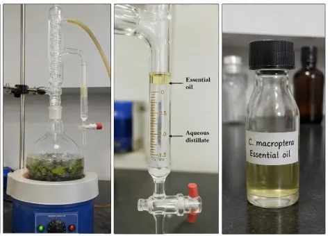
Figure 1: Hydrodistillation Apparatus and Recovered Essential Oil
Table 8: Qualitative Phytochemical Reactions of Essential Oil
| Class | Reported reaction |
|---|---|
| Volatile constituents | +++ |
| Terpenoids | +++ |
| Steroidal constituents | + |
| Phenolic volatile constituents | ++ |
| Flavonoids | − |
| Alkaloids | − |
| Saponins | − |
| Tannins | − |
| Glycosides | − |
| Fixed oil | − |
− absent; + present; ++ moderately present; +++ strongly present. These are qualitative screening observations.
3.4 Antioxidant responses and IC50
DPPH inhibition increased with concentration for both samples. The oil response increased from 38.74% at 6.25 µg/mL to 82.36% at 200 µg/mL. At every matched concentration, the ascorbic acid mean exceeded the oil mean. The profile therefore supports concentration-related radical interaction under the reported assay conditions. DPPH IC50 values of 30.09 µg/mL for ascorbic acid and 54.72 µg/mL for the oil was reported. Nitric oxide inhibition also increased with concentration. The oil exceeded 50% at 5.56 µg/mL, whereas ascorbic acid exceeded 50% at 44.44 µg/mL.
Table 10: Reported Antioxidant IC50
| Assay | Essential oil IC50 (µg/mL) | Ascorbic acid IC50 (µg/mL) |
|---|---|---|
| DPPH | 54.72 | 30.09 |
| Nitric oxide scavenging | 13.18 | 84.56 |
The oil’s reducing-power absorbance rose from 0.238 at 12.5 µg/mL to 0.418 at 400 µg/mL. The only directly shared concentration is 50 µg/mL: oil 0.279 versus ascorbic acid 0.436. This supports reducing capacity [11].
Table 11: Antioxidant Assay Observations
| Assay | Oil concentration (µg/mL) | Oil response | Ascorbic acid concentration (µg/mL) | Ascorbic acid response |
|---|---|---|---|---|
| DPPH | 6.25 | 38.74 ± 0.61 | 6.25 | 48.21 ± 0.27 |
| DPPH | 12.5 | 44.26 ± 0.72 | 12.5 | 51.18 ± 0.78 |
| DPPH | 25 | 52.84 ± 0.91 | 25 | 59.97 ± 1.12 |
| DPPH | 50 | 65.72 ± 0.86 | 50 | 74.65 ± 0.34 |
| DPPH | 100 | 72.48 ± 0.74 | 100 | 79.84 ± 0.71 |
| DPPH | 200 | 82.36 ± 0.88 | 200 | 92.82 ± 1.28 |
| Nitric oxide | 2.78 | 39.84 ± 1.06 | 22.22 | 47.19 ± 0.53 |
| Nitric oxide | 5.56 | 51.72 ± 1.48 | 44.44 | 54.31 ± 0.81 |
| Nitric oxide | 11.11 | 63.46 ± 1.94 | 88.89 | 60.67 ± 1.06 |
| Nitric oxide | 22.22 | 74.18 ± 2.08 | 177.78 | 65.92 ± 1.33 |
| Nitric oxide | 44.44 | 91.26 ± 2.31 | 222.22 | 84.64 ± 1.33 |
| Reducing power | 12.5 | 0.238 ± 0.004 | 10 | 0.243 ± 0.003 |
| Reducing power | 25 | 0.254 ± 0.003 | 20 | 0.278 ± 0.005 |
| Reducing power | 50 | 0.279 ± 0.004 | 30 | 0.362 ± 0.008 |
| Reducing power | 100 | 0.305 ± 0.005 | 40 | 0.413 ± 0.003 |
| Reducing power | 200 | 0.329 ± 0.006 | 50 | 0.436 ± 0.006 |
| Reducing power | 400 | 0.418 ± 0.008 | — | — |
Mean ± SEM of three readings. DPPH and nitric oxide responses are percentage inhibition; reducing-power responses are absorbance at 700 nm.
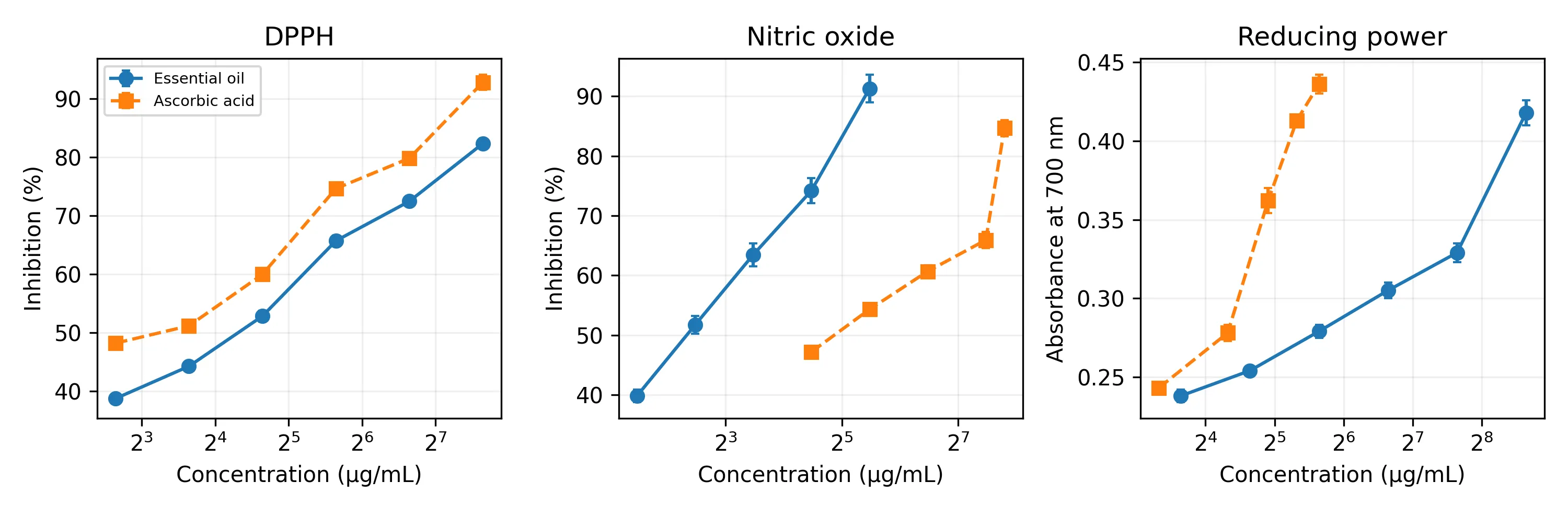
Figure 2: Antioxidant Responses
3.5 Antibacterial screening
The reported oil inhibition zones ranged from 10 ± 0.02 mm against K. pneumoniae to 25 ± 0.04 mm against P. mirabilis. Other comparatively large zones were observed for E. coli and S. aureus. Some oil zones exceed the comparator zones, but different disc loadings and different diffusion characteristics prevent a conclusion of superior antibacterial potency. Earlier studies have investigated plant volatile oils directly and those observations provide context too [19, 20]. The reported disc-associated thresholds were 15 µL/disc for seven organisms and 30 µL/disc for K. pneumoniae.
Table 12: Antibacterial Zones and Disc Associated Inhibitory Thresholds
| Organism | Amikacin zone (mm) | Oil zone (mm) | MIC (µL/disc) |
|---|---|---|---|
| Chromobacterium violaceum | 20 ± 0.01 | 13 ± 0.03 | 15 |
| Escherichia coli | 22 ± 0.05 | 24 ± 0.05 | 15 |
| Klebsiella pneumoniae | 24 ± 0.04 | 10 ± 0.02 | 30 |
| Proteus mirabilis | 26 ± 0.02 | 25 ± 0.04 | 15 |
| Shigella flexneri | 24 ± 0.02 | 13 ± 0.03 | 15 |
| Streptococcus pyogenes | 24 ± 0.06 | 22 ± 0.04 | 15 |
| Staphylococcus aureus | 21 ± 0.00 | 23 ± 0.03 | 15 |
| Pseudomonas aeruginosa | 22 ± 0.08 | 21 ± 0.05 | 15 |
Zones are source mean ± SEM from two readings.
CONCLUSION
Citrus macroptera leaves exhibited characteristic morphology, differentiated mesophyll, collateral vascular bundles and abundant secretory cavities. Reported total ash was 6.82 ± 0.18%, loss on drying 7.18 ± 0.21%, and aqueous extraction yield 13.42% w/w. Essential-oil yield was 0.72% v/w, with refractive index 1.4758, specific gravity 0.8614 and optical rotation +48.6° at 20°C. The oil produced 82.36 ± 0.88% DPPH inhibition at 200 µg/mL, 91.26 ± 2.31% nitric oxide inhibition at 44.44 µg/mL, and reducing-power absorbance of 0.418 ± 0.008 at 400 µg/mL. Its reported DPPH and nitric oxide IC50 estimates were 54.72 and 13.18 µg/mL, respectively. Reported antibacterial zones were greatest for P. mirabilis (25 ± 0.04 mm), E. coli (24 ± 0.05 mm) and S. aureus (23 ± 0.03 mm).
REFERENCES

Vimal Kumar, Satish Kumar Sarankar, Pharmacognostical Characterization and in Vitro Antioxidant and Antibacterial Evaluation of Citrus Macroptera Leaves and Essential Oil, Int. J. Med. Pharm. Sci., 2026, 2 (11), 116-124. https://doi.org/10.5281/zenodo.24420630
 10.5281/zenodo.24420630