

We use cookies to ensure our website works properly and to personalise your experience. Cookies policy
1MDS, Oral & Maxillofacial Surgeon and Medical Director, Suyog Hospital, Mysore
2MDS, Oral Pathologist and Medical Director, Suyog Hospital, Mysore
Fractures of the mandible are among the most common maxillofacial injuries in oral and maxillofacial surgery, and most commonly occur as a result of road traffic accidents, interpersonal violence, falls, and sports trauma. Since the mandible has a horseshoe shape, a single trauma to the mandible will cause fractures at more than one site - either directly at the point of impact or indirectly at the condylar neck, angle, or parasymphysis. We present a clinical case of a combined symphyseal mandibular fracture with bilateral condylar fractures successfully treated with ORIF under general anaesthesia. The symphysis was approached and plated via an intraoral vestibular incision, and both condylar fractures were treated with a retromandibular transmasseteric approach for rigid miniplate fixation. This report describes the diagnostic approach, surgical technique, and rationale for open management of bilateral condylar fractures in conjunction with symphyseal fracture conditions and reviews literature on treatment selection for this fracture pattern.
The mandible is the second most commonly fractured facial bone after the nasal bone, owing to its prominent and unprotected position in the lower third of the face. Mandibular fractures account for a significant proportion of all maxillofacial injuries in trauma series, with the condyle, angle, symphysis/parasymphysis and body being the most frequently affected sites in decreasing or variable order depending on the population studied. The combination of a symphyseal fracture with bilateral condylar fractures is a well-known injury pattern that typically results from a direct anterior blow to the chin and is transmitted bilaterally along the mandibular body to the condylar necks (the thinnest and biomechanically weakest part of the mandible). While the management of condylar fractures is one of the most controversial issues in maxillofacial trauma surgery, it currently has three options: closed reduction with maxillomandibular fixation (MMF) and early mobilisation, open reduction and internal fixation (ORIF) with rigid or semi-rigid osteosynthesis. If condylar fractures happen bilaterally, particularly with a fracture elsewhere in the mandibular arch (the symphysis for instance), the risk of a decrease in posterior facial height, premature posterior occlusal contact, anterior open bite, facial asymmetry and long-term TMJ dysfunction increases considerably if treated by closed means only. However, a growing body of evidence is favouring an open management of displaced and dislocated condylar fractures, especially when they occur bilaterally, in order to better manage ramus height, condylar position and pre-traumatic occlusion. We have presented a case of symphyseal fracture with bilateral condylar fractures treated by ORIF under general anaesthesia and discussed the clinical decision making.
Case Report.
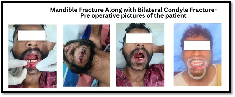
2.1 History and Presenting Complaint. A patient with a history of trauma to the face after a road traffic accident reported to the Department of Oral and Maxillofacial Surgery. The patient had pain and swelling of the lower jaw and chin region, difficulty chewing, limited mouth opening, and a substantial change in the bite. There was no history of loss of consciousness, vomiting, ear or nasal bleed, seizure and the patient was conscious, oriented and haemodynamically stable at the time of presentation. A complete general physical examination was carried out to rule out any systemic and other regional injuries as per standard advanced trauma life support (ATLS) protocol and a focused maxillofacial examination was then performed.
2.2 Clinical Examination. Extraoral examination revealed: • Bilateral preauricular tenderness and swelling in both condylar regions, with restricted and deviated mandibular movements. • Restricted maximal mouth opening (inter-incisal opening reduced compared to normal range), with deviation of the jaw on opening suggestive of bilateral condylar involvement. • Decreased or absent condylar movement on palpation over the preauricular region during mouth opening, more marked on the side with greater displacement. • The step deformity was not consistently palpable at the condylar region, as is typical of high subcondylar/condylar neck fractures, but was clearly palpable at the symphysis. Intraoral examination revealed: • A palpable step deformity and mobility of the fracture segments at the symphysis, with sublingual ecchymosis — a classic sign of an underlying anterior mandibular fracture. • Deranged occlusion with premature posterior tooth contact and an anterior open bite tendency, consistent with bilateral loss of posterior mandibular (condylar/ramal) height. • Mobility of the symphyseal segment on bimanual manipulation, with associated pain. • No obvious mucosal laceration or gross displacement of teeth, although individual teeth in the line of fracture were assessed for mobility and vitality.
2.3 Radiographic and Imaging Findings
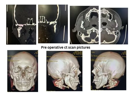
Radiographic evaluation was carried out with an orthopantomogram (OPG) as the initial screening investigation, supplemented by a postero-anterior (PA) view of the mandible and, given the need for precise three-dimensional assessment of the condylar fractures, a computed tomography (CT) scan of the face with axial, coronal, and 3D reconstructions. Imaging confirmed:
Correlating the clinical finding of an anterior open bite with bilateral loss of posterior facial height on imaging confirmed the diagnosis of a symphyseal fracture with bilateral displaced condylar fractures, and open management was planned for both the symphysis and both condyles.
2.4 Treatment Planning
Given (a) the bilaterality of the condylar fractures, (b) the presence of a concomitant fracture elsewhere in the mandibular arch (the symphysis) which removes the buttressing effect an intact contralateral mandible would otherwise provide, and (c) the clinical/radiographic evidence of displacement with derangement of occlusion, the decision was made in favour of open reduction and internal fixation of all three fracture sites (symphysis and both condyles) rather than closed reduction with prolonged maxillomandibular fixation alone. This decision is consistent with the generally accepted indications for open treatment of condylar fractures, which include displacement into the middle cranial fossa, inability to obtain adequate occlusion by closed methods, lateral extracapsular dislocation, bilateral condylar fractures associated with comminuted midface fractures or symphyseal fractures where closed treatment would compromise vertical ramus height, and medical or anatomical contraindications to a prolonged period of maxillomandibular fixation. The patient was planned for surgery under general anaesthesia with nasotracheal intubation to allow uninterrupted intraoperative assessment of occlusion.
2.5 Surgical Procedure
2.6 Post-Operative Course
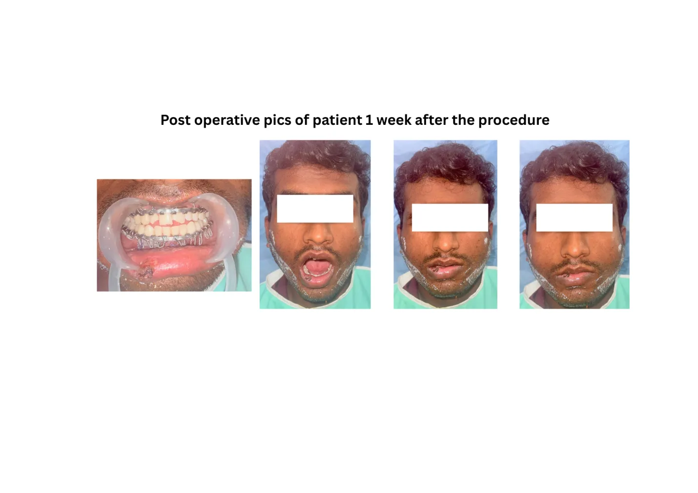
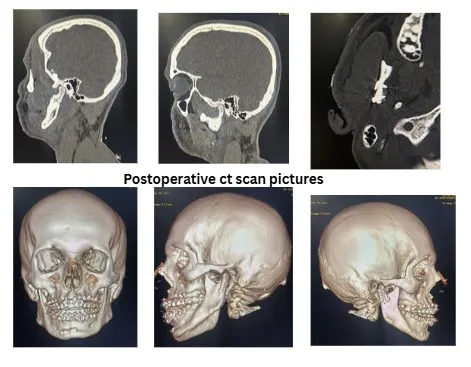
The post-operative period was uneventful. The patient was maintained on intravenous antibiotics and analgesics, and light elastic guidance (rather than rigid MMF) was used for a short initial period to encourage early, guided, physiotherapeutic jaw movement rather than prolonged immobilisation, in keeping with the rationale for rigid internal fixation of condylar fractures — namely, that stable osteosynthesis permits early return of function, which in turn reduces the risk of TMJ ankylosis and myofascial stiffness that can follow prolonged immobilisation, particularly in bilateral condylar injuries. Sutures were removed on the appropriate post-operative day, and the patient was placed on a structured physiotherapy/mouth-opening exercise regimen. At follow-up review, the patient demonstrated a stable, pre-traumatic occlusion, progressively improving maximal inter-incisal mouth opening, symmetrical mandibular movement without deviation, and no clinical evidence of facial nerve weakness. There was no palpable step deformity, no residual malocclusion, and the surgical scars healed favourably. Radiographic review confirmed satisfactory reduction and stable plate position at all three fracture sites with no evidence of hardware failure or non-union.
DISCUSSION
The combination of a symphyseal (or parasymphyseal) mandibular fracture with bilateral condylar fractures is a mechanistically predictable injury pattern: a direct blow to the chin is transmitted as a force vector along both mandibular bodies to the condylar necks, the narrowest and most structurally vulnerable portion of the mandible. This 'guardsman fracture' pattern, classically described in falls and direct anterior trauma, carries a particular risk of loss of posterior facial (ramal) height and consequent anterior open bite if the condylar component is not adequately managed, because both condyles — and therefore both posterior vertical buttresses of the mandible — are compromised simultaneously. The management of condylar fractures continues to generate considerable discussion in the literature, and no single treatment protocol is universally applicable to every condylar fracture. Closed treatment, consisting of a period of maxillomandibular fixation followed by graduated mobilisation, remains a reasonable option for minimally displaced, non-dislocated condylar fractures in patients who can achieve and maintain a stable occlusion, and is generally preferred in the growing patient because of the strong remodelling potential of the paediatric condyle. However, when condylar fractures are bilateral — and more so when they are accompanied by a fracture elsewhere in the mandibular arch that removes the stabilising effect of an otherwise intact mandible, as in the symphyseal fracture described here — closed treatment alone becomes considerably less predictable. In such circumstances the mandible effectively loses its posterior vertical support bilaterally at the same time that its anterior segment is also destabilised, making it difficult to reliably re-establish and maintain occlusal and skeletal relationships without direct visualisation and rigid fixation of at least one, and frequently both, condyles. Several authors have proposed criteria favouring open management of condylar fractures, broadly including: displacement of the condylar fragment into the middle cranial fossa; inability to obtain adequate occlusion by closed reduction; lateral extracapsular displacement of the condyle; presence of a foreign body within the joint; and bilateral condylar fractures in combination with comminuted midface fractures or in edentulous patients where splinting is impractical. Beyond these classical indications, many contemporary maxillofacial surgeons now extend the indication for open treatment to bilateral condylar fractures with significant angulation or shortening even in the absence of the above, on the basis of accumulating evidence — including randomised and prospective comparative studies — suggesting that ORIF produces more predictable restoration of ramus height, mouth opening, and occlusal function, with lower rates of malocclusion and TMJ dysfunction, compared with closed treatment, provided the surgery is performed with an approach that safeguards the facial nerve and achieves stable fixation. The retromandibular transmasseteric approach used in this case is favoured by many surgeons for access to the condylar neck and subcondylar region because it offers a short, relatively direct route to the fracture with a low reported incidence of facial nerve injury when the dissection plane is kept close to the posterior border of the ramus and the branches of the facial nerve are retracted rather than transected. Alternative approaches described in the literature include the preauricular, endaural, submandibular (Risdon), and various transoral approaches (with or without endoscopic assistance), each with its own advantages, limitations, and learning curve; the choice of approach is generally guided by the height of the fracture (condylar head/neck versus low subcondylar), the surgeon's experience, and the need to combine access with fixation of other concurrent mandibular fractures. Fixation of the symphyseal component with a two-miniplate technique, as performed in this case, follows Champy's principle of ideal osteosynthesis lines, wherein a tension band is applied near the alveolar crest to counteract the tensile forces generated during function, complemented by a second, more robust plate near the inferior border to resist the compressive and torsional forces generated in this high-stress region of the mandible. Simultaneous rigid fixation of the symphysis and both condyles allows early, active, physiotherapy-guided mandibular movement rather than prolonged immobilisation, which is particularly advantageous in reducing the risk of TMJ hypomobility/ankylosis — a well-recognised long-term complication of bilateral condylar injuries, especially in patients who have undergone extended periods of MMF. The favourable outcome in this case — restoration of pre-traumatic occlusion, symmetric excursive movements, adequate mouth opening, and absence of facial nerve deficit — underscores the value of a carefully planned, anatomically-directed open approach to combined symphyseal and bilateral condylar fractures, supported by high-resolution imaging for pre-operative classification and by intraoperative maxillomandibular fixation to verify occlusion before definitive plate fixation.
CONCLUSION
Symphyseal mandibular fractures occurring in combination with bilateral condylar fractures represent a biomechanically unstable injury pattern with a substantial risk of malocclusion, facial asymmetry, and TMJ dysfunction if managed by closed reduction alone. Open reduction and internal fixation of both the symphysis and the condylar fractures, performed under general anaesthesia with intraoperative verification of occlusion, allows anatomical reduction of all fracture segments, restoration of posterior mandibular height, and stable fixation that permits early mobilisation and functional rehabilitation. A thorough clinical and radiographic (including CT-based) assessment is essential for accurate fracture classification and surgical planning, and a layered, nerve-sparing surgical technique is key to achieving predictable functional and aesthetic outcomes, as illustrated by the case presented.
5. Conflict of Interest and Source of Funding
The authors declare no conflict of interest. No external funding was received for this case report.
6. Consent
Informed consent was obtained from the patient for publication of this case report and any accompanying clinical images/radiographs.
REFERENCES


R. Rajendra Prasad*, Seema Yoganna, Symphyseal Mandibular Fracture with Bilateral Condylar Fractures: Open Reduction and Internal Fixation under General Anaesthesia — A Clinical Case Report, Int. J. Med. Pharm. Sci., 2026, 2 (9), 235-241. https://doi.org/10.5281/zenodo.22661098
 10.5281/zenodo.22661098