
View Article

  • Identification of Potential Anti-Inflammatory Phytochemicals from Leucas Aspera Using A Computer-Aided Drug Design Approach

  • Department of Pharmaceutical Chemistry, Shantha College of Pharmacy, Peresandra, Chikkabalapur, Karnataka, India

Abstract

Background: Leucas aspera is a medicinal plant traditionally used for the treatment of inflammatory disorders. The present study aimed to evaluate selected phytochemicals from Leucas aspera as potential anti-inflammatory agents using a computer-aided drug design approach. Methods: Eight phytochemicals, namely ursolic acid, oleanolic acid, apigenin, chrysoeriol, catechin, β-sitosterol, acacetin, and β-caryophyllene, were screened against the anti-inflammatory target protein Microsomal Prostaglandin E synthesis-1 (mPGES-1, PDB ID: 5IKR) using molecular docking. Docking studies were performed using AutoDock Vina integrated in PyRx. Protein–ligand interactions were analyzed using Discovery Studio Visualizer. Drug-likeness and pharmacokinetic properties were evaluated using SwissADME. The crystal structure of mPGES-1 (PDB ID: 5IKR) was retrieved from the Protein Data Bank and used as the molecular target for docking studies. Results: Ursolic acid exhibited the highest binding affinity (-9.4 kcal/mol), followed by oleanolic acid (-9.0 kcal/mol) and apigenin (-9.0 kcal/mol). Apigenin, acacetin, catechin, and chrysoeriol demonstrated favorable pharmacokinetic properties with high gastrointestinal absorption and no Lipinski rule violations. Interaction analysis revealed hydrogen bond formation with key amino acid residues including SER143, ASN144, CYS47, ARG376, HIS226, ARG456, LEU171, and GLN372. SwissADME analysis revealed favorable drug-likeness and pharmacokinetic profiles for selected compounds. Conclusion: The study identified ursolic acid, oleanolic acid, and apigenin as promising phytoconstituents with potential anti-inflammatory activity. Further in vitro and in vivo studies are required to validate these findings.

Keywords

Leucas aspera, molecular docking, anti-inflammatory activity, SwissADME, phytochemicals, AutoDock Vina, mPGES-1, Computer-Aided Drug Design

Introduction

× Popup Image

Inflammation is a protective biological response to tissue injury, infection, and other harmful stimuli10,11. Chronic inflammation contributes to numerous diseases including arthritis, cardiovascular disorders, and autoimmune diseases12,13. Although several anti-inflammatory drugs are available, long-term use may result in adverse effects. Medicinal plants represent an important source of bioactive compounds with therapeutic potential4,5. Leucas aspera (Willd.) Link, belonging to the family Lamiaceae, has been traditionally used for the treatment of inflammation, fever, skin disorders, and other ailments7,9. Previous phytochemical investigations have reported the presence of flavonoids, terpenoids, sterols, and phenolic compounds in the plant5,8,9. Computer-aided drug design (CADD) techniques, particularly molecular docking, provide an efficient approach for identifying potential drug candidates by predicting ligand–protein interactions20,22,29. Therefore, the present study was undertaken to evaluate selected phytochemicals from Leucas aspera against an anti-inflammatory target protein using molecular docking and pharmacokinetic analysis. Microsomal prostaglandin E synthase-1 (mPGES-1) is a terminal enzyme involved in prostaglandin E2 biosynthesis and plays a crucial role in inflammatory responses26,28. Selective inhibition of mPGES-1 has emerged as a promising strategy for development safer anti-inflammatory agents. Recent advances in computer-aided drug design (CADD) have accelerated the identification of bioactive compounds through virtual screening molecular docking, and pharmacokinetics prediction.

MATERIALS AND METHODS

1. Selection of Phytochemicals

Eight phytochemicals reported from Leucas aspera were selected:
  1. Ursolic acid
  2. Oleanolic acid
  3. Apigenin
  4. Chrysoeriol
  5. Catechin
  6. β-Sitosterol
  7. Acacetin
  8. β-Caryophyllene

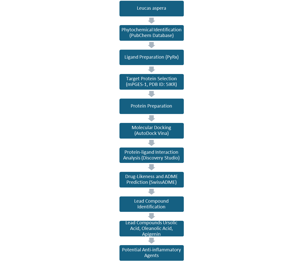
Figure 1. Workflow of the computer-aided drug design (CADD) approach employed for the identification of potential anti-inflammatory phytochemicals from Leucas aspera targeting mPGES-1 (PDB ID: 5IKR).

2. Ligand Preparation

The three-dimensional structure of the selected phytochemicals were obtained from PubChem database in SDF format. Energy minimization and conversion into PDBQT format were performed using PyRx software.

3. Protein Preparation

The crystal structure of the anti-inflammatory target protein (PDB ID: 5IKR) was retrieved from the Protein Data Bank. Water molecules and unwanted heteroatoms were removed before docking.

4. Molecular Docking

Molecular docking was performed using AutoDock Vina implemented in PyRx 0.81,3. The crystal structure of mPGES-1 (PDB ID: 5IKR) was used as the target protein. The docking search was defined with grid center coordinates X=20.9683, Y=20.1647, and Z=33.7710 and dimensions of 59.2686 Å × 66.4320 Å × 77.6215 Å. All selected phytochemicals were docked using the same grid parameters. The binding conformations were ranked according to binding affinity, and the pose with the lowest binding energy was selected for further analysis. Binding affinity values were recorded in kcal/mol.

5. Interaction Analysis

Protein-ligand interactions were visualized and analyzed using Discovery Studio Visualizer to identify hydrogen bonds and hydrophobic interactions.

6. Drug-Likeness and Pharmacokinetics Evaluation

Drug-likeness properties and ADME parameters were predicted using SwissADME2,32,33.

Result and Discussion

  1. Molecular Docking Analysis 

The docking results demonstrated varying binding affinities among the selected phytochemicals.

Table 1. Molecular Docking Results

Rank

Compound

Binding Affinity (kcal/mol)

1

Ursolic acid

-9.4

2

Oleanolic acid

-9.0

3

Apigenin

-9.0

4

Chrysoeriol

-8.6

5

Catechin

-8.6

6

β-Sitosterol

-8.6

7

Acacetin

-8.4

8

β-Caryophyllene

-6.8

Among all compounds evaluated, ursolic acid demonstrated the strongest binding affinity, indicating a greatest tendency to interact with the active site of the target protein.
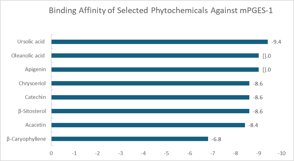
Figure 2. Comparison of molecular docking binding affinities of selected phytochemicals from Leucas aspera against mPGES-1 (PDB ID: 5IKR).
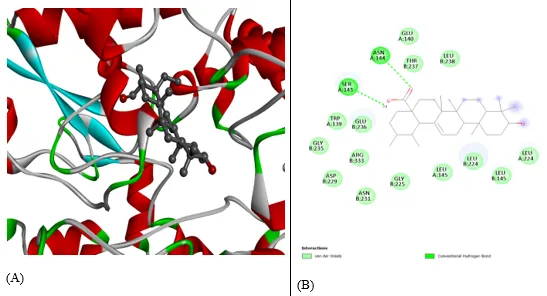
Figure 3. Molecular docking interaction of ursolic acid with 5IKR. (A) Three-dimensional binding pose of Ursolic acid within the active site of microsomal prostaglandin E synthase-1(mPGES-1; PDB ID: 5IKR). (B) Two-dimensional protein-ligand interaction diagram showing the binding interaction of ursolic acid with residue in the active site.

  1. Protein-Ligand Interaction analysis

Table 2. Protein-Ligand Interaction Analysis

Compound

H-Bond Residues

Ursolic acid

SER143, ASN144

Oleanolic acid

ARG456, LEU171

Apigenin

CYS47

Chrysoeriol

ARG376, HIS226

Catechin

ARG376, HIS226

β-Sitosterol

ARG376, HIS226

Acacetin

CYS47

β-Caryophyllene

GLN372

Hydrogen bonding and hydrophobic interactions contributed to ligand stabilization within the binding pocket.
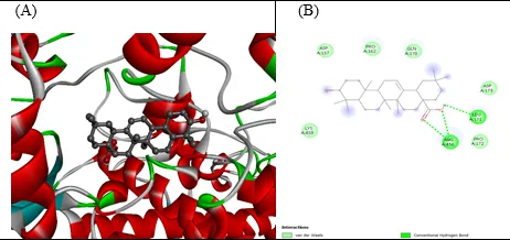
Figure 4. Molecular docking interaction of Oleanolic acid with 5IKR. (A) Three-dimensional binding pose of Oleanolic acid within the active site of microsomal prostaglandin E synthesis-1 (mPGES-1; PDB ID: 5IKR), illustrating its accommodation within the receptor binding pocket. (B)  2D protein-ligand interaction diagram of oleanolic acid docking with 5IKR showing interactions with ARG456 and LEU171 residues.
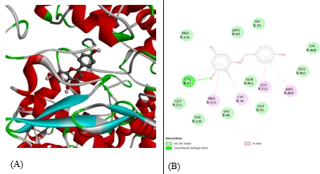
Figure 5. Molecular docking interaction of apigenin with 5IKR. (A) Three-dimensional binding pose of apigenin within the active site of microsomal prostaglandin E synthesis-1 (mPGES-1; PDB ID: 5IKR), illustrating its accommodation within the receptor binding pocket. (B) 2D protein-ligand interaction diagram of apigenin docking with 5IKR showing hydrogen bond interaction with CYS47 residue.

  1.  SwissADME Analysis

Table 3. SwissADME Properties

Compound

Molecular weight

LogP

GI Absorption

Lipinski Violations

Bioavailability Score

Ursolic acid

456.70g/mol

6.43

Low

1

0.85

Oleanolic acid

456.70g/mol

6.60

Low

1

0.85

Apigenin

270.24g/mol

1.53

High

0

0.55

Chrysoeriol

300.26g/mol

1.45

High

0

0.55

Catechin

290.27g/mol

0.70

High

0

0.55

β-Sitosterol

414.71g/mol

7.78

Low

1

0.55

Acacetin

284.26g/mol

1.87

High

0

0.55

β-Caryophyllene

204.35g/mol

4.48

Low

1

0.55

DISCUSSION

The observed docking scores indicate that triterpenoids such as ursolic acid and oleanolic acid possess strong binding affinity toward the selected anti-inflammatory target. Previous studies have reported significant anti-inflammatory effects of these compounds through inhibition of inflammatory mediators and signaling pathways. Apigenin, a well-known flavonoid, has also demonstrated anti-inflammatory activity through modulation of NF-kB and cytokine production18,19,34,37. The present findings are consistent with previous reports and further support the therapeutic potential of phytochemicals present in Leucas aspera7,9,40. Ursolic acid exhibited the highest binding affinity (−9.4 kcal/mol), suggesting stronger interaction with the active site of mPGES-1 compared with the other phytochemicals investigated. The interaction analysis revealed hydrogen bond formation with SER143 and ASN144 residues, which may contribute to stabilization of the protein–ligand complex. Oleanolic acid and apigenin also demonstrated favorable binding energies and interaction profiles, indicating their potential as promising anti-inflammatory lead compounds.

LIMITATIONS

The present investigation is limited to computational predictions and does not include experimental validation. Therefore, in vitro and in vivo studies are required to confirm the anti-inflammatory activity of the identified lead compounds.

FUTURE PERSPECTIVES

Although molecular docking provides valuable insights into protein-ligand interactions, molecular dynamics (MD) simulations are recommended to further evaluate the stability and conformational behaviour of protein-ligand complexes under physiological conditions. Furthermore, in vitro and in vivo studies are necessary to validate the anti-inflammatory efficacy and safety of the identified lead compounds20,21,30.

CONCLUSION

The present study employed molecular docking and pharmacokinetic analysis to evaluate the anti-inflammatory potential of phytochemicals from Leucas aspera against Microsomal Prostaglandin E Synthase-1 (mPGES-1; PDB ID: 5IKR). Among the screened compounds, ursolic acid (-9.4 kcal/mol), oleanolic acid (-9.0 kcal/mol), and apigenin (-9.0 kcal/mol) demonstrated the most favourable binding affinities toward the target protein. SwissADME analysis further revealed promising pharmacokinetic characteristics and drug-likeness profiles for these compounds. Among all the compounds investigated, ursolic acid exhibited the highest binding affinity and may represent the most promising lead candidate for further development. These findings suggest that phytochemicals from Leucas aspera may serve as potential anti-inflammatory lead molecules for future drug development. However, further molecular dynamics simulations and experimental validation through in vitro and in vivo studies are required to confirm their therapeutic efficacy and safety.

Conflict of Interest

The authors declare no conflict of interest.

ACKNOWLEDGEMENTS

The authors acknowledge Shantha College of Pharmacy for providing the facilities required for conducting this study.

REFERENCES

  1. Trott O, Olson AJ. AutoDock Vina: Improving the speed and accuracy of docking. Journal of Computational Chemistry. 2010;31(2):455–461.
  2. Daina A, Michielin O, Zoete V. SwissADME: A free web tool to evaluate pharmacokinetics and drug-likeness. Scientific Reports. 2017; 7:42717.
  3. Morris GM, Huey R, Lindstrom W, et al. AutoDock4 and AutoDockTools4. Journal of Computational Chemistry. 2009;30(16):2785–2791.
  4. Newman DJ, Cragg GM. Natural products as sources of new drugs. Journal of Natural Products. 2020;83(3):770–803.
  5. Harborne JB. Phytochemical Methods. Springer; 1998.
  6. Indian Medicinal Plants Kirtikar KR, Basu BD. Indian Medicinal Plants. 2nd ed. Dehradun: International Book Distributors; 1999.
  7. Mangathayaru K, Lakshmikant J, Shyam Sundar N, Grace XF, Vasantha J. Antimicrobial activity of Leucas aspera flowers. Fitoterapia. 2005;76(7-8):752–754.
  8. Prajapati ND, Purohit SS, Sharma AK, Kumar T. A Handbook of Medicinal Plants. Agrobios; 2003.
  9. Goudgaon NM, Basavaraj NR. Ethnopharmacological review of Leucas aspera and its medicinal importance. Journal of Herbal Medicine. 2018; 12:45–52.
  10. Medzhitov Ruslan Origin and physiological roles of inflammation. Nature.2008;454:428–435.
  11. Nathan Carl Points of control in inflammation. Nature. 2002; 420:846–852.
  12. Serhan CN, Ward PA, Gilroy DW. Fundamentals of Inflammation. Cambridge University Press; 2010.
  13. Libby P. Inflammatory mechanisms in atherosclerosis. Nature. 2002; 420:868–874.
  14. Ikeda Y, Murakami A, Ohigashi H. Ursolic acid: An anti-inflammatory triterpenoid. Molecular Nutrition & Food Research. 2008;52(1):26–42.
  15. Checker R, Sandur SK, Sharma D, et al. Anti-inflammatory effects of ursolic acid. International Immunopharmacology. 2012;14(4):489–496.
  16. Pollier J, Goossens A. Oleanolic acid and its pharmacological properties. Phytochemistry. 2012; 77:10–15.
  17. Liu J. Oleanolic acid and ursolic acid: Research perspectives. Journal of Ethnopharmacology. 2005;100(1-2):92–94.
  18. Salehi B, Venditti A, Sharifi-Rad M, et al. The therapeutic potential of apigenin. International Journal of Molecular Sciences. 2019;20(6):1305.
  19. Ginwala R, Bhavsar R, Chigbu DG, Jain P, Khan ZK. Apigenin as a therapeutic agent. Molecules. 2019;24(24):4537.
  20. Ferreira LG, dos Santos RN, Oliva G, Andricopulo AD. Molecular docking and structure-based drug design strategies. Molecules. 2015;20(7):13384–13421.
  21. Meng XY, Zhang HX, Mezei M, Cui M. Molecular docking: A powerful approach for structure-based drug discovery. Current Computer-Aided Drug Design. 2011;7(2):146–157.
  22. Kitchen DB, Decornez H, Furr JR, Bajorath J. Docking and scoring in virtual screening. Nature Reviews Drug Discovery. 2004;3(11):935–949.
  23. Lipinski CA, Lombardo F, Dominy BW, Feeney PJ. Experimental and computational approaches to estimate solubility and permeability. Advanced Drug Delivery Reviews. 2001;46(1-3):3–26.
  24. Veber DF, Johnson SR, Cheng HY, Smith BR, Ward KW, Kopple KD. Molecular properties influencing oral bioavailability. Journal of Medicinal Chemistry. 2002;45(12):2615–2623.
  25. Egan WJ, Merz KM, Baldwin JJ. Prediction of drug absorption using molecular descriptors. Journal of Medicinal Chemistry. 2000;43(21):3867–3877.
  26. Samuelsson B, Morgenstern R, Jakobsson PJ. Membrane prostaglandin E synthase-1: a novel therapeutic target. Pharmacological Reviews. 2007;59(3):207–224.
  27. Ricciotti E, FitzGerald GA. Prostaglandins and inflammation. Arteriosclerosis, Thrombosis, and Vascular Biology. 2011;31(5):986–1000.
  28. Korotkova M, Jakobsson PJ. Microsomal prostaglandin E synthase-1 as a therapeutic target in inflammatory diseases. Nature Reviews Drug Discovery. 2014;13(5):411–424.
  29. Lionta E, Spyrou G, Vassilatis DK, Cournia Z. Structure-based virtual screening for drug discovery: principles, applications and recent advances. Current Topics in Medicinal Chemistry. 2014;14(16):1923–1938.
  30. Pagadala NS, Syed K, Tuszynski J. Software for molecular docking: a review. Biophysical Reviews. 2017;9(2):91–102.
  31. Ferreira LG, dos Santos RN, Oliva G, Andricopulo AD. Molecular docking and structure-based drug design strategies. Molecules. 2015;20(7):13384–13421.
  32. Daina A, Michielin O, Zoete V. SwissADME: a free web tool to evaluate pharmacokinetics, drug-likeness and medicinal chemistry friendliness of small molecules. Scientific Reports. 2017; 7:42717.
  33. Pires DEV, Blundell TL, Ascher DB. pkCSM: predicting small-molecule pharmacokinetic and toxicity properties using graph-based signatures. Journal of Medicinal Chemistry. 2015;58(9):4066–4072.
  34. Ikeda Y, Murakami A, Ohigashi H. Ursolic acid: an anti- and pro-inflammatory triterpenoid. Molecular Nutrition & Food Research. 2008;52(1):26–42.
  35. Checker R, Sandur SK, Sharma D, et al. Potent anti-inflammatory activity of ursolic acid through inhibition of NF-κB, STAT3 and MAPK signaling pathways. International Immunopharmacology. 2012;14(4):489–496.
  36. Pollier J, Goossens A. Oleanolic acid. Phytochemistry. 2012; 77:10–15.
  37. Liu J. Oleanolic acid and ursolic acid: research perspectives. Journal of Ethnopharmacology. 2005;100(1–2):92–94.
  38. Salehi B, Venditti A, Sharifi-Rad M, et al. The therapeutic potential of apigenin. International Journal of Molecular Sciences. 2019;20(6):1305.
  39. Ginwala R, Bhavsar R, Chigbu DG, Jain P, Khan ZK. Apigenin as a therapeutic agent for neurodegenerative diseases and other disorders. Molecules. 2019;24(24):4537.
  40. Shanmugam S, Annadurai M, Rajendran K. Phytochemical constituents and pharmacological activities of Leucas aspera: a review. Journal of Ethnopharmacology. 2022; 292:115223.

Reference

  1. Trott O, Olson AJ. AutoDock Vina: Improving the speed and accuracy of docking. Journal of Computational Chemistry. 2010;31(2):455–461.
  2. Daina A, Michielin O, Zoete V. SwissADME: A free web tool to evaluate pharmacokinetics and drug-likeness. Scientific Reports. 2017; 7:42717.
  3. Morris GM, Huey R, Lindstrom W, et al. AutoDock4 and AutoDockTools4. Journal of Computational Chemistry. 2009;30(16):2785–2791.
  4. Newman DJ, Cragg GM. Natural products as sources of new drugs. Journal of Natural Products. 2020;83(3):770–803.
  5. Harborne JB. Phytochemical Methods. Springer; 1998.
  6. Indian Medicinal Plants Kirtikar KR, Basu BD. Indian Medicinal Plants. 2nd ed. Dehradun: International Book Distributors; 1999.
  7. Mangathayaru K, Lakshmikant J, Shyam Sundar N, Grace XF, Vasantha J. Antimicrobial activity of Leucas aspera flowers. Fitoterapia. 2005;76(7-8):752–754.
  8. Prajapati ND, Purohit SS, Sharma AK, Kumar T. A Handbook of Medicinal Plants. Agrobios; 2003.
  9. Goudgaon NM, Basavaraj NR. Ethnopharmacological review of Leucas aspera and its medicinal importance. Journal of Herbal Medicine. 2018; 12:45–52.
  10. Medzhitov Ruslan Origin and physiological roles of inflammation. Nature.2008;454:428–435.
  11. Nathan Carl Points of control in inflammation. Nature. 2002; 420:846–852.
  12. Serhan CN, Ward PA, Gilroy DW. Fundamentals of Inflammation. Cambridge University Press; 2010.
  13. Libby P. Inflammatory mechanisms in atherosclerosis. Nature. 2002; 420:868–874.
  14. Ikeda Y, Murakami A, Ohigashi H. Ursolic acid: An anti-inflammatory triterpenoid. Molecular Nutrition & Food Research. 2008;52(1):26–42.
  15. Checker R, Sandur SK, Sharma D, et al. Anti-inflammatory effects of ursolic acid. International Immunopharmacology. 2012;14(4):489–496.
  16. Pollier J, Goossens A. Oleanolic acid and its pharmacological properties. Phytochemistry. 2012; 77:10–15.
  17. Liu J. Oleanolic acid and ursolic acid: Research perspectives. Journal of Ethnopharmacology. 2005;100(1-2):92–94.
  18. Salehi B, Venditti A, Sharifi-Rad M, et al. The therapeutic potential of apigenin. International Journal of Molecular Sciences. 2019;20(6):1305.
  19. Ginwala R, Bhavsar R, Chigbu DG, Jain P, Khan ZK. Apigenin as a therapeutic agent. Molecules. 2019;24(24):4537.
  20. Ferreira LG, dos Santos RN, Oliva G, Andricopulo AD. Molecular docking and structure-based drug design strategies. Molecules. 2015;20(7):13384–13421.
  21. Meng XY, Zhang HX, Mezei M, Cui M. Molecular docking: A powerful approach for structure-based drug discovery. Current Computer-Aided Drug Design. 2011;7(2):146–157.
  22. Kitchen DB, Decornez H, Furr JR, Bajorath J. Docking and scoring in virtual screening. Nature Reviews Drug Discovery. 2004;3(11):935–949.
  23. Lipinski CA, Lombardo F, Dominy BW, Feeney PJ. Experimental and computational approaches to estimate solubility and permeability. Advanced Drug Delivery Reviews. 2001;46(1-3):3–26.
  24. Veber DF, Johnson SR, Cheng HY, Smith BR, Ward KW, Kopple KD. Molecular properties influencing oral bioavailability. Journal of Medicinal Chemistry. 2002;45(12):2615–2623.
  25. Egan WJ, Merz KM, Baldwin JJ. Prediction of drug absorption using molecular descriptors. Journal of Medicinal Chemistry. 2000;43(21):3867–3877.
  26. Samuelsson B, Morgenstern R, Jakobsson PJ. Membrane prostaglandin E synthase-1: a novel therapeutic target. Pharmacological Reviews. 2007;59(3):207–224.
  27. Ricciotti E, FitzGerald GA. Prostaglandins and inflammation. Arteriosclerosis, Thrombosis, and Vascular Biology. 2011;31(5):986–1000.
  28. Korotkova M, Jakobsson PJ. Microsomal prostaglandin E synthase-1 as a therapeutic target in inflammatory diseases. Nature Reviews Drug Discovery. 2014;13(5):411–424.
  29. Lionta E, Spyrou G, Vassilatis DK, Cournia Z. Structure-based virtual screening for drug discovery: principles, applications and recent advances. Current Topics in Medicinal Chemistry. 2014;14(16):1923–1938.
  30. Pagadala NS, Syed K, Tuszynski J. Software for molecular docking: a review. Biophysical Reviews. 2017;9(2):91–102.
  31. Ferreira LG, dos Santos RN, Oliva G, Andricopulo AD. Molecular docking and structure-based drug design strategies. Molecules. 2015;20(7):13384–13421.
  32. Daina A, Michielin O, Zoete V. SwissADME: a free web tool to evaluate pharmacokinetics, drug-likeness and medicinal chemistry friendliness of small molecules. Scientific Reports. 2017; 7:42717.
  33. Pires DEV, Blundell TL, Ascher DB. pkCSM: predicting small-molecule pharmacokinetic and toxicity properties using graph-based signatures. Journal of Medicinal Chemistry. 2015;58(9):4066–4072.
  34. Ikeda Y, Murakami A, Ohigashi H. Ursolic acid: an anti- and pro-inflammatory triterpenoid. Molecular Nutrition & Food Research. 2008;52(1):26–42.
  35. Checker R, Sandur SK, Sharma D, et al. Potent anti-inflammatory activity of ursolic acid through inhibition of NF-κB, STAT3 and MAPK signaling pathways. International Immunopharmacology. 2012;14(4):489–496.
  36. Pollier J, Goossens A. Oleanolic acid. Phytochemistry. 2012; 77:10–15.
  37. Liu J. Oleanolic acid and ursolic acid: research perspectives. Journal of Ethnopharmacology. 2005;100(1–2):92–94.
  38. Salehi B, Venditti A, Sharifi-Rad M, et al. The therapeutic potential of apigenin. International Journal of Molecular Sciences. 2019;20(6):1305.
  39. Ginwala R, Bhavsar R, Chigbu DG, Jain P, Khan ZK. Apigenin as a therapeutic agent for neurodegenerative diseases and other disorders. Molecules. 2019;24(24):4537.
  40. Shanmugam S, Annadurai M, Rajendran K. Phytochemical constituents and pharmacological activities of Leucas aspera: a review. Journal of Ethnopharmacology. 2022; 292:115223.

Photo
Mukthiyar Ahamed
Corresponding author

Department of Pharmaceutical Chemistry, Shantha College of Pharmacy, Peresandra, Chikkabalapur, Karnataka, India

Photo
Manohar KM.
Co-author

Department of Pharmaceutical Chemistry, Shantha College of Pharmacy, Peresandra, Chikkabalapur, Karnataka, India

Photo
Sahana CA.
Co-author

Department of Pharmaceutical Chemistry, Shantha College of Pharmacy, Peresandra, Chikkabalapur, Karnataka, India

Mukthiyar Ahamed*, Manohar KM., Sahana CA., Identification of Potential Anti-Inflammatory Phytochemicals from Leucas Aspera Using A Computer-Aided Drug Design Approach, Int. J. Med. Pharm. Sci., 2026, 2 (8), 534-541. https://doi.org/10.5281/zenodo.21931797

More related articles
Anti-Inflammatory Effect of an Ethanolic Extract o...
V. Venkata Rajesham, Siddartha Goutham Reddy Banda, Prasanna Kuma...
Analgesic and Anti-Inflammatory Activity of Alcoho...
Nimbarge Smita, S. V. Rajendra, Pallavi, Channaveer B. K., Bebi...
A Review on Hibiscus Rosa-Sinensis: Phytochemistry...
Vaishnavi Chavan, Suraj Ujgare, Santosh Ghule, Ronit Bhoir, Nitin...
Formulation and Evaluation of Anti-Inflammatory Ointment Using Ficus Religiosa B...
Kranti Bille, Srushti Harale, Prathamesh Patade, Mayuri Yadkune, Diksha Lohar, Diksha Gawade, Ajay Y...
Guava (Psidium guajava L.) Leaf Bioactives: From Phytochemical Characterization ...
Jannatul Firdaus, Tanlaka Nuradin Fonyuy, Shahnawaz Ahmad, Khushi Rastogi, Anshika Yadav...
Related Articles
Plant-Derived Phytochemicals as Potential Therapeutic Agents Against ESKAPE Path...
Mayank Singh, R. P. Singh, Deepak Kumar Saini, Amita Arjariya...
Analytical Strategies for the Isolation and Identification of Bioactive Boswelli...
Mohammed Shakir Ghouse, Syed Waleed Hussain, Syed Afnanuddin Kirmani, Siddiqui Hajra Yasmeen, Pathan...
Anti-Inflammatory Effect of an Ethanolic Extract of Ficus Dalhousiae Miq Whole P...
V. Venkata Rajesham, Siddartha Goutham Reddy Banda, Prasanna Kumar Lankalapalli, K. Manjeera, Harish...
More related articles
Anti-Inflammatory Effect of an Ethanolic Extract of Ficus Dalhousiae Miq Whole P...
V. Venkata Rajesham, Siddartha Goutham Reddy Banda, Prasanna Kumar Lankalapalli, K. Manjeera, Harish...
Analgesic and Anti-Inflammatory Activity of Alcoholic Extract of Malus Pumila Li...
Nimbarge Smita, S. V. Rajendra, Pallavi, Channaveer B. K., Bebi...
A Review on Hibiscus Rosa-Sinensis: Phytochemistry and Medicinal Uses...
Vaishnavi Chavan, Suraj Ujgare, Santosh Ghule, Ronit Bhoir, Nitin Mahale...
Anti-Inflammatory Effect of an Ethanolic Extract of Ficus Dalhousiae Miq Whole P...
V. Venkata Rajesham, Siddartha Goutham Reddy Banda, Prasanna Kumar Lankalapalli, K. Manjeera, Harish...
Analgesic and Anti-Inflammatory Activity of Alcoholic Extract of Malus Pumila Li...
Nimbarge Smita, S. V. Rajendra, Pallavi, Channaveer B. K., Bebi...
A Review on Hibiscus Rosa-Sinensis: Phytochemistry and Medicinal Uses...
Vaishnavi Chavan, Suraj Ujgare, Santosh Ghule, Ronit Bhoir, Nitin Mahale...