

We use cookies to ensure our website works properly and to personalise your experience. Cookies policy
1Shri Rawatpura Sarkar Institute of Pharmacy, Kumhari, Durg, C.G.
2Rungta Institute of Pharmaceutical Education and Research, Bhilai, C.G.
3Apollo College of Pharmacy, Anjor, Durg, C.G.
The present research is based on formulation of Transethosomal based Capsaicin anti-inflammatory transethosomal patch and determines in-vitro drug release and anti-inflammatory activity of prepared transethosomal patch. Pure drug of Capsaicin was identified by using FTIR spectroscopy and pre formulation study of transethosomal patch was performed by using various methods like determination of lamda max, drug solubility studies, melting point of pure drug etc. The present formulation is based on transethosome and HPMC polymer and other key ingredient and excipients which were use preparation of capsaicin based transethosomal patch. Evaluated the prepared transethosomal patch by their physical appearance, percent moisture content & uptake, weight uniformity, thickness of the patch, microscopic studies, Drug content, % drug release and anti-inflammatory activity. In all formulation batch, F2 batch formulation was give satisfactory result like that drug content 98.5%, cumulative drug release 92.9%. Consequently, the findings of this study clearly demonstrated that a transethosomal patch loaded with capsaicin offers a potential substitute for the conventional dosage form. To assess the efficacy of this method, however, more clinical studies are necessary. After taking into account everything mentioned above, it was determined that the current research study's goal could be effectively met.
The chronic autoimmune illness known as rheumatoid arthritis (RA) mostly affects synovial joints, such as those in the hands, knees, and feet, where the body's immune system unintentionally attacks its own joints, resulting in inflammation, discomfort, and destruction. The cause is not known, but genes and environmental factors are thought to be involved. RA is a condition that affects about 0.5% to 1% of the population worldwide. [1] It is more frequent in women than in men, with a frequency ratio of nearly 3:1. It is typically initiated in middle life but may also start at any age. The highest age of onset is from 40 to 60 years of age. The prevalence of RA differs geographically, with more prevalent rates in developed nations than in developing nations. There is a genetic susceptibility to RA, and some HLA types predispose individuals to it. Other environmental factors, including infection and smoking, also contribute to the disease's development. [2] The immune system of individuals with RA mistakenly targets synovial membrane tissues which triggers inflammatory reactions. The abnormal tissue growth known as pannus develops through synovial cell proliferation which damages both cartilage and bone structures. Joint injury and persistent inflammation are caused by inflammatory cytokines including interleukin-6 and tumour necrosis factor-alpha, as well as immune cells like T and B cells. The long-lasting inflammation causes cartilage and bone destruction which produces joint deformities together with significant functional impairment of the affected joints. [3][4] Rheumatoid arthritis (RA) is defined by long-lasting inflammation in joints which causes pain along with swelling. The application of capsaicin in cream or patch form helps reduce joint pain through its effect on nerve endings located within rheumatoid arthritis-affected areas. The main pain-relieving function of capsaicin is supported by limited evidence which indicates weak anti-inflammatory effects through its impact on pro-inflammatory mechanisms although these effects remain secondary to pain relief. The local application of capsaicin minimizes its systemic adverse effects because it affects only the region where it is used unlike oral RA treatment drugs such as NSAIDs or corticosteroids. [5][6] Transdermal drug delivery systems are one way to distribute drugs via the skin for systemic effect (TDDS). The medication is usually applied topically as a patch or film, where it enters the bloodstream and is dispersed throughout the body. [8][9] Drugs are gradually and carefully given over an extended period of time using TDDS, which may improve therapy efficacy and patient compliance. TDDS is potentially easier for people to use and less painful than injections because it doesn't involve penetration through the skin. By avoiding patches going through the liver and gastrointestinal tract, transdermal drug delivery may be able to increase medication bioavailability by avoiding the first pass metabolism. Patches are easier to administer and might make people more likely to stick with treatment regimens compared to pills that are taken on a daily basis. TDDS can reduce the risk of peak-and-trough peaks and related side effects by providing a constant drug level. [10] [11] Drugs contained in transdermal patches are usually in reservoir or matrix forms and are within an adhesive layer. The medication diffuses into the skin from the patch once it has been applied. The outermost layer of skin, the stratum corneum, is made up of densely packed dead keratinised cells that provide a strong barrier. A drug must penetrate this barrier using one of three primary pathways. The first method, transcellular route, allows the drug to go through individual skin cells. Another option, intercellular route, allows the drug to diffuse through the fat-rich spaces between the cells. [12] [13] A less common method called appendageal route lets the drug enter through hair and sweat gland openings. The efficiency of permeation is controlled by the drug's physicochemical properties, including molecular size, lipophilicity, polarity, and the ability to cross the stratum corneum. [14] Once the medication has penetrated the stratum corneum, it will go on to the dermis and epidermis. The medicine keeps diffusing through the epidermis, which is mainly made of live cells. The medicine enters the capillaries in the dermis because it is more vascularised there. medication absorption into blood capillaries and subsequent systemic circulation occurs after the medication reaches the dermis. [15] [16] The medication then travels to various parts of the body to work its magic. Typically, the duration of medication release via transdermal patches ranges from a few hours to several days. To do this, the drug's diffusion through the patch's matrix or reservoir may be controlled. To maintain a constant pharmaceutical supply, some patches utilise rate-controlling membranes to control the release of the medicament.
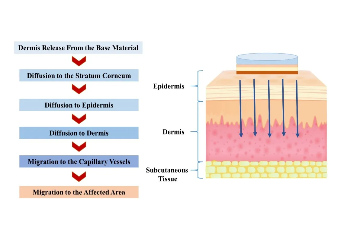
Figure 1. Transdermal Drug Delivery System (TDDS)
Although there are certain advantages to transdermal medication delivery systems, there are also some drawbacks that affect how well and how often they work. There are a lot of obstacles to overcome when trying to administer medications through the skin. The stratum corneum, which is part of the skin barrier, is very resistant to drug absorption. Transdermal administration isn't the best option for molecules with a large molecular weight or low water solubility since these substances have a harder time penetrating the skin's layers. [18] Another factor of concern is the risk of inducing skin irritation or allergy, especially with prolonged use of patches, which may affect patient compliance. Further, adhesion problems may occur, especially in those regions of the body that are more susceptible to movement, perspiration, or friction, with resultant variable drug delivery. Lastly, production cost for transdermal patches tends to be more expensive than the traditional oral or injectable dosage forms, potentially restricting their extensive applications in some therapeutic categories. [19] By delivering medication to inflamed joints through the skin, transdermal patches offer a strategic method for pain management in rheumatoid arthritis (RA). When opposed to systemic treatment, local administration frequently provides better symptom alleviation since the medicine is positioned at the site of inflammation and discomfort. The drug's ability to evade the digestive system and first-pass metabolism means that it has few if any of the common oral adverse effects of NSAIDs, including gastric discomfort. [20] [21] Additionally, the controlled-release matrix used in the patch's delivery mechanism keeps the plasma concentration constant, resulting in continuous analgesia with reduced need for re-dosing. Common formulations of these benefits are seen in the widespread usage of lidocaine patches for local anaesthesia of superficial joint pain, fentanyl patches for prolonged opioid analgesia of severe, chronic pain, and diclofenac patches for bringing anti-inflammatory and analgesic effects together to moderate both pain and inflammation in RA-involved joints. Together, these systems demonstrate how transdermal technology can improve efficacy, safety and patient compliance with RA pain management. [22] Transethosomes are novel vesicular systems with drug-delivery applications, especially transdermal. Transethosomes are derivations of conventional ethosomes, where the strengths of liposomes, ethosomes, and transfersomes are coalesced. [23] Transethosomes consist of phospholipids, ethanol, water, and an edge activator (a surfactant), making them extremely adaptable and facilitating enhanced penetration across biological barriers, especially the skin. The inclusion of ethanol in the composition is surprising in its effect on drug solubility and skin permeability, and membrane elasticity is improved by the edge activator. When used together, these two properties make transethosomes an excellent vehicle for the transdermal transport of hydrophilic and lipophilic medicines across biological membranes. [24] Different vesicular carriers have been engineered to improve drug delivery, each with different structural constituents. Transferosomes consist of phospholipids mixed with edge activators, making them more deformable and capable of penetrating the skin to a deeper extent. Liposomes, which are possibly the most studied vesicular carriers, are comprised of phospholipids and cholesterol in bilayered vesicles that can encapsulate lipophilic and hydrophilic drugs. [25] [26] Niosomes are analogous to liposomes but are prepared from non-ionic surfactants and cholesterol with better stability and economy. Ethosomes contain phospholipids and high levels of ethanol, which increase skin penetration by breaking the lipid structure of stratum corneum. Phytosomes are created by complexing phytoconstituents with phospholipids, thus enhancing bioavailability of phytopolymers. Finally, there are pharmacosomes, which are phospholipid vesicle carriers where the drug exists as phospholipids only and is typically prepared through the conjugation of drugs with phospholipids for increased solubility and absorption. All of these systems are tailored to maximize drug delivery by changing the vesicle composition for meeting specific therapeutic needs. [27] [28] Phospholipids are the primary components forming the bilayer structure of transethosomes, mimicking biological membranes, and ensuring compatibility with the skin. Ethanol disrupts the skin's lipid structure, enhancing penetration. It also improves the solubility of hydrophobic drugs and contributes to the vesicle's fluidity. [29] A non-ionic or anionic surfactant is used to impart flexibility to the vesicles, allowing them to deform and squeeze through narrow channels in the skin. Water acts as a medium to hydrate the phospholipids and form vesicles. Traditional systems, like creams or ointments, often face the challenge of limited penetration through the skin. Transethosomes, due to their composition, can effectively penetrate deeper layers of the skin, delivering drugs more efficiently. [30] Because of its ability to encapsulate hydrophilic and lipophilic medicines, transethosomes have several potential applications in drug delivery. Ethanol and surfactants work together to make the medicine more stable in the vesicle, which means it won't break down too soon. [31] [32].
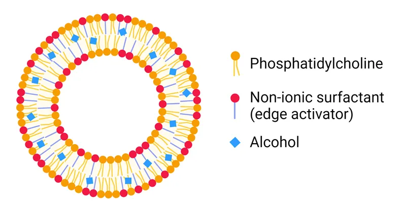
Figure 2. Structure of Transethosome
By allowing for regulated and prolonged medication release, transethosomes can improve patient compliance and decrease administration frequency. Patients experience less pain and discomfort with transethosome medication administration compared to injectable or oral drug delivery systems. [33]
Formulation And Development
MATERIAL
The standard raw material active pharmaceutical ingredient (Drug) of Capsaicin is procurement from West-Coast Pharmaceutical Ltd., Plot no.C3, opposite M.R. Industries, Gota Railway crossing, Ahmadabad (Gujarat), 3824. All chemicals and solvents used in this study, including soya lecithin, Span 80, polyvinyl alcohol (PVA), polyethylene glycol 400 (PEG 400), propylene glycol, hydroxypropyl methylcellulose (HPMC), chloroform, potassium bromide (KBr), ethanol, and methanol, were obtained from the central chemical repository of Ravatpura Sarkar Institute of Pharmacy, Kumhari, Durg. All reagents were of analytical grade and used without further purification.
Table 1. Instrument used:
|
S. No. |
Instrument Name |
Company Name |
|
1. |
Digital Weighing Balance |
Keyroy Pvt. Ltd Varanasi, India |
|
2. |
Water Bath |
Servo Enterprises |
|
3. |
Franz Diffusion Cell |
Nishant Enterprises |
|
4. |
UV-Visible Spectrophotometer |
Simadzu, Japan |
|
5. |
Refrigerator |
LG |
|
6. |
Electron Microscope |
Atico Medical Pvt.Ltd |
|
8. |
FTIR Spectrophotometer |
PerkinElmer Spectrum IR Version 10.7.2 |
|
10. |
Thermometer |
H.L. Scientific Industry |
|
11. |
Zeta Potential Analyzer |
Litesizer500 |
|
12. |
Transmission Electron Microscope (TEM) |
FE-TEM(JEM-3100F), CIL Punjab university |
Preformulation Studies
Rotational development of a drug's dosage form begins with preformulation studies, which involve testing. To create a dosage form that is safe, stable, powerful, bioavailable, and effective, it is necessary to study the physiochemical characteristics of the novel pharmacological ingredient both on its own and in combination with the excipients. [34]
Organoleptic character
The study of the organoleptic properties of the drug ingredient included tasting, seeing, smelling, and other similar features. [35]
Solubility
The amount of a material that can dissolve in water at a given temperature and pressure is called its solubility. The highest solute concentration in a solvent at equilibrium is used as a unit of measurement. Saturated solution is the name given to the resultant liquid.1 milligram of the medicine was dissolved in a variety of solvents. [36] [37]
Selection of solvent
The drug's solubility was tested in a range of solvents, both polar and non-polar, in accordance with the IP standard. It was discovered that ethanol is the most frequent and stable solvent for capsaicin. [38]
Melting point
We used a Thiele tube to find Capsaicin's melting point. Half of the capsaicin medication sample was placed in a sealed capillary tube, which was then submerged with a thermometer that was connected to the tube via thread. We started heating the Capsaicin medicine sample and saw it melt at different temperatures. [39]
Process of making capsaicin standard stock solutions
After precisely weighing 100 milligrams of API drug, it was transferred to 100 milliliters of ethanol in a volumetric flask to create the standard stock solution of capsaicin. Make a 100 millilitre ethanol solution by diluting 10 millilitres of the solution. To achieve a concentration of 10µg/ml, the solution was used to further dilute the standard stock solution. [40]
Determination of λ max:
Between 200 and 800 nanometres, the standard Capsaicin solution (20µg/mL) was scanned. Following correcting for baseline, in comparison to an ethanol solvent designed as a blank. The absorbance peak was recorded at 293 nanometres. Various criteria for work were created ranging from 10 to 50µg/ml. After experimenting with other ranges of wave lengths, we settled on 293–300 nm as the sweet spot, thanks to the linear relationship between area, concentration, and the horizontal axis. By inputting the wavelength range that has to be determined, the horizontal axis was chosen. The selected wavelength ranges of 280-300 nm demonstrated a strong linear relationship between concentration and area under the curve. [41] [42]
Calibration curve of pure drug
For the purpose of plotting the calibration curve, various concentrations of the medication were found on the x-axis, and the absorbance of capsaicin was found on the y-axis. The concentration was plotted against the peak area absorbance, and the data was then submitted to linear regression analysis on the maximum absorbance (λ max). [43]
FTIR studies
Fourier Transform Infrared Spectroscopy (FTIR) was used to identify the substance capsaicin. A mortar and pestle were used to triturate a drug sample with IR grade potassium bromide (KBr) in a ratio of 1:100. The exact amount was then transferred to the sample chamber and placed in the sample holder. As an alternative to potassium bromide, nizole was used for the liquid sample. The material underwent 45 scans at 4000-400 cm-1. The spectra were captured using a DLATGS detector and an IR solution from Simadzu, version 1.50. Both the reference spectrum and the standard reference spectra were used to match the spectrum and major peaks, respectively. [44] [45]
Preparation of Capsaicin Loaded Transethosomal Patch
COLD Method
Beaker A contains ethanol and phosphatidylcholine, whereas Beaker B contains water and an edge stimulant. The combination of these two stages is heated to 30ºC. For ten minutes, an aqueous and organic component are combined while being stirred consistently at a temperature of 30 degrees Celsius. Finally, a probe sonicator sonicates the formulation. [46]
Method for preparation of Transethosomes of Capsaicin
Transethosomes were created using a cold method. First, all required materials and chemicals were selected and carefully weighed. Beaker A contains ethanol and phosphatidylcholine, whereas Beaker B contains water, an edge stimulant, and the medication capsaicin. The combination of these two stages is heated to 30ºC. Following heating, the organic and aqueous components are combined, stirred consistently for ten minutes, and the temperature is maintained throughout. Following the completion of the procedure, the formulation is sonicated for 15 minutes using a sonicator and allowed to sit at room temperature for two hours in order to form intact vesicles. [47] [48] (Table 2) describes the components in formulating the transethosomes of capsaicin.
Table 2. Formulation Table for Drug loaded Transethosome
|
Formulation code |
F1 |
F2 |
F3 |
|
Soya Phospholipid(W/V) |
2.0% |
2.5% |
3.0% |
|
Ethanol (V/V) |
25% |
30% |
35% |
|
Span 60 (W/V) |
0.8% |
1% |
1.5% |
|
Drug(Capsaicin) (W/V) |
0.5% |
0.5% |
0.5% |
|
Distilled water (V/V) |
Upto 100% |
Upto 100% |
Upto 100% |
Preparation of The Patch
Procedures:
Matrix-type transdermal patches consist of varying ratios of HPMC, ethanol, distilled water, and propylene glycol, manufactured using solvent evaporation procedures utilising bangles. The base of the bangle was encased in aluminium foil, onto which a backing membrane was formed by pouring a 4% w/v aqueous polyvinyl alcohol (PVA) solution, subsequently dried at 50°C for 4 hours. The drug matrix was created by dissolving the necessary quantity of the drug and HPMC in a mixture of ethanol and water. Propylene glycol was added into this solution and mixed well. The uniformly dispersed material was applied on a PVA backing membrane and cured at ambient temperature for 24 hours. The desiccated films were extracted and encased in aluminium foil. [49] [50] (Fig. 3, Table 3.) describes the components in formulating the transdermal patches.
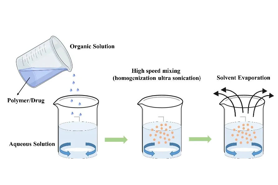
Figure 3. Solvent Evaporation technique
Table 3. Composition of patch
|
Formulation code |
Ingredients |
|||
|
HPMC |
Propylene glycol |
Methanol |
Water |
|
|
F1 |
10% |
25% |
25% |
upto100% |
|
F2 |
10% |
25% |
25% |
upto100% |
|
F3 |
10% |
25% |
25% |
upto100% |
Incorporation of Capsaicin Loaded Transethosomes In the Patch:
The capsaicin-loaded transethosomal formulation was gradually incorporated into the HPMC patch base with moderate agitation. The transethosomal patch was subsequently blended with a mechanical stirrer for five minutes. [51] Formulated capsaicin loaded transethosmal patch is shown in (Fig. 4).
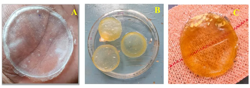
Figure 4. (A) Polymer based patch (B) & (C) Capsaicin loaded Transethosomal patch
Evaluation and Characterization
Physical Appearance
A visual examination was performed on each of the transdermal patches to determine their color, clarity, flexibility, and softness. [52]
Digital Microscope
Digital microscopes equipped with advanced imaging techniques can provide high- resolution image to study the size, shape and uniformity of transethosome vesicles. These factors are crucial as the influence drug encapsulation efficiency and penetration capacity. [53]
Entrapment Efficiency
The drug entrapment efficiency determined how much of the drug was wrapped into formed spherical vesicles. Simple indirect analysis technique was used to calculate the percentage of EE. First, a clear supernatant solution was obtained by centrifuging 1 mL of tranethosome was used to dilute the collected supernatant. Three times this procedure was carried out. UV spectrophotometry was used to eventually analyse the supernatant (test sample) at 293 nm. [54] The formula used to calculate the percentage (%) of EE is as follows:
% EE = Total amount of drug- Amount of free drug × 100 / Total amount of drug
Zeta Potential Measurement
Zeta potential, which is known to have an impact on stability, is a measurement of the strength of the electrostatic or charge repulsion or attraction between particles. Its measurement can be used to enhance the formulation of transethosomes and provides comprehensive insight into the reasons behind dispersion, aggregation, or flocculation. Nearly every macroscopic or particulate substance that comes into contact with a liquid develops an electrical charge on its surface. In general, when the absolute value of the zeta potential is more than 30 mV, particles may be distributed steadily. Furthermore, the zeta potential exhibits fast aggregation below 5 mV and limited stability below 20 mV. Nonetheless, a number of investigations have documented that the transethosome formulations' zeta potentials varied from -10 to -30 mV. LiteSizer500 was used to measure the transethosomal formulation's zeta potential at 250C. [55]
Transmission Electron Microscopic Structure Studies:
TEM is a method that employs an electron beam to image an nano particle sample, offering a significantly higher resolution than is possible with light-based imaging techniques. [56]
Thickness of Patch
At various locations along the transdermal film, a traveling microscope, dial gauge, screw gauge, or micrometer is used to measure the film's thickness. A screw gauge was used to measure the thickness of a small number of randomly chosen patches at three separate locations. The average thickness of a single patch was then calculated. [57]
Folding endurance
One film was folded repeatedly in the same place until it broke in order to ascertain this. The value of folding endurance was determined by how many times the film could be folded in the same spot without breaking or cracking. [58]
Weight Uniformity
Five randomly chosen patches are weighed individually, and the average weight is determined in order to study weight fluctuation. The average weight and the individual weight shouldn't differ much. [59]
Percentage Moisture Content
After being individually weighed, the produced films are stored for 24 hours at room temperature in a desiccator filled with calcium chloride. After a predetermined amount of time, the films are weighed once more until their weight remains constant. The following formula is used to determine the percentage moisture content: [60]
% moisture content = (Initial weight– Final weight) / Final weight × 100
Percentage Moisture Uptake
After a 24-hour period at room temperature, a weighted film was removed from the desiccator and exposed to 84% relative humidity, which is a saturated solution of aluminum chloride, until the film's weight remained constant. The difference between the final and initial weights in relation to the initial weight was used to compute the percentage of moisture uptake. [61]
% moisture uptake = (Final weight–initial weight)/ initial weight × 100
Drug Content Uniformity
To determine the drug content, three patches of each formulation were taken, and 100 ml of methanol was added (separately) while being continuously stirred for two hours to dissolve the entire patch. After filtering and appropriately diluting the solutions, they were examined at 293 nm using a UV spectrophotometer. Three films' average drug content (as a percentage) was recorded. [62]
In Vitro Release Study
Using a Franz diffusion cell and a semi-permeable membrane, in vitro release was performed. The two chambers that make up the cell serve as the donor and receptor compartments. The upper end of the donor compartment was open, allowing air to enter. The receptor compartment had a sampling port, and the temperature was kept at 37±0.5°C. (Fig. 5) The hydro-alcoholic solution's diffusion medium. A semi-permeable membrane maintained the drug-containing film in the donor compartment and kept it apart from the receptor compartment. A clamp was used to hold the donor and receptor compartments together. To avoid the development of a concentrated drug solution underneath the semi-permeable membrane, the receptor compartment containing the hydroalcoholic solution was kept at 37±0.5°C and agitated using a magnetic stirrer. At prearranged intervals, 1 ml samples were taken and replaced with new solutions of the same. Using spectrophotometry, the drug's concentration was measured at 293 nm. [63]
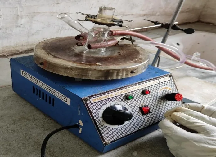
Figure 5. In-vitro drug release studies by Franz diffusion cell
RESULT & DISCUSSION
Preformulation Studies
Organoleptic Characteristics:
Capsaicin, the active component in chili peppers, is known for its unique organoleptic properties, which relate to the sensory characteristics we perceive through our senses of taste, smell, and touch. Organoleptic properties of capsaicin showed in (table 4).
Table 4. Organoleptic characteristics of Drug
|
S. No. |
Property |
Description |
|
1. |
Taste |
Pungent, Spicy |
|
2. |
Odor |
Odorless or faint peppery smell |
|
3. |
Texture |
Crystalline, colorless or pale yellow |
|
4. |
Sensory Perception |
Burning sensation on the tongue or skin |
Solubility
Capsaicin's solubility in a pure drug sample was determined to be showed in (table 5).
Table 5. Solubility of capsaicin
|
S. No. |
Solvent |
Solubility of Capsaicin |
|
1 |
Ethanol |
Very soluble |
|
2 |
Acetone |
Very soluble |
|
3 |
Chloroform |
Soluble |
Melting Point:
By using the Thiele tube method, the melting point of both the pure medication and the excipients was determined. (Fig. 6) Capsaicin was determined to have a melting point of 640 degrees Celsius. Melting Point determination data is shown in (Table 6).
Table 6. Melting point of Drug
|
S. No. |
Temperature when solid completely melted |
Average |
|
1 |
630C |
64 |
|
2 |
640C |
|
|
3 |
660C |
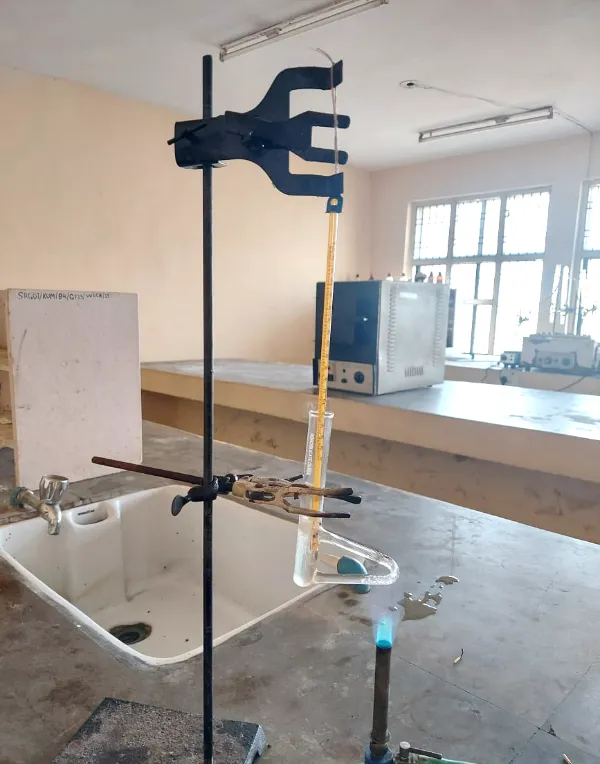
Figure 6. Melting Point of Drug
Determination of λ Max:
The spectra of the pure substance were scanned from 200 to 800 nanometres using 20μg/ml of ethanol. It was discovered that the maximum wavelength of the pure medicine, which is capsaicin, is 293 nanometres. Determination of λ Max graph of absorption is showm in (Fig. 9)
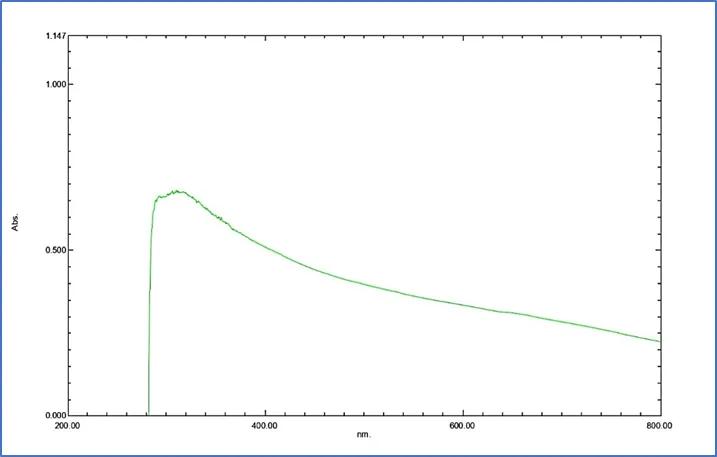
Figure 9. Graph of Absorbent maxima λ max of capsaicin
Preparation of Calibration Curve of Pure Drug
In order to draw the calibration curve, multiple concentrations of the medication were used on the x-axis, and absorbance was used on the y-axis. The graph displays the calibration curve. By utilising the concentration and absorbance data, a beerlumbert's plot was successfully completed. With regard to capsaicin, the value of the coefficient of correlation (R2) was shown to be 0.998. At a wavelength of 293 nm, the calibration curve was determined to have a maximum value of λ is shown in (Table 7, Figure 7).
Table 7. Absorbance of capsaicin
|
S. No. |
Concentration(μg/ml) |
Absorbance |
|
1 |
10 |
0.273 |
|
2 |
20 |
0.348 |
|
3 |
30 |
0.593 |
|
4 |
40 |
0.676 |
|
5 |
50 |
0.764 |
Figure 7. Graph of Standard calibration curve data of Capsaicin
Fourier Transform Infrared Spectroscopy Studies
The drug is considered pure since the peak generated by performing FTIR on a pure drug was determined to be between the range of primary principal peaks previously reported as the theoretical range. The structure of the medication molecules was found to be consistent with these results. (Figure 8).
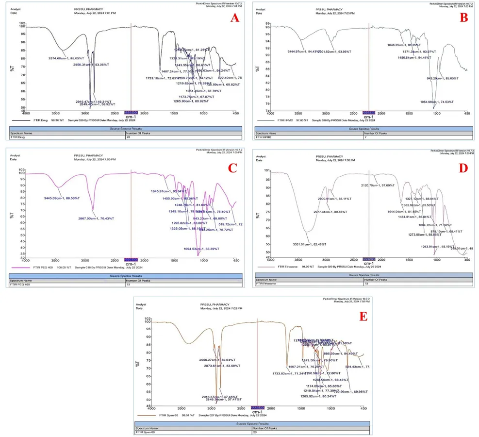
Figure 8. (a) FTIR of Capsaicin (b) FTIR of HPMC (c) FTIR of PEG 400 (d) FTIR of Transethosome (e) FTIR of Span 60
Evaluation Of Transethosome
Physical Appearance:
There was no greasiness or grittiness apparent in the formulation, which had a light-yellow tint to the naked eye. (Fig. 9) For the purpose of further assessment, the transethosome that has been generated with a good look, smoothness, and homogeneity is utilised.
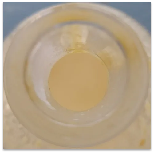
Figure 9. Physical appearance of transethosome
Entrapement Efficiency
The transethosomal preparations' percentage of drug entrapment efficiency was measured using the centrifugation technique. F2 had the highest rate of drug entrapment at 80% and F1 the lowest at 60%. According to the findings shown in (Table 8), the entrapment rose up to a certain concentration before beginning to decrease, suggesting that concentrations relative to the formulation reflect this crucial value.
Table 8. Entrapement efficiency of different transethosomal formulation
|
Transethosomal formulation |
Entrapement efficiency (%) |
|
F1 |
60 |
|
F2 |
80 |
|
F3 |
70 |
Surface Morphology
Digital microscopy and transmission electron microscopy (TEM) were utilised in order to ascertain the form and surface morphology of the transethosome droplet.
Digital Microscope
The micrograph showed round droplets, the dispersed of particle due to the lipid nature of the carriers the lipid purity affects the particle shape. (Fig. 10) Digital microscope study was done from SRIP Kumhari.
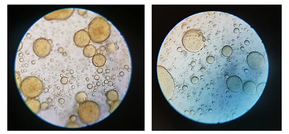
Figure 10. (a) Digital microscope of capsaicin loaded transethosom
Transmission Electron Microscopy
Transethosomal vesicles were characterised using Transmission Electron Microscopy, which provides details on the size, shape, and internal morphology of the vesicle. The aggregation size was found to be 100-500 nanometres in the optimised formulation F1 transethosome. In contrast, F2 exhibited no aggregates and was uniformly and discretely shaped, measuring 50-100 nm in diameter. It was discovered that the F3 transethosome was 1-2μm in size. There was no aggregation or clustering seen in the image taken using F2 Transmission Electron Microscopy. (Fig 11) Optimisation of formulation parameters, including lipid content and composition, can be aided by this finding. Investigations were conducted using transmission electron microscopy at PUNJAB UNIVERSITY.
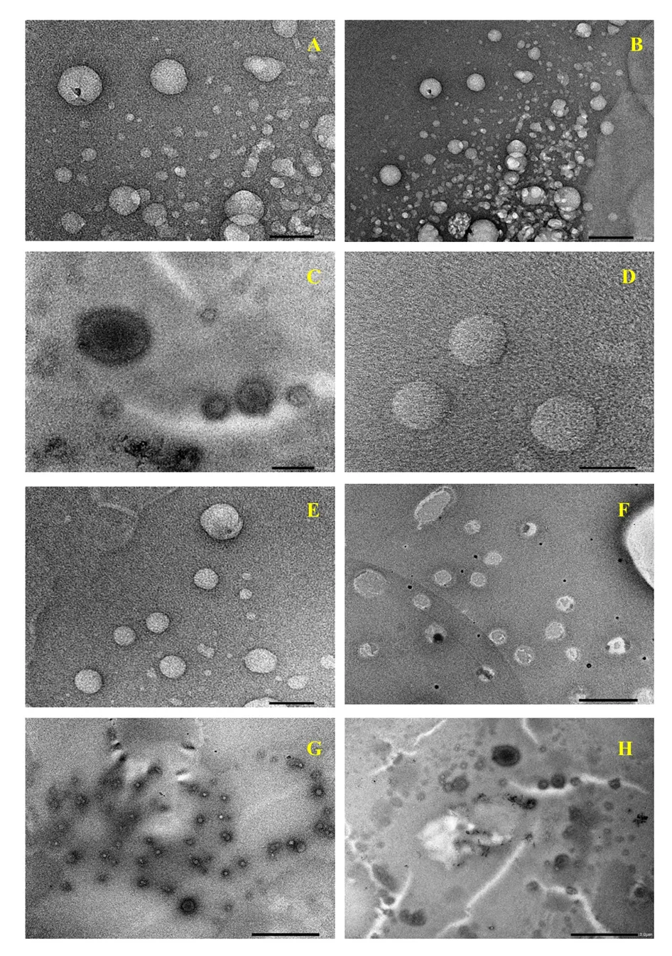
Figure 11. Transmission Electron Microscopy analysis of Transethosomal formulation
F1 (A) 100nm F1 (B) 200nm F1 (C) 500nm F2 (D) 50nm F2 (E) 100nm F3 (F) 1μm F3 (G) 2μm F3 (H) 2μm
Zeta Potential
As seen in the figure, the values of the Zeta Potential were negative and varied from -1.3 to -10.6 mV. (Table 9) The presence of repulsive interactions between scattered vesicles is indicated by the presence of a negative and high value of the potential of F2, which allows for stability to be achieved with a minimal of the possibility of vesicles aggregating. (Fig. 12) Studies on the zeta potential that were conducted at PUNJAB UNIVERSITY.
Table 9. Result of zeta potential measurement
|
Mean zeta potential |
-10.6 mv |
|
Standerd deviation |
29.5 mv |
|
Distribution Peak |
-12.0mv |
|
Conductivity |
2.379mS/cm |
|
Mean intensity |
385.5 kcounts/s |
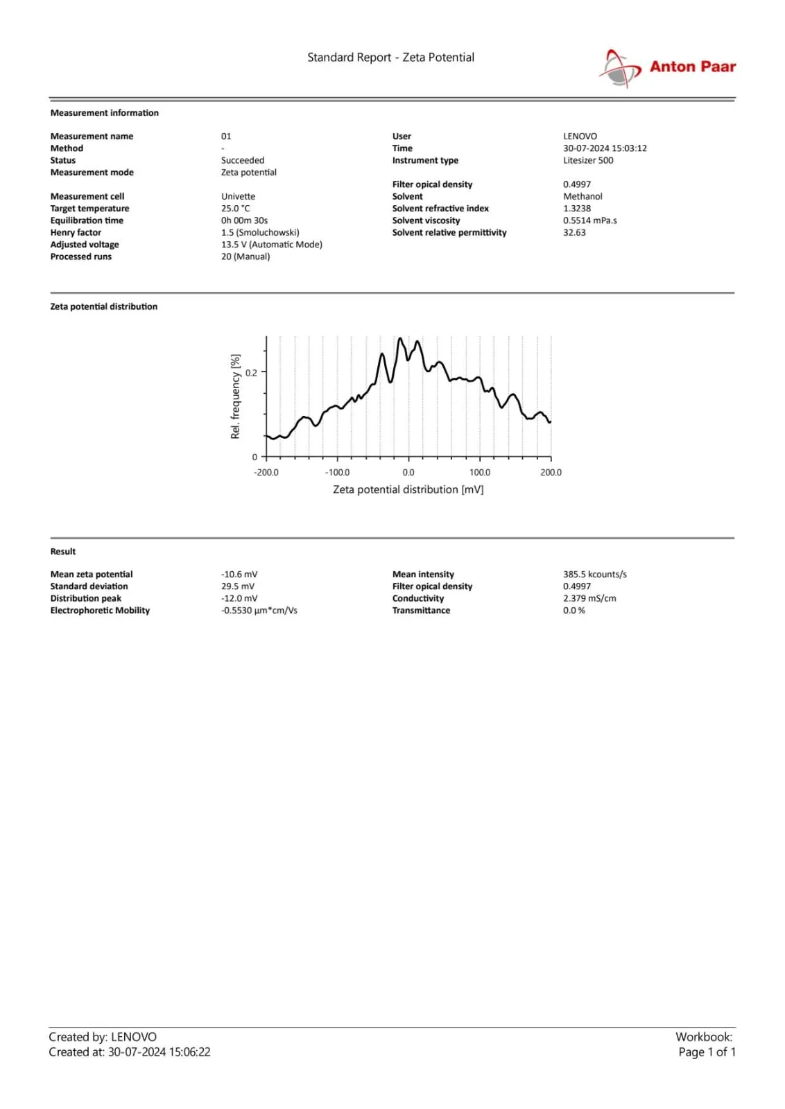
Figure 12. Zeta potential measurement of transethosomal formulation
Evaluation of Transethosomal Patch
It is clearly thin, flat, and transparent (as indicated in Fig. 13) in the process of preparation.The prepared film having a fine finish and good look is used to carry out additional analysis.Among all the formulations, Formulation F2 possessed superior physicochemical and mechanical properties and was thus the most ideal for transdermal drug delivery. It exhibited optimum patch thickness of 0.36 mm with a compromise of adequate drug loading and elasticity, which are required for superior mechanical properties. Long-term skin adhesion without breakage requires adequate mechanical strength and flexibility, as explained in terms of folding endurance of 210 folds. Its 98.9% drug loading ensured accurate dosing and reliable medication delivery during the patch. Moreover, F2's 4.5% content of water is adequate to ensure stability of the patch and avoid microbial deterioration. Its 9.4% absorption of moisture was also within acceptable parameters, showing that the patch shall be long lasting irrespective of any humidity level. Uniform weights of (149 mg) are reflections of uniform action and outstanding formulation reproducibility. In overall, the findings affirm that Formulation F2 is the most stable, consistent, and efficient transdermal delivery patch among the ones being tested. Various parameter result displayed in (Table 10).
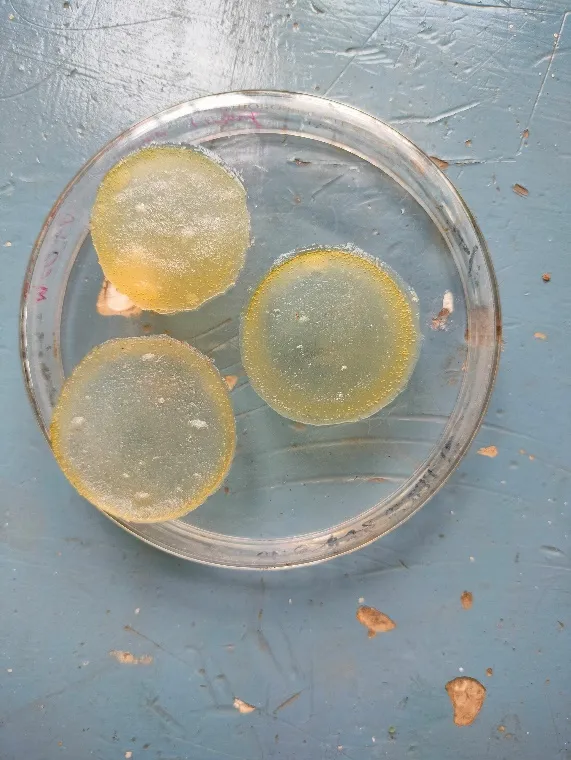
Figure 13. Physical appearance of capsaicin loaded transethosomal patch
Table 10. Evaluation Results of Different Transdermal Patch Formulations
|
Formulation Code |
Patch Thickness (mm) |
Folding Endurance |
Weight Uniformity |
Moisture Content (%) |
Moisture Uptake (%) |
Drug Content (%) |
|
F1 |
0.42 ± 0.01 |
185 ± 5 |
155 ± 2.4 |
6.1 ± 0.3 |
11.8 ± 0.6 |
96.2 ± 0.8 |
|
F2 |
0.36 ± 0.02 |
210 ± 4 |
149 ± 1.9 |
4.5 ± 0.2 |
9.4 ± 0.5 |
98.9 ± 0.4 |
|
F3 |
0.40 ± 0.01 |
198 ± 6 |
152 ± 2.1 |
7.2 ± 0.4 |
12.6 ± 0.7 |
95.5 ± 1.1 |
|
Standerd Range |
0.2-0.5 mm |
≥ 200 |
±5% of mean |
2% –10% |
3% –15% |
95% –105% |
Kinetics of Drug Release
The Franz diffusion cell technique was utilised in order to ascertain the in-vitro drug release profile of the produced formulations. A transethosomal patch loaded with capsaicin is developed according to the Korsmeyer-Peppas Model (Anomalous kinetics). A phosphate buffer with a pH of 6.8 was used for the drug release process, which lasted for a total of eight hours. Result datat showed in (Table 11,12). A calculation was made to determine the cumulative percent of medication release across all formulations. In the formulation that has the highest possible amount of drug entrapment, the highest possible percentage of drug release was discovered. The dissolution of drugs in immediate release and modified release dose forms is described by a number of theories and kinetics models. [64]
Table 11. In vitro release study of capsaicin loaded transethosomal patch formulation
|
S No. |
Time (hr) |
Cumulative %drug release |
Log cumulative %drug release |
Cumulative % drug remains |
Log cumulative % drug remain |
Square root of time |
Log time |
|
1 |
0 hr |
0 |
0 |
0 |
0 |
0 |
0 |
|
2 |
1 hr |
11.8 |
1.071 |
88.2 |
1.946 |
1 |
0 |
|
3 |
2 hr |
22.6 |
1.354 |
77.4 |
1.888 |
1.414 |
0.301 |
|
4 |
3 hr |
34.3 |
1.535 |
65.7 |
1.817 |
1.732 |
0.477 |
|
5 |
4 hr |
46.4 |
1.666 |
53.6 |
1.730 |
2 |
0.602 |
|
6 |
5 hr |
55.7 |
1.746 |
44.3 |
1.646 |
2.236 |
0.699 |
|
7 |
6 hr |
68.3 |
1.834 |
31.7 |
1.500 |
2.449 |
0.778 |
|
8 |
7 hr |
79.2 |
1.899 |
20.8 |
1.318 |
2.646 |
0.845 |
|
9 |
8 hr |
92.9 |
1.967 |
7.1 |
0.851 |
2.828 |
0.903 |
Table 12. Kinetics study of drug release by different model
|
S.no. |
Model name |
Equation |
R2 |
|
1. |
Zero |
11.939x + 6.2387 |
0.9866 |
|
2. |
First |
0.0671x + 1.4723 |
0.9909 |
|
3. |
Higuchi |
20.205x +8.7015 |
0.9976 |
|
4. |
Korsmeyer Peppas |
0.9895x + 1.0644 |
0.9995 |
Zero-order kinetics -
It signifies an optimal release profile wherein the drug is dispensed at a uniform pace, irrespective of its concentration. This model is characterized by a linear correlation between the cumulative proportion of drug released and time. (Table 13,14) It is generally denoted by the equation: [65]
Qt = Q0 + K0t
Where,
Qt is the amount of drug released in time t,
Q. is the initial amount of the drug in the solution
K0 is the zero order release constant
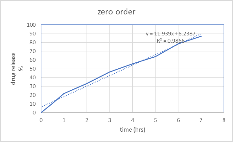
Figure 14. Zero order model plot
The data follows zero-order release kinetics if the plot is linear and shows the total percentage drug release over time. The slope of the line is equal to K0.
First order kinetics-
Drug release that is proportionate to the amount of drug still inside it is known as first order release; this means that the amount of drug released decreases with each passing unit of time. (Table 13,14) [66] The model is expressed using the following equation:
log Qt log Qo + K1 t/2.303
Where,
Qt is the amount of drug released in time t
Q is the initial amount of drug in the solution and
K1 is the first order release constant.
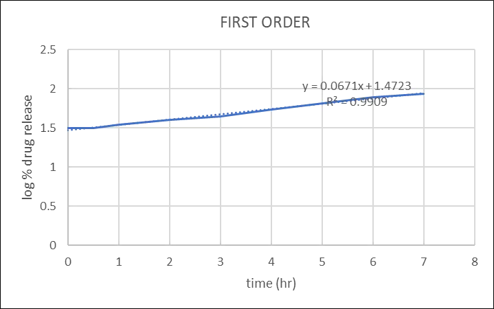
Figure 15. First order model plot
The release follows first order kinetics, as seen by the straight line that results from plotting the data as cumulative percent medication remaining vs time. Multiplying 2.303 by the slop value yields the constant K.
Higuchi's Model
It is among the oldest and most used models for explaining how drugs are released from solid and semi-solid matrices. It is predicated on the idea that drug release is a diffusion-controlled process and is based on Fick's law of diffusion. [67] The following equation can be used to illustrate the model:
Qt = Kh 1/2
Where,
Qt is the amount of drug released in time t
Kh is Higuchi's dissolution constant
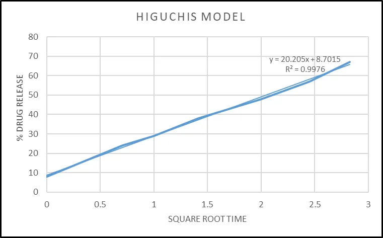
Figure 16. Higuchi model plot
Plotting the data using the equation, which measures cumulative drug release against the square root of time, produces a straight line, signifying that the drug was released through a diffusion mechanism. K is equivalent to the slop. (Table 13,14)
Korsmeyer-Peppas Model
A mathematical model called the Korsmeyer-Peppas model is frequently used to explain the kinetics of drug release from a polymeric matrix, particularly in situations where the release mechanism is unclear (such as in systems where both diffusion and erosion processes are at play). (Table 13,14) In pharmaceutical sciences, the model is especially helpful for describing how medications release from different dose forms, including pills, gels, and transdermal patches. [68] The following equation describes the model:
Mt/M∞=ktn
Where,
Mt/M∞ is the fraction of the drug released at time t, K is the release rate constant (which incorporates structural and geometric characteristics of the drug delivery system), n is the release exponent, which indicates the drug release mechanism, t is time.
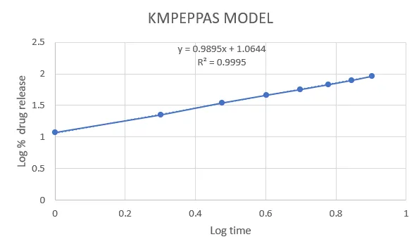
Figure 17. KMPeppas model plot
CONCLUSION:
A unique and effective method for treating rheumatoid arthritis has been developed through the creation of a transethosomal patch that contains capsaicin. Phospholipid, ethanol, and an edge stimulant are the components that make up transethosomes. Phospholipids forms the bilayer of the transethosomes, helping to encapsulate and protect the capsaicin. Also improves skin penetration due to its lipid compatibility with biological membranes. The lipid layer becomes more flexible and the size of the vesicles decreases when exposed to ethanol. Skin pore distortion and permeability can be assisted by an edge stimulator. Because of their fluidity and extremely small particle size, transethosomes are able to pass through many layers of skin. This delivery system significantly enhances the skin penetration of capsaicin while reducing its associated side effects like irritation. The formulation successfully achieves sustained drug release, providing prolonged therapeutic effects. The study’s findings suggest that transethosomes can be an effective carrier for capsaicin, making the patch a promising alternative to conventional topical treatments. The produced formulation was thoroughly examined for its physical appearance, drug content, entrapment efficiency, zeta potential, transmission electron microscopy, and in-vitro drug release studies. Results from this study clearly indicate that capsaicin loaded transethosomal patch is a viable substitute for the conventional dosage form. However, in order to determine whether or whether this method is helpful, more clinical study is required. After taking into account everything mentioned above, it was determined that the current research study's goal could be effectively met
REFERENCES

Ajay Sahu, Astha Verma, Chiranjeev Sahu, Madhuri Baghel, Girish Sahu*, Development & Evaluation of Capsaicin-Loaded Transethosomes for Enhanced Anti-Inflammatory Action in Rheumatoid Arthritis, Int. J. Med. Pharm. Sci., 2026, 2 (9), 653-675. https://doi.org/10.5281/zenodo.23020571
 10.5281/zenodo.23020571